Volume:16 Issue: 18 | Bio-protocol

Current Issue in 2026

Volume: 16, Issue: 18

left

Sep

20

Sep

5

Aug

20

Aug

5

Jul

20

Jul

5

Jun

20

Jun

5

May

20

May

5

Apr

20

Apr

5

Mar

20

Mar

5

Feb

20

Feb

5

Jan

20

Jan

5

right

Biochemistry

How to Perform a Tracer Displacement BRET Assay for the TRPML1 Ion Channel

How to Perform a Tracer Displacement BRET Assay for the TRPML1 Ion Channel

MC Micael R. Cunha
CC Carolina M. C. Catta-Preta
KM Katlin B. Massirer
RC Rafael M. Couñago
85 Views
Sep 20, 2026
The transient receptor mucolipin subtype 1 (TRPML1) is a ubiquitously expressed ion channel involved in lysosomal homeostasis. Recent pharmaceutical interest in developing agonist ligands has emerged due to beneficial effects in neurodegenerative diseases. The major high-throughput screening techniques to investigate this ion channel involve fluorescent calcium imaging and electrophysiology. Despite their high capacity for screening compounds, it is well known that both methods face hurdles, such as the need for expensive, specialized equipment. Here, we present a novel technique to screen for ligands of TRPML1 using a bioluminescence resonance energy transfer (BRET) assay. This assay consists of a target engagement assay in live cells, which permits the determination of binding constants between ligands and the target of interest in equilibrium or time-dependently. We employ a full-length TRPML1 C-terminally tagged with the small bioluminescent protein nanoluciferase. This ensures the correct localization of the ion channel in the lysosomal membrane and an optimal placement of the luciferase in the cytoplasm. We also developed a cell- and lysosome-permeable fluorescent BRET tracer that gives a BRET signal only when bound to the ion channel. This new protocol allows researchers worldwide to screen compounds that would interact with TRPML1 by using any plate reader with luminescent and fluorescence filters.
A SPAAC-Based Bioorthogonal Method for Verifying Protein Palmitoylation

A SPAAC-Based Bioorthogonal Method for Verifying Protein Palmitoylation

YZ Yaoyu Zhang
JL Jiahui Li
XP Xinxin Pei
TM Tengfei Ma
YW Yi Wang
BZ Buchang Zhang
CX Changzhi Xu
31 Views
Sep 20, 2026
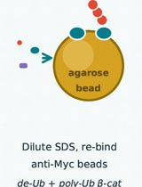
Palmitoylation is a crucial post-translational modification, and bioorthogonal chemistry based on azide-alkyne cycloaddition is typically used to verify protein palmitoylation. Traditional copper-catalyzed click chemistry (CuAAC) proceeds with fast kinetics and is widely used, but it requires a copper catalyst and suffers from copper-induced toxicity and nonspecific labeling. By contrast, strain-promoted click chemistry (SPAAC) has slower kinetics but is catalyst-free, offering high specificity, low cytotoxicity, and simple operation. However, SPAAC is mostly applied to live-cell labeling and imaging of known palmitoylated proteins in the field of palmitoylation, and its use in identifying novel palmitoylated proteins is still limited. Here, we present a SPAAC-based method for detecting endogenous protein palmitoylation. Compared with CuAAC, this method eliminates the need for copper catalysts and reducing agents, thereby simplifying the procedure and reducing reagent usage.

Bioinformatics and Computational Biology
Digital Quantification of Membrane DAB Immunohistochemical Staining in FFPE Cervical Cancer Tissues Using an Open-Source CellProfiler Pipeline

Digital Quantification of Membrane DAB Immunohistochemical Staining in FFPE Cervical Cancer Tissues Using an Open-Source CellProfiler Pipeline

KK Kripa Krishnakumar
NR Nuha Rasheed
NN Nadira Nithyanandan
RM Roopak Murali
LJ Leena Dennis Joseph
GV Ganesh Venkatraman
Rajesh Kumar Gandhirajan Rajesh Kumar Gandhirajan
101 Views
Sep 20, 2026
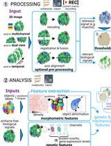
Immunohistochemistry (IHC) is a highly specific and widely used laboratory technique for assessing protein localization and expression in tissue samples. Interpretation of 3,3’ diamino benzidine (DAB)-based IHC is often based on observer-dependent manual scoring or traditional imaging software, which may show variability in DAB staining quantification. Furthermore, conventional image analysis tools often face limitations in precisely defining cell boundaries and quantifying membrane-specific signals. In this study, we present a standardized image analysis workflow using CellProfiler, an open-source software for image analysis for the quantification of membrane staining intensity in IHC images captured from slides prepared using formalin-fixed paraffin-embedded (FFPE) human cervical cancer tissue sections. The image analysis workflow was demonstrated using ASCT2 (SLC1A5), a membrane-localized amino acid transporter, as a representative biomarker for membrane-associated protein expression. This protocol involves image preprocessing, object identification, segmentation, and intensity measurement modules to distinguish cell membranes from cytoplasmic regions, enabling automated quantification of membrane intensity signals. The CellProfiler pipeline demonstrated improved accuracy in cell boundary identification and quantification of membrane-specific staining intensity. This is a rapid quantification process, since processing of each image only takes a few seconds; therefore, the analysis for 100 images can be performed within 10–15 min. This segmentation and quantification strategy is applicable to other membrane-based biomarkers after appropriate optimization of segmentation parameters. Following further minor modifications to the object identification modules, this pipeline can be used to detect and quantify cytoplasm- or nuclei-localized DAB-IHC markers across different tissue types. Overall, this protocol provides a standardized, user-friendly, and reproducible workflow for quantitative IHC image analysis that can be broadly applied to the study of protein biomarkers of different localizations, such as nuclei, cytoplasm, and cell membranes from different tissue types.

Cancer Biology

Homogeneous Time-Resolved Fluorescence-Based Assay to Screen ADP-Ribosyl Hydrolase Inhibitors

Homogeneous Time-Resolved Fluorescence-Based Assay to Screen ADP-Ribosyl Hydrolase Inhibitors

AY Alice Yu
DW Duo Wu
45 Views
Sep 20, 2026
ADP ribosylation (ADPr) is a crucial post-translational modification that plays a vital role in DNA damage repair. Catalyzed by ADP ribose polymerases using NAD+ as a substrate, ADPr activates DNA repair pathways rapidly, thereby maintaining genomic integrity. The involvement of ADP ribose hydrolases in this process is significant, as they hydrolyze PAR chains, facilitating the release of ADPr-modified proteins from DNA or other proteins, which is essential for subsequent DNA repair steps. This protocol outlines a high-throughput screening method for identifying inhibitors of ADP ribose hydrolases, utilizing His-Tb-conjugated and ADPr-modified His-ADP ribose polymerase as the signal donor, and GST-d2-conjugated GST-XRCC1 as the signal receptor. The detection of time-resolved fluorescence signals enables efficient evaluation of compounds with potential therapeutic activity against cancer.

Developmental Biology

Protocol for Mouse Embryonic Aorta–Gonad–Mesonephros (AGM) Region Frozen Sectioning and Immunofluorescence

Protocol for Mouse Embryonic Aorta–Gonad–Mesonephros (AGM) Region Frozen Sectioning and Immunofluorescence

YJ Yan Jia
SH Sixuan Huo
ML Mei Li
47 Views
Sep 20, 2026
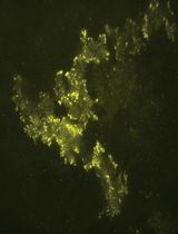
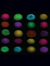
The aorta–gonad–mesonephros (AGM) region is the site where hematopoietic stem cells (HSCs) first emerge during development, and is therefore widely used to study in vivo hematopoiesis and to discover novel regulatory mechanisms. The endothelial-to-hematopoietic transition (EHT) process can be directly observed via immunofluorescence on frozen sections of the AGM region. However, the mouse AGM region is extremely delicate and lies deep within the embryo, between the notochord and the somatic mesoderm. Here, we present a step-by-step protocol covering embryo collection, fixation, dehydration, and embedding with a defined orientation, followed by frozen sectioning, immunofluorescence staining, and confocal imaging. The protocol is highly reproducible and easy to follow and provides clear instructions on orienting the embryo and anatomically locating AGM. By filling a technical gap, the protocol can enable researchers to reliably study HSC emergence and EHT in the mouse embryonic AGM.

Microbiology

Purification of MNase for Use in Ribosomal Profiling of High-Salinity Extremophiles

Purification of MNase for Use in Ribosomal Profiling of High-Salinity Extremophiles

PG Pavlina Gregorova
MI Matthew F. Isada
JD Jocelyne DiRuggiero
LS Peter L. Sarin
55 Views
Sep 20, 2026
Nucleases are key tools in molecular biology, enabling controlled nucleic acid digestion for applications such as ribosome profiling. Micrococcal nuclease (MNase) from Staphylococcus aureus is widely used as a tool in molecular biology and biochemistry, but its reduced activity under high-salt conditions necessitates higher enzyme input to achieve efficient digestion, increasing costs in studies of halophilic organisms. Here, we present an optimized protocol for the heterologous expression and purification of the recombinant staphylococcal MNase. The procedure enables reproducible production of a highly active, stable enzyme and incorporates an enzymatic activity assay to standardize batches to minimize variability. The resulting MNase exhibits robust activity in high-salt environments and remains stable during storage, providing a cost-effective and reliable alternative to commercial nucleases for ribosome profiling and related applications.
A NanoLuc-Based Protease Biosensor for Highly Sensitive Detection of Intracellular Protease Activity: Applications to Apoptosis and Coronavirus Infection

A NanoLuc-Based Protease Biosensor for Highly Sensitive Detection of Intracellular Protease Activity: Applications to Apoptosis and Coronavirus Infection

MA Masashi Arakawa
EM Eiji Morita
40 Views
Sep 20, 2026
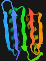
FlipNanoLuc is a highly sensitive protease biosensor based on the β-strand-flipping principle of NanoLuc luciferase, which is derived from Oplophorus gracilirostris. In the inactive configuration, one β-strand of NanoLuc is repositioned, thereby suppressing luciferase activity. Upon cleavage of the embedded protease recognition sequence by a target protease, the flipped β-strand is released, and luciferase activity is reconstituted. Incorporation of the LgBiT fragment (NanoBiT technology) yields strong luminescent output once the reporter is reconstituted, whereas the CL1-PEST1 degradation tag lowers background luminescence by promoting the degradation of the uncleaved, inactive form; together, these two modifications widen the dynamic range. A firefly luciferase normalization cassette connected via a P2A self-cleaving peptide is encoded in the same reporter plasmid, thereby eliminating the need for separate co-transfection. Because the readout directly reports intracellular protease activity in living cells, the system is suitable for detecting protease activation during apoptosis or viral infection and, in principle, for evaluating protease inhibitors and antiviral compounds. This protocol describes the following: (1) generation of HEK293T cells stably expressing FlipNanoLuc by retroviral transduction; (2) validation of reporter activity by protease overexpression; (3) detection of apoptosis using staurosporine; (4) detection of human coronavirus OC43 infection; and (5) detection of SARS-CoV-2 infection in BHK cells stably expressing hACE2. All luminescence assays employ dual-luciferase normalization and are compatible with standard 384-well plate readers.

Neuroscience

Proximity Labeling in Caenorhabditis elegans to detect Neuronal Proteins During Memory Formation

Proximity Labeling in Caenorhabditis elegans to detect Neuronal Proteins During Memory Formation

AR Aelon Rahmani
YC Yee Lian Chew
105 Views
Sep 20, 2026
Memory is a fundamental process, regulated by protein–protein interactions within neuronal proteome networks. Learning-dependent changes in specific brain regions important for memory have been detected by mass spectrometry, by comparing proteins from animals trained to learn with mock-trained controls. Detection through this method relies on relative protein abundance; brain dissection is readily available for macroscopic animals to spatially control protein identification by mass spectrometry. In the nematode C. elegans, however, such spatial control is limited due to its microscopic size, hindering its utilization in proteomics. A protocol to address this limitation would strengthen an already excellent model to study memory, given that many proteins for learning are evolutionarily conserved in the worm and single-cell expression is uniquely defined across all 302 neurons. We modified existing protocols to enable (i) proximity labeling detection of neuronal proteins in C. elegans and (ii) high-throughput enrichment of these proteins from >3,000 whole worm bodies simultaneously, to assess trained vs. mock-trained proteomes. This involved the biotin ligase enzyme TurboID, which promiscuously labels nearby proteins with its substrate biotin. Enzyme expression was transgenically restricted to the nervous system, and biotin supplementation was limited to the training (or mock training) period in a classical (gustatory) conditioning paradigm. Labeled proteins were enriched by pull-down using streptavidin, which has a high binding affinity to biotin, and then processed for mass spectrometry runs and qualitative data analysis. This protocol is uniquely advantageous in that it minimizes proteins present before a temporal window of interest (training/mock training), improving the detection of lowly abundant proteins from a specific tissue in the worm (neurons). We have demonstrated that the protocol can sufficiently detect novel learning regulators, thus providing a useful framework to interrogate proteomes in microscopic brains.
An In Vitro Model to Study Drugs That Affect Macrophage Adhesion to Murine Brain Endothelial Cells After Proinflammatory Insults of LPS and Pilocarpine

An In Vitro Model to Study Drugs That Affect Macrophage Adhesion to Murine Brain Endothelial Cells After Proinflammatory Insults of LPS and Pilocarpine

AR Abriel J. Rivera-Rivera
PG Paola N. Gracia-Ayala
AV Arot L. Velázquez Pulliza
AM Antonio H. Martins
YF Yancy Ferrer-Acosta
60 Views
Sep 20, 2026
Neuroinflammation disrupts blood–brain barrier (BBB) integrity, promoting leukocyte recruitment into the central nervous system and contributing to the progression of neurological disorders. This protocol describes a reproducible macrophage adhesion assay to evaluate interactions between immune cells and brain endothelial cells and to screen compounds with potential anti-inflammatory activity. Murine brain endothelial cells (bEnd.3) were cultured to confluency and exposed to inflammatory stimuli, such as lipopolysaccharide (LPS) or pilocarpine, a cholinergic muscarinic receptor agonist reported to induce inflammatory responses through seizure-associated neuroinflammatory mechanisms, in the presence or absence of candidate therapeutic compounds. In these studies, the natural flavonoid quercetin and the synthetic alkyl-lysophospholipid edelfosine were tested for their effects on macrophage adhesion. After 48 h of treatment, fluorescently labeled murine macrophages (RAW 264.7) were added to the endothelial monolayer, and adherent cells were quantified by fluorescence microscopy. The assay was validated using dexamethasone as an anti-inflammatory control and inflammatory stimulation with LPS or pilocarpine. As expected, dexamethasone reduced macrophage adhesion, whereas both LPS and pilocarpine significantly increased adhesion, demonstrating the assay's sensitivity to changes in endothelial inflammatory status. Overall, this protocol provides a reliable and accessible platform for investigating endothelial–immune cell interactions under neuroinflammatory conditions and for evaluating therapeutic compounds that may preserve BBB function and reduce inflammatory cell recruitment in neurological disease models.
Protecting Against Cytoplasmic Protein Aggregates with Cytoplasmic PML Variants

Protecting Against Cytoplasmic Protein Aggregates with Cytoplasmic PML Variants

JZ Jia-Xin Zhu
YX Yuchen Xia
JL Jiaqi Liu
LC Li Cao
SH Steven X. Hou
YW Yang Wang
43 Views
Sep 20, 2026
Cytoplasmic protein aggregation is a defining feature of multiple neurodegenerative diseases, including amyotrophic lateral sclerosis, frontotemporal dementia, Huntington’s disease, and certain forms of motor neuron disease. Recent evidence indicates that promyelocytic leukemia protein (PML) and engineered PML-derived variants can act as versatile aggregate-remodeling factors. In particular, cytoplasmically redirected PML variants recognize pathological cytoplasmic inclusions and promote their clearance. Here, we describe a protocol to generate and validate two engineered cytoplasmic PML variants: full-length mPML, which is redirected to the cytoplasm by disruption of its nuclear localization sequence, and the truncated mPMLΔRBC variant, which lacks the RING, B-box, and coiled-coil domain but retains aggregate-reducing activity. The protocol integrates fluorescence-based imaging, bimolecular fluorescence complementation, detergent-soluble/insoluble fractionation, and validation in primary rat cortical neurons. This workflow provides a practical platform for assessing cytoplasmic aggregate burden and for comparing the aggregate-remodeling activities of PML-derived constructs. It can also be adapted to other disease-associated aggregation-prone proteins, including TDP-43, SOD1, FUS, tau, polyGA, and polyQ-expanded proteins.

Plant Science

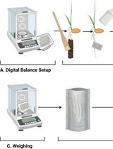
A Protocol for Accelerating Homozygous Line Screening in Arabidopsis thaliana

A Protocol for Accelerating Homozygous Line Screening in Arabidopsis thaliana

QZ Qiuyu Zhang
YW Yifan Wang
CJ Chengqiao Jiang
XZ Xueqin Zhou
WZ Wenqing Zhong
GY Guanghui Yu
88 Views
Sep 20, 2026
Obtaining homozygous mutant and transgenic lines is a critical yet time-consuming step in Arabidopsis thaliana research. Conventional breeding procedures require seeds to undergo complete maturation and natural desiccation before harvest, followed by cold stratification to overcome seed dormancy. This process substantially prolongs generation turnover and delays genetic screening. Here, we describe a rapid germination strategy based on the use of partially dehydrated seeds collected approximately 15 days after pollination (DAP). At this developmental stage, embryos have reached physiological maturity, while the seeds have not yet entered deep desiccation-induced dormancy. After surface sterilization and short-term cold treatment (2–3 days at 4 °C), these seeds readily germinate on 1/2 MS medium and develop into normal seedlings. By bypassing the prolonged maturation and dormancy phases associated with naturally dried seeds, this protocol shortens each generation cycle by approximately 1–2 weeks. The method is particularly useful for accelerating the propagation of transgenic materials and the identification of homozygous mutant lines in Arabidopsis.

Stem Cell

Isolation of Human Umbilical Cord Blood Hematopoietic Stem Cells and Directed Differentiation Into Megakaryocytes

Isolation of Human Umbilical Cord Blood Hematopoietic Stem Cells and Directed Differentiation Into Megakaryocytes

WX Wenjun Xia
ZM Zeqing Miao
WZ Weiwei Zhang
ZL Zhixia Liu
HW Huang Wu
YF Yahan Fan
59 Views
Sep 20, 2026
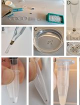
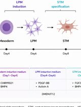
Platelets originate from megakaryocytes, whose generation involves a series of biological processes including directed differentiation, proliferation, polyploidization, and maturation of hematopoietic stem cells. Abnormalities in megakaryocyte development and maturation can lead to quantitative and functional defects in platelets, thereby contributing to hemostatic or thrombotic disorders as well as the development of malignancies. Investigating megakaryocyte development and maturation and platelet production can provide important theoretical foundations for the diagnosis and treatment of thrombocytopenia, thrombotic diseases, and myeloproliferative neoplasms. Currently, there are three main clinical sources of hematopoietic stem cells (HSCs): bone marrow (BM), peripheral blood (PBSC), and umbilical cord blood (UCB). Among these, umbilical cord blood (UCB)-derived HSCs, due to their higher differentiation efficiency and stronger proliferative capacity, are the preferred starting cell source for studying megakaryocyte (MK) development and maturation and the mechanisms of platelet production. This article describes a detailed protocol covering all necessary steps for isolating CD34+ hematopoietic stem cells from umbilical cord blood, followed by in vitro induction culture with stem cell factor (SCF) and thrombopoietin (TPO) to generate mature megakaryocytes that highly express early megakaryocyte markers (CD41a, CD61) and late maturation markers (CD42a, CD42b). This protocol provides an effective tool for studying megakaryocyte development and platelet production and holds potential value for application in research on megakaryocyte-related diseases.
Probing the Luminal Compartment of 3D Organoids via Particle Tracking Microrheology

Probing the Luminal Compartment of 3D Organoids via Particle Tracking Microrheology

Katrina N. Lyon Katrina N. Lyon
BS Barkan Sidar
CD Cameron Dudiak
GJ Grace Jordan
DB Diane Bimczok
56 Views
Sep 20, 2026
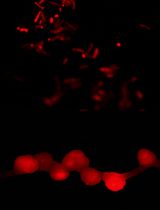
The mucus layer lining the human stomach is a critical barrier that protects the underlying epithelium from gastric acid and harmful pathogens such as Helicobacter pylori. The efficacy of this barrier relies on the structural integrity of the mucus, which is determined by various biochemical and biophysical features. Human gastric organoids—3D cellular models that resemble the stomach—contain mucus and have been used to investigate gastric disease. The luminal compartment of three-dimensional epithelial organoids represents a physiologically relevant but experimentally inaccessible microenvironment. In gastric organoids, luminal accumulation of mucus creates a confined viscoelastic hydrogel that mimics native gastric mucus. However, the small volume and topological confinement of organoids preclude conventional bulk rheometry. Here, we describe a particle tracking microrheology (PTM) protocol to measure the viscoelastic properties of the mucus within intact organoid lumina following microinjection of fluorescent microspheres. High-speed fluorescence imaging and particle trajectory analysis enable the quantification of viscous and elastic properties of the mucus through calculation of mean squared displacement (MSD), diffusive scaling exponent (alpha), and frequency-dependent storage (G’) and loss (G’’) moduli. This method enables rheological measurements in nanoliter-scale compartments without disrupting organoid architecture. We further discuss the impact of mucus heterogeneity and microstructure on scale-dependent mechanical behavior. This protocol is broadly applicable to other organoid systems and can be adapted to Transwell or organ-on-chip platforms for in situ luminal measurements.