Improve Research Reproducibility A Bio-protocol resource

- Home
- Protocols

-

Preprint
Human Cytomegalovirus (HCMV) infection of CD14+ monocytes and reactivation
Last updated date: Mar 3, 2021 Views: 1047 Forks: 0
Infection
- CD14+ monocytes purified by MACS purification from primary PBMCs, are grown in concentration of 1-2*106 cells/ml, in x-vivo-15 media in 15ml round bottom tubes (Falcon 352059). The volume should be 0.5ml-3ml per tube and tubes should be placed at a 45⁰ angle, to reduce clumping.
Cells may be incubated over-night at 37⁰C following purification, prior to infection. - Before infection: pipette the cells gently to remove from tube walls and to dislodge clumps and count the cells.
- Spin the cells (300xg, 10min) and remove most of the media (leaving up to 0.5ml). Thaw an appropriate amount of HCMV immediately before infection, add the virus to the cells and mix gently and thoroughly by pipetting.
The use of a GFP tagged virus allows for convenient tracking of plaques following reactivation. - Incubate the cells with the virus for 3 hours. During this incubation tap the tube gently every 1hr to mix the cells and reduce cell clumping.
- Wash the virus at least 2 times by filling the tube with media, pelleting the cells (300g, 10min) and aspirating most of the sup. Be careful not to aspirate the cells.
- After the last wash, bring the cells to a concentration of 1*106 cells/ml with fresh media, and place 0.5ml-3ml in each tube. Mix the cells by gently pipetting 20 times.
- Incubate at 37⁰C (keep the tubes tilted at a 45⁰ angle).
HCMV reactivation
Reactivation by co-culture with fibroblasts:
- Count cells and plate 105 cells per well of 96 well plate. Let cells adhere for 3 hours, and replace media with 100µl of x-vivo-15 media.
- For negative control: at the same time cells are taken for reactivation, set aside 105 infected monocytes in 100µl media, to be used as negative control. Lyse cells by snap freezing and keep in (-80⁰C).
- Add to each well 104 human fibroblasts, in 100µl DMEM media (10% FBS, 1%pen-Strep, 1% L-glutamine).
- For negative control: plate 104 human fibroblasts in empty well, let cells adhere and add the latent cells lysate (described in step 2) as negative control.
- On the next day, partially replace the media by taking out 100µl and adding 100 µl of fresh DMEM. Over the next few days monitor the co-culture to ensure fibroblast viability and gradually replace by replacing 100µl of media with fresh DMEM every 2-3 days.
- Over the next 10-14 days, view the co-culture under a fluorescent microscope and monitor for appearance of GFP positive plaques.
Differentiation to DCs:
- Count cells and plate 105 cells per well of 96 well plate in 200µl of x-vivo-15 media supplemented with:
- granulocyte-macrophage CSF (Peprotech) at 1,000 U/ml
- interleukin-4 (Peprotech) at 1,000 U/ml
- Incubate cells for 5 days.
- Stimulate cells by replenishing media and adding 500 ng/ml of LPS (Sigma), incubate for 48 hours.
- Overlay infected cells with fibroblasts and monitor for appearance of GFP positive plaques (as described in steps 3-6 of the previous section).
Reagents used
| Reagent | Reagent type | Source | Cat# |
| X-vivo-15 | Cell media | Lonza | BE02-060F |
| granulocyte-macrophage CSF | cytokine | Peprotech | 300-03-20 |
| interleukin-4 | cytokine | Peprotech | 200-04-20 |
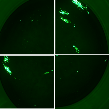
Example of plaques formed on fibroblasts co-cultured with HCMV infected CD14+ monocytes (TB40 strain with a GFP reporter, 3), following differentiation to DCs.
Copyright: Content may be subjected to copyright.
How to cite:
Readers should cite both the Bio-protocol preprint and the original research article where this protocol was used:
- Shnayder, M, Stern-Ginossar, N and Schwartz, M(2021). Human Cytomegalovirus (HCMV) infection of CD14+ monocytes and reactivation. Bio-protocol Preprint. bio-protocol.org/prep902.
- Shnayder, M., Nachshon, A., Rozman, B., Bernshtein, B., Lavi, M., Fein, N., Poole, E., Avdic, S., Blyth, E., Gottlieb, D., Abendroth, A., Slobedman, B., Sinclair, J., Stern-Ginossar, N. and Schwartz, M.(2020). Single cell analysis reveals human cytomegalovirus drives latently infected cells towards an anergic-like monocyte state. eLife. DOI: 10.7554/eLife.52168
Category
Do you have any questions about this protocol?
Post your question to gather feedback from the community. We will also invite the authors of this article to respond.
0 Q&A
This protocol preprint was submitted via the "Request
a Protocol" track.