
- Home
- Protocols

-

Measurement of thickness of the inner mucus layer
Last updated date: Feb 24, 2021 Views: 934 Forks: 0
1. Collect the large intestine containing luminal contents by ligating ileum and rectum.
2. Samples were fixed in Carnoy's solution (FUJIFILM Wako Junyaku) for 2 hours, transferred into 100% ethanol, embedded in paraffin, and sectioned (8 micron).
3. Paraffin-embedded sections were dewaxed and hydrated.
4. Sections were incubated with Target Retrieval Solution Citrate pH6 (DAKO) for 20 minutes at 105 degrees Celsius using autoclave.
5. Rinse with phosphate-buffered saline (PBS) twice.
6. Remove PBS and add goat serum in Block Ace (DS Pharma Biomedical) for 15 minutes at room temperature.
7. Rinse with PBS.
8. Incubate with primary antibodies (Abs) in Tris-buffered Saline (TBS) overnight at 4 degrees Celsius.
9. Wash three times with PBS.
10. Incubate with secondary Abs in TBS for 1 hour at room temperature in the dark.
11. Remove secondary Abs and rinse three times with PBS.
12. Nuclear staining was done with 1 μg/mL of 4',6-diamidino-2-phenylindole (DAPI: Dojindo Laboratories) for 15 minutes at room temperature in the dark.
13. Rinse with PBS.
14. Secure with cover glass.
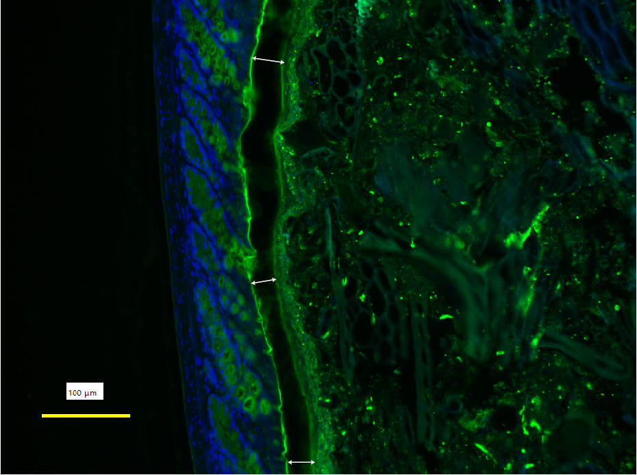
15. Images of inner mucus layer was acquired at 10 different sites for each sample, using Keyence BZ-X700 fluorescence microscope (Keyence)
16. Thickness of inner mucus layer was measured at three different points for each image. We made efforts to choose the sites where the inner mucus layer was preserved as much as possible, and three measure points were set to maintain some distance each other (Figure). Finally, the average of thirty measurement values of each sample was used as the thickness of inner mucus layer.
Primary Antibody: Rabbit Anti-mucin2 polyclonal antibodies (clone H-300: Santa Cruz Biotechnology)
Secondary Antibody: AlexaFluor 488-conjugated goat Anti-Rabbit IgG antibodies (A11034; Invitrogen)
- Ara, T, Hashimoto, D and Teshima, T(2021). Measurement of thickness of the inner mucus layer. Bio-protocol Preprint. bio-protocol.org/prep871.
- Ara, T., Hashimoto, D., Hayase, E., Noizat, C., Kikuchi, R., Hasegawa, Y., Matsuda, K., Ono, S., Matsuno, Y., Ebata, K., Ogasawara, R., Takahashi, S., Ohigashi, H., Yokoyama, E., Matsuo, K., Sugita, J., Onozawa, M., Okumura, R., Takeda, K. and Teshima, T.(2020). Intestinal goblet cells protect against GVHD after allogeneic stem cell transplantation via Lypd8 . Science Translational Medicine 12(550). DOI: 10.1126/scitranslmed.aaw0720
Do you have any questions about this protocol?
Post your question to gather feedback from the community. We will also invite the authors of this article to respond.