Improve Research Reproducibility A Bio-protocol resource

- Home
- Protocols

-

Preprint
Collection of in vivo capacitated sperm
Last updated date: Jan 19, 2021 Views: 1225 Forks: 0
Collection of in vivo capacitated sperm
- Timed mating. For timed mating, the hyperstimulated (super-ovulated) or non- hyperstimulated female mice can be used. Hyperstimulation procedure increases the probability of the successful timed mating. Alternatively, the vaginal smears can be used to determine the female cyclephase to increasethe probability of the timed mating success.
- Introduce a male to a femalemouse in the single cage during the last dark hour of the 12 hours long dark cycle. Avoid extensive animals light exposure.
- Check the presence of vaginal plugs as soon as the light cycle begins. Females with plugs can be used for experiment, females without plugs can be used in another timed mating runs.
- Sperm isolation. Sacrifice the female mice with the confirmed vaginal plugs after desired time of sperm presence (capacitation) inside the female reproductive tract (e.g., 3h or 8h post-coitus).
- Dissect the oviducts and placed them to 0.3 ml dropletof M2 medium (individually) and further dissect them carefully to ampullar, middle isthmus and UTJ part.
- Immediately place each part to an individual 100ul droplet in mini-Petri dish and subject them to gently release sperm.
- Slides preparation. Observe the droplets (optimally by 3 persons) under microscopes (e.g. for 10 min) and pick individual sperm by glass micropipette and placed to 10ul PBS droplet on the fibronectin coated 8-well Teflon slides (Figure 1; on average about 10 sperm per sample shouldbe obtained at least). If too much tissue material was transferred by micro pipette, the PBS washing step can be added.
- Subject the rest of the material to finer dissection, remove macroscopic pieces manually, mix the rest of the material with PBS (material/PBS 1:10) and smear it on the fibronectin coated 3-well Teflon slides (Figure 2; ~30-50ul per a well).
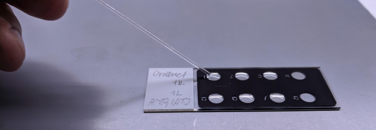
Figure 1: Sperm transfer by glass micro pipette into 8 well Teflon micro droplet slides (Thermo Scientific™ PTFE Diagnostic Slides #10727951).
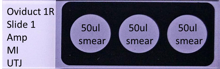
Figure 2: 3 well Teflon smeared slides setup (Thermo Scientific™ PTFE Diagnostic Slides #10632391).
Copyright: Content may be subjected to copyright.
How to cite:
Readers should cite both the Bio-protocol preprint and the original research article where this protocol was used:
- Ded, L and Chung, J(2021). Collection of in vivo capacitated sperm. Bio-protocol Preprint. bio-protocol.org/prep763.
- Ded, L., Hwang, J. Y., Miki, K., Shi, H. F. and Chung, J.(2020). 3D in situ imaging of the female reproductive tract reveals molecular signatures of fertilizing spermatozoa in mice. eLife. DOI: 10.7554/eLife.62043
Category
Do you have any questions about this protocol?
Post your question to gather feedback from the community. We will also invite the authors of this article to respond.
0 Q&A
This protocol preprint was submitted via the "Request
a Protocol" track.
