
- Home
- Protocols

-

APEX2 reaction
Last updated date: Dec 2, 2020 Views: 986 Forks: 0
1. Incubate HEK293FT cells (ideally in low passage) for seven splits with the respective “Heavy” or “Light” SILAC media (SILAC Protein quantification Kit, Thermo Scientific,
#A33969).
- Note: is important that the media is amino acid free and the FBS is dialyzed.
- Note: The “Light” group will be exposed to L-Lysine-2HCl and L-Arginine-HCl, while the “Heavy” group will be exposed to 13C6 L-Lysine-2HCl and 13C6 L-Arginine-HCl.
- Note: It is recommended to check the “Heavy” amino acid incorporation using mass spectrometry after this step.
2. Coat T150 cm flasks with Matrigel
3. Cells were plated to two T150 cm flasks per condition (25 million cells to “Light”, 35 million cells to “Heavy” in each flask) and transfected with 3xFLAG-APEX2-NES and 3xFLAG-APEX2-LRRK2
- 8.5 mg of NES and 17 uL Lipofectamine2000 /T150 "Light
- 34 mg of LRRK2 and 68 uL Lipofectamine2000/ T150 “heavy”
4. 24 hours later cells were treated with biotin-phenol 500 uM in 30 mL of complete media for 30 min
5. Add 1 mM H2O2 quickly and agitate for 1 min at RT
6. Remove medium and wash 2x with quencher solution (TBS supplemented with 1 mM CaCl2, 10 mM sodium ascorbate, 1 mM sodium azide, and 1 mM Trolox) for 1 min each
7. Wash 2x with PBS
8. Was 1x with quencher
9. Add 10 mL of quencher and collect cells by pipetting
10. Centrifuge at 3000g for 10 min at 4oC
11. Resuspend cells with RIPA lysis buffer (plus protease inhibitor and quenchers), 800 uL/ each. Incubate on ice for 10 min
12. Clear lysate by centrifuging at 15000g for 10 min, at 4oC
13. Measure protein concentration with the "660 assay” and add 1:1 per replicate.
14. Wash 500 uL Magnetic Streptavidin beads/ replicate with 1 mL RIPA
15. Incubate combined Heavy and Light 1:1 per replicate in the beads, rotate for 1 h at RT
- Note: the combined amount of protein is around 5 mg per replicate.
16. Remove supernatant and wash with RIPA (2x)
17. Wash sequentially with KCl (1M), Na2CO3 (0.1M) and Urea (2M) in 0.1M TRIS buffer
18. Wash with RIPA (2x)
19. Elute the biotinylated proteins by boiling the beads in 45µl of 2x protein loading buffer supplemented with 2 mM biotin and B-Mercaptoethanol for 10 min.
20. Vortex briefly, cool the tubes and briefly spin them down
21. Run a 12-well gel, loading L&H (1:1) per lane, 150V for 1h
22. Stain with GelCode Blue Stain for 30 min
23. Wash with pure water overnight
24. Submit samples for mass spectrometry
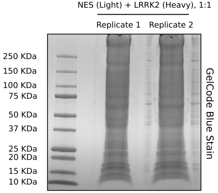
Image of the GelCode Blue Stain of the two replicates before submitting the gel to mass spectrometry.
- Bonet-Ponce, L and Cookson, M(2020). APEX2 reaction. Bio-protocol Preprint. bio-protocol.org/prep669.
- Bonet-Ponce, L., Beilina, A., Williamson, C. D., Lindberg, E., Kluss, J. H., Saez-Atienzar, S., Landeck, N., Kumaran, R., Mamais, A., Bleck, C. K. E., Li, Y. and Cookson, M. R.(2020). LRRK2 mediates tubulation and vesicle sorting from lysosomes. Science Advances 6(46). DOI: 10.1126/sciadv.abb2454
Category
Do you have any questions about this protocol?
Post your question to gather feedback from the community. We will also invite the authors of this article to respond.