
- Home
- Protocols

-

Proliferation assay in vivo
Last updated date: Nov 27, 2020 Views: 1227 Forks: 0
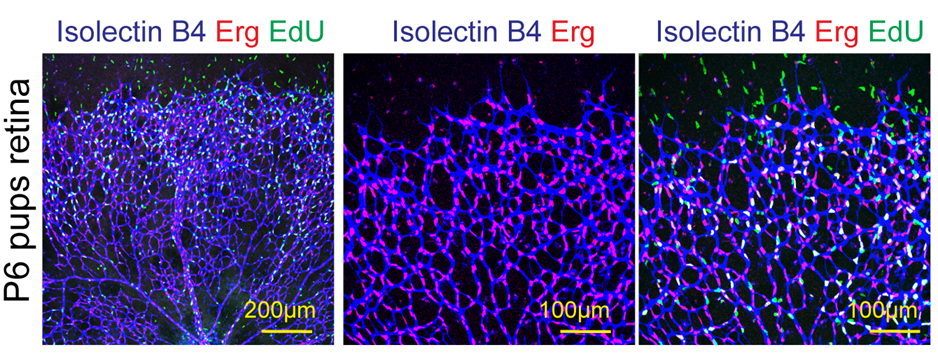
EdU (5-ethynyl-2'-deoxyuridine) based cell proliferation assay in vivo (Sivaraj et al., 2020)
1. For retina, P6 pups received 100 µg of EdU in 50 µl (2mg/ml) per 3g of body weight intraperitoneally for 2 hr.
2. P6 pups were sacrificed and whole eyes were harvested and fixed in ice cold 4% PFA for 2 hr.
3. After fixation, retinas were dissected and processed as described previously (Pitulescu et al., 2010).
4. After two washes with ice-cold PBS, samples were incubated in blocking buffer for 1 hr on rotating shaker.
5. Next, blocking buffer was replaced by Pblec buffer.
6. Isolectin-B4 (IB4; Vector, Cat#Ab200839) or rabbit monoclonal anti-ERG (Abcam, Cat# ab22552) were diluted in Pblec buffer and each retina was incubated in 100 μl of solution overnight at 4°C.
7. Next, samples were washed five times in incubation buffer (diluted blocking buffer 1:1 in PBS) and incubated with the appropriate Alexa Fluor488, 546 and 594-conjugated secondary antibodies for 2 hr at room temperature.
8. Retinas were washed three times with ice-cold PBS.
9. Retinas were incubated with Click-it EdU stock solutions for 30 minutes at room temperature, protected from light.
10. Retinas were washed three-five times with ice-cold blocking buffer.
11. Later, retinas were incubated with DAPI (4′,6-diamidino-2-phenylindole) (1 µg/ml) in blocking buffer for 10 minutes at room temperature, protected from light.
12. Finally, retinas were washed three- five times with ice-cold PBS and mounted under a stereomicroscope.
Solutions:
1. EdU (5-ethynyl-2'-deoxyuridine) 1mg/ml (i.e. 2mg EdU, dissolve in 100ul DMSO by vortexing, add 900ul of PBS to a final volume of 1ml).
2. Blocking buffer (1%BSA, 1% Triton X-100, 3% heat inactivated donkey serum in PBS).
3. Pblec buffer (1 mM CaCl2, 1 mM MgCl2, 0.1 mM MnCl2, 0.1% Triton X-100 in PBS).
4. Click-it EdU stock solutions (for 100ul of staining solution mix):
| 10x Click-it EdU reaction buffer | 10 µl |
| CuSO4 | 4 µl |
| Alexa Fluor Azide 647 | 0.25 µl |
| 10x Click/it EdU buffer additive | 10 µl |
| water | 75.75 µl |
| Click-it EdU stock solutions | 100 µl |
References
1. Pitulescu, M.E., Schmidt, I., Benedito, R., and Adams, R.H. (2010). Inducible gene targeting in the neonatal vasculature and analysis of retinal angiogenesis in mice. Nature protocols 5, 1518-1534.
2. Sivaraj, K.K., Dharmalingam, B., Mohanakrishnan, V., Jeong, H.W., Kato, K., Schroder, S., Adams, S., Koh, G.Y., and Adams, R.H. (2020). YAP1 and TAZ negatively control bone angiogenesis by limiting hypoxia-inducible factor signaling in endothelial cells. eLife 9.
- Sivaraj, K and Adams, R(2020). Proliferation assay in vivo. Bio-protocol Preprint. bio-protocol.org/prep658.
- Sivaraj, K. K., Dharmalingam, B., Mohanakrishnan, V., Jeong, H., Kato, K., Schröder, S., Adams, S., Koh, G. Y. and Adams, R. H.(2020). YAP1 and TAZ negatively control bone angiogenesis by limiting hypoxia-inducible factor signaling in endothelial cells. eLife. DOI: 10.7554/eLife.50770
Category
Do you have any questions about this protocol?
Post your question to gather feedback from the community. We will also invite the authors of this article to respond.