
- Home
- Protocols

-

WB analysis
Last updated date: Nov 12, 2020 Views: 1236 Forks: 0
Western Blot for Detecting the Activation of GSDMD
Abstract
Gasdermin D (GSDMD), which mediates a regulated lytic cell death mode termed pyroptosis,
is identified as a substrate for murine caspase-1 and caspase-11 and human caspase-1, caspase-4, and caspase-5. Pyroptosis is a type of proinflammatory programmed cell death mediated by caspase-1–cleaved GSDMD (N-terminal fragment), which binds to the membrane to form membrane pores and promote the release of proinflammatory mediators, notably IL-1b.
Materials and Reagents
- C57BL/6J Mice (The animal center of Wuhan University)
- Mouse MCSF (Peprotech, catalog number: 315-02-50)
- Penicillin (50 U/ml)/Streptomycin (50 mg/ml) (Thermo Fisher Scientific, GibcoTM, catalog number: 15140-122)
- DMEM (Gibco, catalog number: C11995500BT)
- RPMI 1640 medium (Gibco, catalog number: C11875500BT)
- 70% ethanol
- Fetal bovine serum (FBS) (Gibco, catalog number: 10099141)
- Rabbit polyclonal antibody anti-GSDMD (This antibody was a gift from Dr. Feng Shao, National Institute of Biological Sciences, Beijing, China)
- Goat Anti-Rabbit IgG (H+L), HRP conjugate antibody (Proteintech, catalog number: SA00001-2)
- Phosphate buffered saline (PBS)
- PMSF (Roche, catalog number: 10837091001)
- RIPA Lysis Buffer (Beyotime, catalog number: P0013D)
- BCA Protein Assay Kit (Beyotime, catalog number: P0012S)
- Precision plus protein dual color standards (Bio-rad, catalog number: 1610394)
- Methanol (Sinopharm Chemical Reagent Co.,Ltd, catalog number: CFSR-80080418)
- Tris base (Sigma, catalog number: T1503)
- Tween-20 (Sigma, catalog number: P9416)
- SDS (Sigma, catalog number: 436143)
- TEMED (Sigma, catalog number: T9281)
- Glycine (Sigma, catalog number: G8898)
- PVDF membrane (Millipore, 0.22mm, catalog number: IPVH00010)
- WesternBright ECL kit (Advansta, K-12045-D50)
- ACK lysis buffer (see Recipes)
- 5x Loading buffer (see Recipes)
- 1x Tris-Glycine running buffer (see Recipes)
- 1x TBST buffer (see Recipes)
- Transfer buffer (see Recipes)
- Blocking buffer (see Recipes)
Equipment
- Microcentrifuges (Eppendorf, model: 5702/ 5424)
- 37 °C, 5% CO2 incubator (Thermo Fisher Scientific, model: BB150)
- UVP E3 Image System
- Bio-Rad TC10 Automated Cell Counter
- Counting Slides (Bio-Rad, catalog number: 145-0011)
- Equipment for SDS-PAGE and Western blot
Software
UVP software (Version: 8.0.1)
Procedure
A. Bone marrow derived macrophages preparation and bacterial infection
- Euthanize mouse by CO2 and cervical dislocation in accordance with IACUC protocol. Sterilize within 70% ethanol.
- Remove femur and tibia from mouse and remove excess muscles while maintaining bone integrity in a biosafety cabinet, then put bones in 60-mm cell culture dish. Keep bones in 70% ethanol for 2 min, discard 70% ethanol and add RPMI 1640 medium (without FBS) to dilute residual ethanol. Then transfer them to the 10-cm cell culture dish containing 10 ml 1640 RIPM medium (without FBS).
- For each femur and tibia, cut each end using sterile scissors. Cut approximately 1 mm off of each head of the bone.
- Prepare a 1 ml syringe with needle, fill syringe with 0.5 ml of 1640 RIPM medium, hold the bone with forceps and insert needle into the opening of the bone. Repeat this step until bone marrow expel completely from bone, and pipet bone marrow to single cells.
- Filter the solution with a 40 μm cell strainer into a fresh 15 ml conical tube to make single cell suspension.
- Centrifuge cells at room temperature at 300 x g for 5 min, and remove supernatant.
- Add 1 ml of ACK lysis buffer and incubate for 1min. Immediately dd 9 ml of RPMI 1640 medium.
- Centrifuge cells at room temperature at 300 x g for 5 min.
- Remove media and resuspend cells in approximately 10 ml of sterile PBS.
- Cell concentration was measured by cell counter.
- Centrifuge cells at room temperature at 300 x g for 5 min, discard PBS and cell concentration are adjusted to 1 x 10⁶ cells/ ml by suspension in complete DMEM medium (containing 10% FBS) supplemented with 30 ng/ml of M-CSF and penicillin (50 U/ml)/Streptomycin (50 mg/ml) (10 ml in 10-cm cell dish). Allow cells to differentiate in 37 °C 5% CO2 incubator. Consider this step as Day 1.
- On Day 3, replace fresh complete DMEM medium (containing 10% FBS) supplemented with 30 ng/ml of M-CSF and penicillin (50 U/ml)/Streptomycin (50 mg/ml).
- On Day 5, ~90% adherent macrophages will be observed (Figure 1). Scrape the BMDMs very gently with cell scraper and pipet to single cell.
- Centrifuge cells at room temperature at 300 x g for 5 min.
- Remove media and resuspend cells in 10 ml of complete DMEM containing 30 ng/ml per well of mouse M-CSF and penicillin (50 U/ml)/Streptomycin (50 mg/ml).
- Count cells using cell counter.
- Seed a 6-well cell culture treated plate at 1 x 10⁶ cells per well in a total volume of 2 ml per well.
- The BMDMs were infected at 10 multiplicity of infection (MOI) with Mycobacterium bovis BCG for different times.
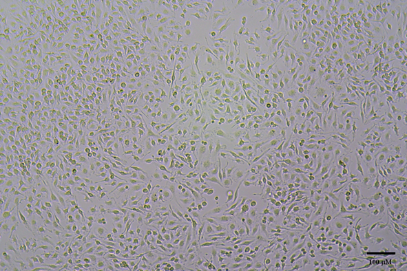
Figure 1. Characterization of mouse BMDMs. Bone marrow cells were cultured in the presence of M-CSF. On Day 5, the macrophage population was largely adherent. Scale bar = 100 µm.
B. Assessment of GSDMD activation in Mycobacterium bovis BCG -infected BMDMs lysates by Western Blotting
- Aspirate media from cultures; wash cells with 1x PBS; aspirate.
- Lyse cells by adding RIPA lysis buffer containing 1 mM PMSF (~200 mL/ well of 6-well cell culture).
- Incubate on ice for 2 min. Transfer the extract to a microcentrifuge tube.
- Microcentrifuge for 10 min at 13,000 rpm.
- Transfer supernatant to a clean microcentrifuge tube.
- Measure the protein concentration using the BCA Protein Assay Kit.
- Mix samples with 5 ´ loading buffer at 1´ final concentration and heat sample to 95-100 °C for 5-10 min; spin the samples for 10 s at room temperature at 2,000 ´ g to collect the sample to the bottom of the tube.
- Load 15-20 μg per lane onto 12% polyacrylamide SDS-PAGE gel (Table 1) and load 5 μl precision plus protein dual color standards to determine molecular weights.
- Electrophoresis at constant 80 Volts in 1 ´ Tris-Glycine running buffer until the protein dye reaches the bottom of the gel.
- Using Wet/Tank Blotting Systems and transfer the proteins from the gel to a PVDF membrane in transfer buffer (constant 150 mA for 2 h at 4 °C).
- Incubate membrane in 25 ml of blocking buffer in a plastic container at room temperature with gentle agitation for 1 h.
- Wash three times for 10 min each with 10 ml of TBST.
- Incubate membrane and rabbit polyclonal antibody anti-GSDMD (1:1,000) in 5 ml TBST with gentle agitation overnight at 4 °C.
- Wash three times for 10 min each with 15 ml of TBST.
- Incubate membrane with the 10 ml TBST containing 1:10,000 HRP-conjugated goat anti-rabbit secondary antibody with shaking at room temperature for 1 h.
- Wash three times for 10 min each with 15 ml of TBST.
- Incubate membrane with 5 ml ECL for 1 min at RT, detect by UVP E3 Image System and the data analysis show in Figure 2.

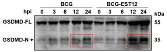
Figure 2. BMDMs infected with BCG strains were detected for GSDMD activation by western blot. BMDMs were infected with Mycobacterium bovis BCG and recombinant EST12 BCG for different time periods, and lysates were prepared and blotted with the GSDMD antibody, and the activation of N fragment (~35 kDa) of GSDMD were detected as indicated in figure.
Recipes
- ACK lysis buffer
8.3 g of NH4Cl
1.0 g of KHCO3
200 μl of 0.5 M EDTA
Add ddH2O to 1 L, adjust to pH 7.2-7.4 and filter sterilize - 5x Loading buffer
0.25 M Tris-HCl (pH 6.8)
0.5 M Dithiothreitol
50% glycerol
10% SDS
0.25% bromophenol blue - 1x Tris-Glycine running buffer
3.03g of Tris-HCl
14.4 g of glycine
1 g of SDS
Add ddH2O to 1 L. - Transfer buffer (store at 4 °C)
3.03g of Tris-HCl
14.4 g of glycine
20% methanol
Add ddH2O to 1 L. - 1x TBST buffer
2.48 g of Tris base
8.8 g of NaCl
0.5 ml Tween 20
Add ddH2O to 1 L, adjust to pH 7.0-7.4. - Blocking solution
5% nonfat milk (w/v) in 1x TBST buffer
Acknowledgments
We acknowledge F. Shao (National Institute of Biological Sciences, Beijing, China) for providing rabbit polyclonal antibody anti-GSDMD.
References
- Shi, J., Zhao, Y., Wang, K., Shi, X., Wang, Y., Huang, H., Zhuang, Y., Cai, T., Wang, F., Shao, F. (2015). Cleavage of GSDMD by inflammatory caspases determines pyroptotic cell death. Nature 526(7575): 660–665.
- He, W. T., Wan, H., Hu, L., Chen, P., Wang, X., Huang, Z., Yang, Z. H., Zhong, C. Q., Han, J. (2015). Gasdermin D is an executor of pyroptosis and required for interleukin-1b secretion. Cell Res 25(12): 1285–1298.
- Sharma, D., Kanneganti, T. D. (2016). The cell biology of inflammasomes: Mechanisms of inflammasome activation and regulation. J Cell Biol 213(6):617-29.
- Kayagaki, N., Stowe, I. B., Lee, B. L., O'Rourke, K., Anderson, K., Warming, S., Cuellar, T., Haley, B., Roose-Girma, M., Phung, Q. T., Liu, P. S., Lill, J. R., Li, H., Wu, J., Kummerfeld, S., Zhang, J., Lee, W. P., Snipas, S. J., Salvesen, G. S., Morris, L. X., Fitzgerald, L., Zhang, Y., Bertram, E. M., Goodnow, C. C., Dixit, V. M. (2015). Caspase-11 cleaves gasdermin D for non-canonical inflammasome signalling. Nature 526(7575):666-71.
- Qu, Z(2020). WB analysis. Bio-protocol Preprint. bio-protocol.org/prep621.
- Qu, Z., Zhou, J., Zhou, Y., Xie, Y., Jiang, Y., Wu, J., Luo, Z., Liu, G., Yin, L. and Zhang, X.(2020). Mycobacterial EST12 activates a RACK1–NLRP3–gasdermin D pyroptosis–IL-1β immune pathway . Science Advances 6(43). DOI: 10.1126/sciadv.aba4733
Category
Do you have any questions about this protocol?
Post your question to gather feedback from the community. We will also invite the authors of this article to respond.