
- Home
- Protocols

-

Skeletal stain followed by micro-computed tomography
Last updated date: Oct 18, 2020 Views: 1030 Forks: 0
Alizarin Red skeletal staining and micro-computed tomography – zebrafish skull analysis
- Euthanize zebrafish with 250-300 mg/L tricaine overdose
- Fix samples in tubes with 4% PFA overnight at 4°C
*** If bone staining by Alizarin Red S is not desired, then wash samples in PBS 3 x 10 minutes on a nutator in lieu of steps 3-9. *** - Wash samples in 0.5% KOH for at least 30 minutes on a rocker
- Keeping the lid off, bleach samples with 3% H2O2 in 0.5% KOH for several hours until pigmentation is cleared. At the end of this step, the eyes should have turned into a light brown color.
- Wash samples with 35% NaBO4 for at least 30 minutes on a rocker
- Clear samples with 1% trypsin in 35% NaBO4 for several hours on a rocker (gentle rocking) until tissue is reasonably cleared. By the end of this step, the body should be a translucent white color. Note that over- clearing in this step will cause the sample to be extremely fragile, with possibility of the skeleton falling apart.
- Wash samples with 10% glycerol in 0.5% KOH for at least one hour on a rocker
Stain samples with 0.02% Alizarin Red S pH 7.5 in 10% glycerol and 0.5% KOH overnight on a rocker. This solution is a clear/yellow color at low (acidic) pH levels, while purple at high (basic) pH levels. Thus, residual solution from step 7 will shift the pH higher and cause the stain to be more purple than red.
- Wash samples with 50% glycerol in 0.5% KOH on a rocker until residual dye is removed, may take overnight; change to fresh solution as needed
- Wash samples in 50% ethanol for 20 minutes on a nutator
- Dehydrate samples in 100% ethanol 2 x 20 minutes on a nutator
- Embed heads in paraffin to transport to scanner
- Remove paraffin and glue individual heads to the tips of glass pipettes. A glue gun will work well.
- Mount entire fixture onto the scanner specimen holder
- Scan heads on a Nikon Metrology Xt S 225 ST with the following parameters for phenotypic investigation:
- energy at 120 kV
- current at 26 µA
- 3141 projections at two frames/second and averaging two frames
- no filter
- 6 µm resolution
Render three-dimensional reconstructions using Arivis or the software of your choice
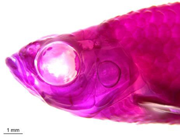
Adult zebrafish stained with Alizarin Red S.
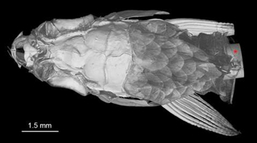
Three dimensional rendering of adult zebrafish micro-computed tomography scan. Glass pipette tip is visible at the back of the head, as denoted by the red asterisk.
- Teng, C S, Maxson, R and Crump, J(2020). Skeletal stain followed by micro-computed tomography. Bio-protocol Preprint. bio-protocol.org/prep551.
- Teng, C. S., Ting, M., Farmer, D. T., Brockop, M., Maxson, R. E. and Crump, J. G.(2018). Altered bone growth dynamics prefigure craniosynostosis in a zebrafish model of Saethre-Chotzen syndrome. eLife. DOI: 10.7554/eLife.37024
Category
Do you have any questions about this protocol?
Post your question to gather feedback from the community. We will also invite the authors of this article to respond.