Improve Research Reproducibility A Bio-protocol resource

- Home
- Protocols

-

Preprint
Fluorescence quenching assay
Last updated date: Jul 15, 2020 Views: 1169 Forks: 0
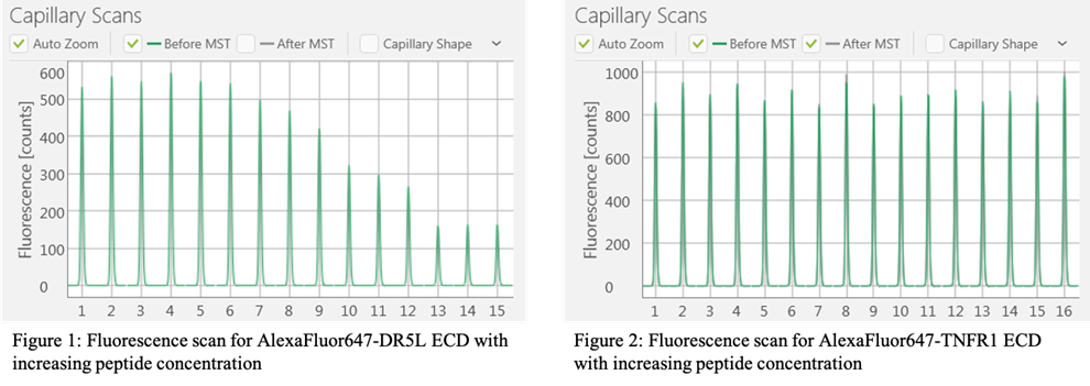
Measuring fluorescence quenching using capillary scanning function of the Monolith NT.115 Instrument
Set-up for this assay was adapted from the MST starting guide by Nanotemper Technologies.
- Prepare 2-fold dilutions of 2x unlabeled peptide into 20 mM HEPES pH 7.2, 100 mM KOAc, 0.1% Tween-20, 1% DMSO
- Distribute 10 ul of 2x unlabeled peptide into PCR tubes
- Prepare working stock of 400 nM AlexaFluor647-labeled ECD protein in 20 mM HEPES pH 7.2, 100 mM KOAc, 0.1% Tween-20
- Add 10 ul of the labeled ECD protein for a final concentration of 200 nM
- Incubate samples for 30 min at RT in the dark
- Load samples by capillary action onto Premium capillaries (Nanotemper Technologies, cat. #MO-K025).
- Scan for fluorescence on a Monolith NT.115 Instrument (NanoTemper Technologies, Germany) at 25°C, medium power.
Important factors to avoid non-specific fluctuations in fluorescence:
- Keep % of DMSO constant throughout all samples
- Use 0.1% Tween-20 in the final sample to prevent aggregation of peptide
- Make serial dilutions from a singular working stock of peptide
- Use Premium capillaries (#MO-K025)
Copyright: Content may be subjected to copyright.
How to cite:
Readers should cite both the Bio-protocol preprint and the original research article where this protocol was used:
- Lam, M(2020). Fluorescence quenching assay. Bio-protocol Preprint. bio-protocol.org/prep397.
- Lam, M., Marsters, S. A., Ashkenazi, A. and Walter, P.(2020). Misfolded proteins bind and activate death receptor 5 to trigger apoptosis during unresolved endoplasmic reticulum stress. eLife. DOI: 10.7554/eLife.52291
Category
Do you have any questions about this protocol?
Post your question to gather feedback from the community. We will also invite the authors of this article to respond.
0 Q&A
This protocol preprint was submitted via the "Request
a Protocol" track.