
- Home
- Protocols

-

Dephosphorylation assay
Last updated date: Jul 10, 2020 Views: 2438 Forks: 0
Cell Lysate Dephosphorylation Assay
1. Culture 293FT cells to approximately 90% confluence on 35 mm polystyrene tissue culture plates.
2. Aspirate or decant media and wash cells gently once with 1 ml of ice cold PBS. Aspirate excess PBS.
3. Add 1 ml of PBS to each plate and scrape the cells with a cell scrapper. Transfer the cells to a microtube.
4. Spin down the cells at 900 x g for 5 min at 4°C.
5. Remove PBS and add Lysis Buffer supplemented with 1X EDTA-free Protease Inhibitor Cocktail.
(Do not add phosphatase inhibitors, such as sodium orthovanadate, since they inactivate CIP.)
6. Incubate the cell lysate on ice for 20 min.
7. Centrifuge at 20,000 x g for 20 min at 4°C.
8. Collect the supernatant into new microtubes.
9. Determine protein concentration by the Bradford assay.
10. Add 30 µg of the cell lysate into each microtube and keep the sample tubes on ice.
11. Add 10X CIP Buffer and 1X EDTA-free Protease Inhibitor Cocktail to each sample tube and mix well.
12. Add 3 µl of CIP (10,000 U/ml) to each sample tube (use 10 U of CIP for each 10 µg of the cell lysate).
Add 3 µl of distilled water to the control sample tube.
Bring up the total volume of each reaction mixture to 30 µl with Lysis Buffer.
13. Incubate the reaction mixtures for 12 hr at 37°C.
(reduce the incubation time if protein degradation is a problem).
14. Add 5X SDS sampling buffer to stop the reaction.
15. Boil samples for 5 min at 95°C.
16. Save 10 µl of each reaction mixture for Phos-tag gel analysis and store the remaining samples at -80°C.
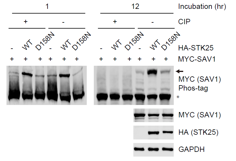
MYC-SAV1 was co-transfected with HA-STK25 WT (wild-type) or D158N (kinase-dead) into 293FT cells.
The cell lysates were treated with or without CIP (Calf Intestinal Alkaline phosphatase) for 1 hour or 12 hours. CIP-treated and non-CIP-treated lysates were subjected to Phos-tag gel analysis and immunoblot.
Asterisk and arrow designate non-phosphorylated SAV1 and phosphorylated SAV1, respectively.
- Bae, S and Luo, X(2020). Dephosphorylation assay. Bio-protocol Preprint. bio-protocol.org/prep381.
- Bae, S. J., Ni, L. and Luo, X.(2020). STK25 suppresses Hippo signaling by regulating SAV1-STRIPAK antagonism. eLife. DOI: 10.7554/eLife.54863
Category
Do you have any questions about this protocol?
Post your question to gather feedback from the community. We will also invite the authors of this article to respond.