Improve Research Reproducibility A Bio-protocol resource

- Home
- Protocols

-

Preprint
Sample preparation and Western blot assay
Last updated date: Apr 3, 2020 Views: 1681 Forks: 0
Sample preparation and Western blot assay
- Cells were trypsinized by 0.05% Trypsin-EDTA, blocked with feeder medium and collected by centrifugation at 4 °C. The pellet was gently washed by cold PBS and collected again.
- Cell pellet was resuspended and lysed in cold RIPA buffer (Sigma, R0278) with freshly added protease inhibitors (Sigma, 11697498001) for 30 min on ice (50 μL RIPA buffer was used for 1 million cells). Vortex the sample every 10 min.
- After centrifuge at 14,000 g for 15 min at 4 °C, the supernatant was collected as soluble fraction and the pellet was solubilized using 8M urea with 2% SDS and 50 mM pH7.6 Tris-HCl buffer as insoluble fraction. The protein concentration of the soluble fraction was measured by Pierce™ BCA Protein Assay Kit (Thermo Scientific, CAT# 23227). The amount of urea solution used to solubilize the pellet was decided by the whole protein volume in the soluble fraction. For every 1 μg of protein in the soluble fraction, 1 μL of urea solution was used for the insoluble fraction. The pellet was resuspended in the 8M urea solution and incubated at 37 °C for 2 hours and pipetted until solubilized thoroughly (try to avoid foaming when pipette).
- To store the protein samples, ethanol and dry ice were used for snap-freezing before transferring to -80 °C freezer. To thaw the samples, soluble fraction was thawed on ice and insoluble fraction was thawed by incubation at 37 °C for 5 min.
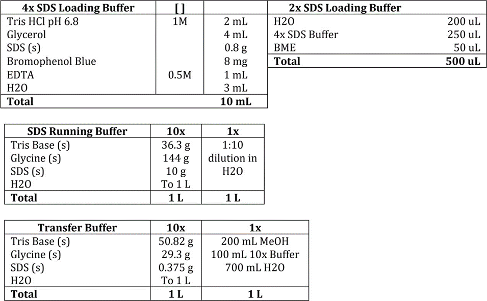
- 5-15 μg protein sample from soluble fractions and equivalent volumes of insoluble fractions were separated by 8-16% SDS-PAGE (Bio-Rad, 456-1105) and transferred to PVDF membranes (Thermo Fisher, 88518) by wet transfer with 100V voltage for 75 min at 4 °C. In detail, samples were mixed with 2X or 3.3X SDS loading buffer. Mixed soluble samples were boiled at 100 °C for 5 min in the thermal block and mixed insoluble samples were incubated for 20 min at RT before loading to SDS-PAGE gel.
- The membrane was blocked with 5% Bovine Serum Albumin (Sigma, A9647) for 1h at room temperature, incubated with primary antibodies overnight at 4 °C, washed in TBST X3, incubated with HRP-conjugated mouse and rabbit secondary antibodies (Amersham ECL Western Blotting Detection Reagent, VWR, 95038-560) for 1h at room temperature, washed in TBST X3 and then developed using ECL Western Blotting Detection System (Fisher Scientific, RPN2108) and X-ray films (Thermo Fisher, 34091).
Buffer preparation:
Copyright: Content may be subjected to copyright.
How to cite:
Readers should cite both the Bio-protocol preprint and the original research article where this protocol was used:
- An, D(2020). Sample preparation and Western blot assay. Bio-protocol Preprint. bio-protocol.org/prep262.
- An, D., Fujiki, R., Iannitelli, D. E., Smerdon, J. W., Maity, S., Rose, M. F., Gelber, A., Wanaselja, E. K., Yagudayeva, I., Lee, J. Y., Vogel, C., Wichterle, H., Engle, E. C. and Mazzoni, E. O.(2019). Stem cell-derived cranial and spinal motor neurons reveal proteostatic differences between ALS resistant and sensitive motor neurons. eLife. DOI: 10.7554/eLife.44423
Category
Do you have any questions about this protocol?
Post your question to gather feedback from the community. We will also invite the authors of this article to respond.
0 Q&A
This protocol preprint was submitted via the "Request
a Protocol" track.