
- Home
- Protocols

-

HSPC and MPP2 isolation, barcoding, EPO treatment, and transplantation
Last updated date: Feb 7, 2024 Views: 1078 Forks: 0
Protocol for lentiviral barcoding of EPO treated hematopoietic stem and progenitor cells: Cell purification, barcoding, and transplantation
Study overview:
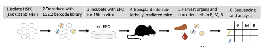
Figure 1: Overview of the experimental design (a) HSPCs were sorted from the bone marrow of donor mice, lentivirally barcoded, cultured ex vivo with or without 1000 ng/ml EPO for 16 hr, and transplanted into sublethally irradiated mice. At week 4 post-transplantation, the erythroid (E), myeloid (M), and B-cells
Material:
A full list of reagents and material used in this protocol is provided in the original article in the ‘Key resources’ table
Step 1- progenitor isolation
A. BM isolation: Bone collection and ckit enrichment
- Sacrifice mice
- 6 BL6N D45.1 Males aged 8 weeks
- Recover BM (tibia, femur, iliac)
- Take some cells for unstained and single stain controls
B. Flushing bones
- Put bones and medium into a 10 cm petri dish.
- Prepare the scissors and tweezer by rinsing with EtOH.
- Rinse before taking bone in petri dish medium to dilute EtOH.
- Prepare a 15 ml tube or 50 ml with 4-5 ml/ mouse cold PBS+ to collect the cells.
- Prepare a 2ml syringe with a long green needle (21G 50mm)
- fill it with 2ml cold PBS+ from the collection tube
- Put needle into tibia or non-star like end of femur.
- For iliac bone at V shaped end at the tip of the V.
- Take care to use pointiest part of needle.
- Put entire bone into cold PBS+ and eject 1ml of cold PBS+.
- For tibia and femur, turn the bone ¼ without detaching it, for iliac bone just put deeper into bone (should feel that is breaking a membrane), and eject other 1ml into the tube.
- If bone is not white yet, restart, cold PBS+ taken is from the collection tube!
- Empty bones can be collected in lid of petri dish.
- Between each bone, dissociate the red pulp by pipetting up and down with the syringe and ejecting against the tube wall.
- Never put tweezer into collection tube. If lose bone try to get back with needle.
- Pass the cell through a 100um cell strainer with the pipette
- Spin down for 5 mins at 4°C at 1700 rpm
- Rinse the petri dish and the Falcon with medium.
C. cKit enrichment
Cell preparation
- Pool cells from 3 mice into a single samples (this will give 2 samples of 3 mice)
- Centrifuge cells at 1700rpm 5 min 4°C
- Take off the supernatant and leave the last drop
- Add 25 ul of a-CD117 beads in each tube, mix, add 125ul of cold 10%RPMI, mix.
- Incubate for 20 min on ice covered by aluminium
- Add 5ml of cold 10%RPMI to the cells
- Pass through a 100 um cell strainer into 50 ml tube.
- Wash tube and cell strainer with 5 ml of cold medium.
- Take off the last drop of cells under the cell strainer with an 1000ul pipette.
- Centrifuge cells at 1700 rpm 6min 4°C
- Resuspend in 3ml cold medium.
MACS for c-kit+ cells
- Wash the LS column with 3ml of cold 10%RPMI
- Mix cells with an insulin syringe with short 25G needle
- Eject cells against the LS column wall.
- Wash the tube and column with 3 ml of medium. [end of the ckit- collection]
- Detach the column, put into a new 15mL falcon (ckit+ collection tube) and fill up with medium (5mL)
- Insert the plunger and eject the cells [end of the ckit+ collection]
- Count cells
D. Sorting
| Staining for | Sca1(1:200, clone = D7) c-kit (1:100, clone = M1/70) flt3 (1:100, clone = A2F10) CD150 (1:100, clone = TC15-12F12.2) |
| Staining in | 10% RPMI |
| Incubation time | 30 minutes on ice |
| Washing | directly through filter FACS tubes |
| Resupension | in 10% RPMI |
| Expected sorting yield | MPP: 100,000 cells HSC: 10,000-14,000 cells |
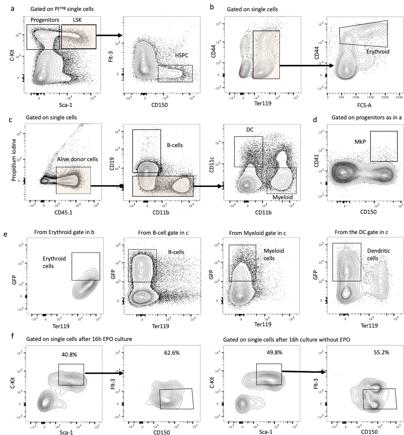
Figure 1: Gating strategies and hematopoietic stem and progenitor cell (HSPC) marker expression after lentiviral transduction and ex vivo culture with or without erythropoietin (EPO). (a) HSPCs were gated as propidium iodide-negative single C-Kit+ Sca-1+ Flt3- CD150+ cells of C-Kit+-enriched bone marrow cells. (b) Erythroblast cells were gated as Ter119+ CD44+ FSChi cells on Ter+ enriched cells. (c) Gating strategy for B-cells (CD19+ CD11b-), dendritic cells (CD19 CD11b- CD11c+), and myeloid cells (CD119- CD11c- CD11b+) on Ter119- live single-donor cells. (d) Gating for MkP (C-Kit+ Sca-1- CD150+ CD41+) from C-Kit+-enriched bone marrow cells. (e) Sort gating for GFP+ erythroid, myeloid, B-, and dendritic cells, respectively, used for barcoding analysis. (f) Representative flow cytometry plots of sorted HSPC pool after 6 hr lentiviral transduction and 16 hr ex vivo incubation with or without EPO.
Step 2-Introducing barcodes-transduction
- Centrifuge the cells and remove the supernatant
- Resuspend in 100uL of Stem Span + 50ng/mL mSCF + 1,3uL of virus corresponding to the LG2.2 lentivirus library. The precise amount of virus was titrated to achieve a transduction efficiency of 10%. This can be reduced to 1% to further reduce multiple barcode usage.
- Mix with the pipette
- Centrifugate at RT 90 min at 300g (brake 2-2)
- Remove the plate and put in the incubator at 37°C 5% CO2 during 4.5h
- Wash twice the cells with complete RPMI
- Resuspend in 500uL of Stem Span + 50ng/mL mSCF
Step 3-EPO treatment
After transduction, cells were incubated with or without human recombinant EPO (Eprex, erythropoietin alpha, Janssen) in Stem Span culture medium supplemented with 50ng/mL mSCF (SS+) at a final concentration of 1000 or 160 ng/ml or PBS for 16 hr at 37°C.
- Coat a 1,5 ml Eppendorf tube with 1 ml 10% RPMI.
- Transfer transduced cells into coated Eppendorf tubes
- Add 1 ml of 10% RPMI, centrifuge 6 min 1700 rpm, 22°C.
- Take off supernatant with 1 ml pipette.
- Resuspend in 400µl SS+ or 420 if transduction control and mix well.
- Take transduction control 20 ul in extra well on plate and add 100 ul or more of SS+.
- Take 200µl for the non-EPO condition and place it into a well.
- Add EPO on the rest and place it into well.
- Culture the cells for 16h at 37°C.
Step 4-injection of barcoded cells into irradiated recipients
After EPO incubation, barcoded MPP2 and unbarcoded CD48- HSPCs were mixed at a ratio of 32/45 (to be as close as possible to the original ratio of both populations in the HSPCs). On average, 2600 cells (mean 2684 cells ± 175 cells) were injected in the tail vein of each mouse in a volume of 100 microliters. Three hours prior to tail vein injection recipient mice (8 week old BL6N D45.1/1 male mice) were 6 Gy sublethally irradiated on a CIXD irradiator.
- Coat a 1,5 ml Eppendorf tube with 1 ml 10% RPMI.
- Mix incubated cells well and transfer into coated Eppendorf tube.
- Wash wells again with 200 ul of 10%RPMI.
- Centrifuge 6 min 1700 rpm 22°C.
- Remove supernatant and wash again in DPBS.
- Resuspend cells in number of mice*100 ul+20 ul and take fraction (1%) for flow cytometry check -> transfer into FACS tubes with 100 ul (or 50ul) antibody mix (as previous day, without CD34) and keep for 35 min (flt3) or 15 (without flt3) in fridge.
- Keep all samples at 4°C till injection or flow cytometry check.
- Perié, L, Cosgrove, J and Eisele, A(2024). HSPC and MPP2 isolation, barcoding, EPO treatment, and transplantation. Bio-protocol Preprint. bio-protocol.org/prep2564.
- Eisele, A. S., Cosgrove, J., Magniez, A., Tubeuf, E., Tenreira Bento, S., Conrad, C., Cayrac, F., Tak, T., Lyne, A., Urbanus, J. and Perié, L.(2022). Erythropoietin directly remodels the clonal composition of murine hematopoietic multipotent progenitor cells. eLife. DOI: 10.7554/eLife.66922
Do you have any questions about this protocol?
Post your question to gather feedback from the community. We will also invite the authors of this article to respond.