
- Home
- Protocols

-

EdU assay zebrafish embryos
Last updated date: Sep 15, 2023 Views: 674 Forks: 0
EdU assay – Giulia Boezio (doi: 10.7554/eLife.57603)
- Incubate embryos/larvae in 0.5 mM EdU (Thermo Fisher)/0.5% DMSO dissolved in PTU water at 28C from 24 to 36 hpf (12h) or 48 to 72 hpf (24h), according to the experiment.
- Do not treat more than 12 animals/4 ml in 6-well plates.
- As we observed different efficiencies of cell labeling with different batches of EdU, all the samples of all the replicates for each experiment were treated with the same batch of EdU.
- Embryo or larvae were then euthanized with 0.2% Tricaine, followed by fixation with 4% PFA for 2h at room temperature.
- Embryos were then washed in PBS several times and the yolk was removed using forceps to facilitate the penetration of the antibody.
- Permeabilize embryos/larvae with 10 μg/ml Proteinase K for a time dependent on the developmental stage (10-12 min at 36hpf and 30min at 72hpf).
- Wash >4 times in PBDT. PBDT: 0.5% Triton X-100, 1% DMSO, 1% BSA, 1X PBS
- Block in PBDT + 5% goat serum.
- Incubate with primary antibody overnight at 4°C.
- Wash extensively (minimum 4x20 minutes) in PBDT.
- Incubate with secondary antibody + 1 μg/ml DAPI overnight at 4°C.
- On the following day, wash 4 × 15 min in PBS + 0.1% TritonX
- Following these 4 washes post-second antibody incubation, the Click-iT reaction was performed, following the Click-iT EdU Cell Proliferation Kit for Imaging, Alexa Fluor 647 dye manufacturer’s instructions (see below).
- Prepare 1X Click-iT® EdU buffer additive by diluting the 10X stock solution 1:10 in deionized water. Prepare this solution fresh and use the solution on the same day.
- Prepare Click-iT® reaction cocktail: 500ul for a 2ml Eppendorf tube containing max 20 embryos.
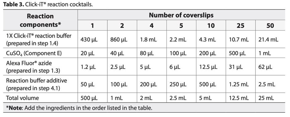
It is important to add the ingredients in the order listed in the table; otherwise, the reaction will not proceed optimally. Use the Click-iT® reaction cocktail within 15 minutes of preparation.
- Wash the embryos/larvae in each well twice with 3% BSA in PBS for 5 min. Remove the wash solution.
- Add 0.5 mL of Click-iT® reaction cocktail to each tube and incubate the plate for 30 minutes at room temperature, protected from light.
- Remove the reaction cocktail, then wash each tube once with 3% BSA in PBS for 5min. Remove the wash solution.
- Embryos/larvae were then mounted in agarose and genotyped after imaging.
Primary antibodies dilutions used are as follows: GFP (AvesLab, 1:400), Elastin2 (1:100), tRFP (Evrogen, 1:200), anti-dsRed (Takara Bio Clontech, 1:200), anti-Alcama/Dm-Grasp (DSHB, 1:50), anti-p-Smad3 (Abcam, 1:100) and anti-pSmad1/5/8 (Cell Signaling Technology, 1:100). Secondary antibodies (Secondaries Alexa FluorTM 488-568-647 IgG (H+L), Thermo Fisher Scientific) were used at 1:500 concentration. Elastin2 antibody was purified from the serum stock described previously (Miao et al., 2007).
- Boezio, G, Helker, C and Stainier, D(2023). EdU assay zebrafish embryos. Bio-protocol Preprint. bio-protocol.org/prep2421.
- Boezio, G. L., Bensimon-Brito, A., Piesker, J., Guenther, S., Helker, C. S. and Stainier, D. Y.(2020). Endothelial TGF-β signaling instructs smooth muscle cell development in the cardiac outflow tract. eLife. DOI: 10.7554/eLife.57603
Do you have any questions about this protocol?
Post your question to gather feedback from the community. We will also invite the authors of this article to respond.