
- Home
- Protocols

-

High-resolution 3D tissue imaging
Last updated date: Jul 14, 2023 Views: 1027 Forks: 0
High-resolution 3D tissue imaging protocol in mice
Lu Mu1, Xueqiang Xu2, Xuebing Yang1, Yan Zhang1, Hua Zhang1*
- State Key Laboratory of Agrobiotechnology, College of Biological Sciences, China Agricultural University, Beijing 100193, China.
- Biomedical Pioneering Innovation Center, School of Life Sciences, Peking University, Beijing 100871, China; Peking-Tsinghua Center for Life Sciences, Academy for Advanced Interdisciplinary Studies, Peking University, Beijing 100871, China.
*Corresponding author: Hua Zhang, huazhang@cau.edu.cn
Detailed protocol
This protocol realized a whole-mount imaging system to visualize ovarian blood vessels with a single-cell resolution. This was achieved by combining a vascular endothelium–specific endogenous dual-fluorescent tracing mouse model [tyrosine kinase (Tek)–Cre;mTmG mice] with a modified 3D tissue-transparency technique, followed by scanning the transparent tissues under a high-resolution spinning-disc confocal microscope.
Tissues collection
- Perfusion with cold PBS to remove the blood (perform this step selectively depending on the content of blood in the tissues).
- Collect the tissues in 4% PFA in the dark for 24 hours at 4℃.
Clearing method of tissues (modified according to Li et al., Proc. Natl. Acad. Sci. USA. 2017 Aug 29;114(35):E7321-E7330. doi: 10.1073/pnas.1708981114.)
- Stock N-methylacetamide (M26305, Sigma-Aldrich) was prepared by diluting melted N-methylacetamide to 40% (v/v) in phosphate-buffered saline (PBS), which was then used to dissolve Histodenz (D2158, Sigma-Aldrich) to 86% (w/v) concentration.
- Triton X-100 (0.1% v/v) and 1-thioglycerol (M1753, Sigma-Aldrich) (0.5% v/v) were added to the Histodenz solution to be the final clearing solution.
- The tissues were collected and washed with PBS containing 0.2% Triton X-100 and 1-thioglycerol (0.5%) in the dark for 24 hours at room temperature to remove blood cells.
- The tissues were placed in the clearing medium (1:50 v/v) and incubated in the dark at room temperature on a rotor for 72 hours.
High-resolution 3D tissue imaging
- The cleared tissues were embedded in a 35-mm dish with 14-mm glass bottom (D35-14-1-N, Cellvis) containing fresh clearing solution and tightly covered by a coverslip.
- Confocal imaging was performed on an inverted Leica (DMi8) and Andor Dragonfly spinning-disc confocal microscope, a scientific complementary metal-oxide semi-conductor (sCMOS) camera (Andor Zyla 4.2), and using either a 20× 0.8 numerical aperture (NA) 650-μm working distance or a 40× 1.3 NA 250-μm working distance objective.
- A pixel density of 2048 × 2048 was used, and Z-step 0.5 to 1.0 μm for 650 μm (20× objective) or 0.3 to 0.6 μm for 250 μm (40× objective). The 488-nm (mG) and 568-nm mTomato (mT) lines of the Andor Integrated Laser Engine (ILE) system with a spinning-disc confocal scan head (Andor Dragonfly 500) were imaged. Images were acquired by Fusion 2.1 software (https://andor.oxinst.com/products/dragonfly#fusion).
Image processing
- Images were processed by ImageJ (http://rsbweb.nih.gov/ij/) for projection of all z stacks and merged color channels.
- The 3D vascular structure of tissues and the rotary 3D movie was processed by Imaris (https://imaris.oxinst.com/) software.
Materials
- 35-mm dish with 14-mm glass bottom (D35-14-1-N, Cellvis)
- N-methylacetamide (M26305, Sigma-Aldrich)
- Histodenz (D2158, Sigma-Aldrich)
- 1-thioglycerol (M1753, Sigma-Aldrich)
Images
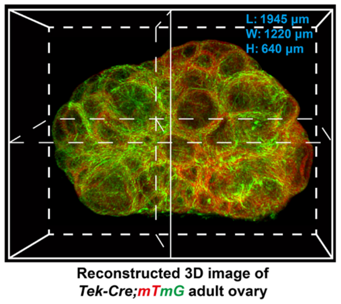
Xu et al., Sci Adv. 2022 Jan 14;8(2):eabi8683. doi: 10.1126/sciadv.abi8683.
The transparent ovaries were scanned by high-resolution confocal microscopy to reconstruct a 3D image of the ovarian blood vessels.
- Zhang, H, Mu, L, Xu, X, Yang, X and Zhang, Y(2023). High-resolution 3D tissue imaging. Bio-protocol Preprint. bio-protocol.org/prep2368.
- Xu, X., Mu, L., Li, L., Liang, J., Zhang, S., Jia, L., Yang, X., Dai, Y., Zhang, J., Wang, Y., Niu, S., Xia, G., Yang, Y., Zhang, Y., Cao, Y. and Zhang, H.(2022). Imaging and tracing the pattern of adult ovarian angiogenesis implies a strategy against female reproductive aging. Science Advances 8(2). DOI: 10.1126/sciadv.abi8683
Do you have any questions about this protocol?
Post your question to gather feedback from the community. We will also invite the authors of this article to respond.