
- Home
- Protocols

-

In vitro kinase assay using immunoprecipitated Cdk5 from SCN
Last updated date: Feb 20, 2020 Views: 1363 Forks: 0
SCN extraction
-Mice 2 months old were kept under light: dark (12:12) conditions.
-7 mice for each time point corresponding to ZT 0, 4, 8, 12, 16, 20 (0-12 light on, 12-20 light off) were sacrificed and brain extracted. During the night phase, killing and tissue extraction was performed under red light.
-Brains were frozen in N2 liquid and pooled together for each appropriate time point and samples stored at -80°C.
-The Optic chiasm was removed using forceps and SCN extracted from the Brain.
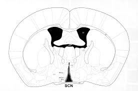
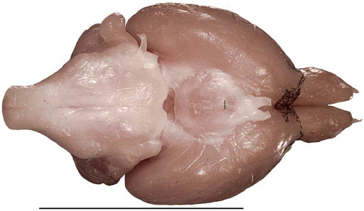
A B
A (Image taken Easton at al., 2004). B The mouse brain library
-7 SCN for each time point were pooled together in the same tube and left on ice or stored at -80°C.
Protein Extraction
-Brain Lysis Buffer + protease inhibitors (50 mM Tris-HCl pH 7.4, 150 mM NaCl, 0.25% SDS, 0.25% sodium deoxycholate, 1 mM EDTA) was added to the frozen samples (2 times the tissue volume) and samples were homogenized using the blue pestle.
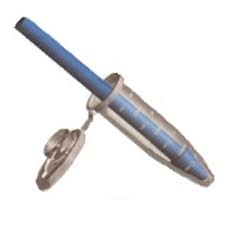
(Image taken from Universal Medical)
-Samples were vortexed and put on ice for 20’ (samples were vortexed each 5’)
-Samples were sonicated for few seconds at 10-30 % of maximal amplitude, in order to eliminate all the residual pieces of tissue not homogenized.
-Samples were centrifuged at 4°C for 20’ at 12000 x g and supernatant was collected (total protein extract).
-Protein were quantified using Pierce Rapid Gold BCA Protein Assay Kit (Thermo fisher) and finally quality of extract was evaluated running samples on a SDS/PAGE 10% gel stained with Coomassie Blue.
-Subsequently the accumulation of the protein of interest (CDK5) around the clock was detected by Western Blot (Rabbit anti-CDK5 cell signaling 1:1000 over night at 4°C) and compared to loading control protein (i.e. Tubulin; Rabbit anti-tubulin 1:1000 Abcam 3 hours Room Temperature)
Immunoprecipitation of CDK5 around the clock
-A protein amount corresponding to between 400 and 800 µg of total extract for each time point discussed before was diluted with Brain Lysis Buffer in a final volume of 250 µL.
-5 ul of CDK5 antibody were added to the mix (antibody ratio 1:50).
-Immunoprecipitation reaction was performed over night at +4°C on a rotary shaker.
-The day after, samples were captured with 80 µL of 50% (w/v) protein-A agarose beads (Roche) and the reaction was kept at 4°C for 3 hours on a rotary shaker. Prior to use, beads were washed three times with the appropriate protein buffer and resuspended in the same buffer (50% w/v).
-The beads were collected by centrifugation (1000 x g) and washed three times with NP-40 buffer (100 mM Tris-HCl pH7.5, 150 mM NaCl, 2 mM EDTA, 0.1% NP-40). After the final wash, each sample was split in 2 different tubes (circa 40 ul of beads for each tube called “tube IP” and “tube KA”, acronyms for Kinase Assay.
IP for Western Blot
-Samples from “tube IP” were resuspended in 2% SDS 10%, glycerol, 63 mM Trish-HCL pH 6.8 and proteins were eluted for 15 min at RT.
-Laemmli buffer was finally added, samples were boiled 5 min at 95°C and loaded onto 10% SDS-PAGE gels.
-Western Blot was performed (Rabbit anti-CDK5 cell signaling 1:1000 over night at 4°C) to detect the amount of CDK5 immunoprecipitated at each time point.
In Vitro Kinase assay
-Reaction was performed adding to samples from “tube KA” a mix containing 5X Kinase buffer ( 30 mM Hepes pH 7.2, 10 mM MgCl2, 1 mM DTT in 25ul of reaction, so that the final concentration is 1X), 1 µg of histone H1 (Sigma-Aldrich), [γ-32P] ATP (10 Ci) ( final volume mix 20 ul + beads), and incubating the samples at 30°C for 1 Hour
-Reaction was stopped with laemmly buffer and samples were boiled at 90°C.
-Samples were loaded on a 15% SDS/PAGE.
-Gel was stained 10’ in Coomassie Blue (20% methanol, 10% Acetic Acid, Coomassie blue)
-Gel was distained in 20% Methanol, 10% Acetic Acid
-Gel was washed 3 times with distilled water
-Gel was equilibrated in drying solution containing 20% Methanol, 10% Glycerol for 1 Hour.
-Gel was dried in a specific gel dryer at 80°C for 1h 30’ (vacuum pump active).
- Finally, the phosphorylation was detected by autoradiography.
Kinase assay evaluation
- CDK5 IP signal obtained from the WB was quantified by optical densitometry (Imaje J / Fijj)
- Phosphorylation signal obtained from the In vitro Kinase assay was quantified in the same way
- The ratio CDK5 IP / [γ-32P] ATP gives the final quantification about the effective kinase activity for each time point.
- Brenna, A and Albrecht, U(2020). In vitro kinase assay using immunoprecipitated Cdk5 from SCN. Bio-protocol Preprint. bio-protocol.org/prep216.
- Brenna, A., Olejniczak, I., Chavan, R., Ripperger, J. A., Langmesser, S., Cameroni, E., Hu, Z., De Virgilio, C., Dengjel, J. and Albrecht, U.(2019). Cyclin-dependent kinase 5 (CDK5) regulates the circadian clock. eLife. DOI: 10.7554/eLife.50925
Do you have any questions about this protocol?
Post your question to gather feedback from the community. We will also invite the authors of this article to respond.