Improve Research Reproducibility A Bio-protocol resource

- Home
- Protocols

-

Preprint
Protocol for imaging the same Class IV neurons at different stages of development
Last updated date: Oct 27, 2022 Views: 397 Forks: 0
Protocol for imaging the same Class IV neurons at different stages of development
This protocol for the preparation of agar pads to mount and image Drosophila larvae is adapted from Monica Driscoll's method (https://www.wormatlas.org/agarpad.htm). This detailed protocol is associated with “analysis of the elongation of internal branches.”
Preparation of agar pads:
- Prepare 5% agar solution in water, melt it and keep at 65 °C on a heat block to maintain its molten state.
- Cut the tip of a 1000 ml pipette tip using scissors and pipette 100-200 ml of agar solution onto the center of a clean glass slide.
- Cover the agar with another clean slide perpendicular to sandwich the agar drop and press gently so that the agar drop flattens to a circle. While pressing the agar drop, avoid getting air bubbles.
- Follow Monica Driscoll's agar pad method using an additional two taped slides if you are not confident enough to get a flat agar pad. An uneven agar pad is unsuitable for larvae imaging as they get stuck or do not get flattened or curled up on pressing with the coverslip.
- After solidifying the agar, gently separate the slides so that the agar pad adheres to one of them.
- The Agar pad should be prepared just before use to avoid it from drying out.
- After the agar pad is prepared, put a drop of halocarbon 700 (sigma) on the agar pad and spread it out. The prepared agar pad was kept at 4° C for 5 min before mounting larvae.
Mounting of live larvae:
- For larvae imaging, larvae were washed with 20%, followed by 5%, sucrose solution.
- The larvae were gently placed with their dorsal side up on a 5% agar bed adhered to a glass slide and imaged in a drop of 50% phosphate-buffered saline, 50% Halocarbon oil 700 (Sigma-Aldrich).
- Larvae were further immobilized by gently pressing them with a 22 mm–by–22 mm coverslip lined with Vaseline or vacuum grease.
- This whole setup was again kept at 4° C for 5 min.
- Their movement was further minimized by imaging at 4°C for 2 to 5 min using an OKO lab temperature control module connected to a spinning disk confocal microscope (Nikon). Although this step is not necessary.
- Larvae were imaged on the spinning disk confocal using 20X and 40X objective as described previously in Shree et al., Science Advances, 2022.
- After imaging, larvae were returned to the apple agar plate (that has a dollop of yeast paste in the center) in the Darwin chamber at 25° C and imaged again after 24 hours. This method allowed us to image same class IV neuron throughout larvae development without using an anesthetic.
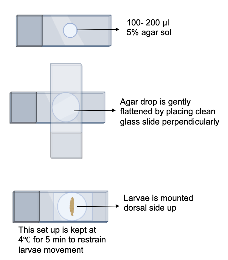
Figure 1: Schematic for preparation of agar pads and mounting of larvae.
Copyright: Content may be subjected to copyright.
How to cite:
Readers should cite both the Bio-protocol preprint and the original research article where this protocol was used:
- Shree, S and Howard, J(2022). Protocol for imaging the same Class IV neurons at different stages of development. Bio-protocol Preprint. bio-protocol.org/prep2012.
- Shree, S., Sutradhar, S., Trottier, O., Tu, Y., Liang, X. and Howard, J.(2022). Dynamic instability of dendrite tips generates the highly branched morphologies of sensory neurons. Science Advances 8(26). DOI: 10.1126/sciadv.abn0080
Do you have any questions about this protocol?
Post your question to gather feedback from the community. We will also invite the authors of this article to respond.
0 Q&A
This protocol preprint was submitted via the "Request
a Protocol" track.