
- Home
- Protocols

-

Quantification of phenotypes
Last updated date: Jan 7, 2020 Views: 1144 Forks: 0
Measurement of commissure size
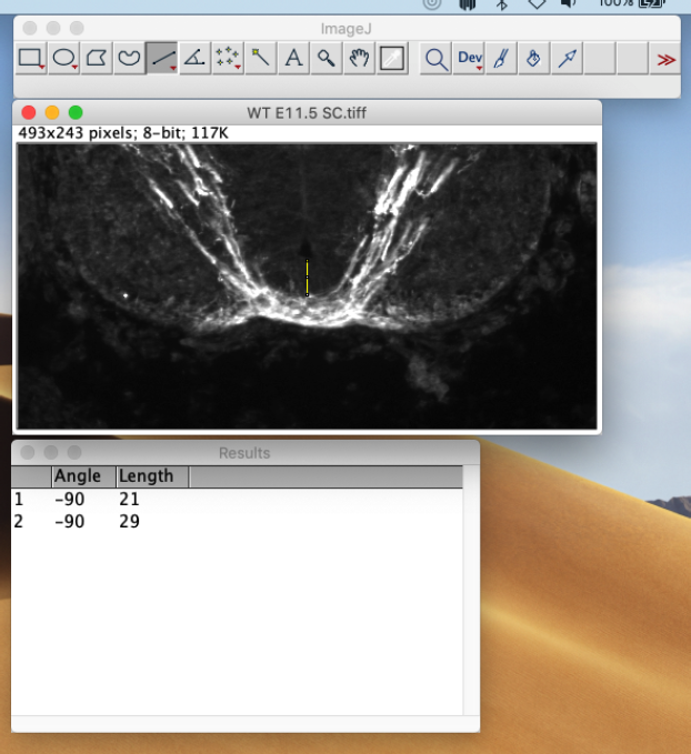
Open the image to be quantified in ImageJ, adjust the brightness/contrast as needed (select Image then Adjust then Brightness/Contrast) so that the immunolabeled commissural axons are clearly distinguished from the background. Zoom in and/or crop the image if necessary (select Image then Zoom or Image then Crop) to focus on the midline area (see the example image below).
Using the Straight command, draw a straight line across the commissure bundle at the midline. Select Analyze and then Measure. The thickness of the commissure will be shown as Length in the Results window. Repeat the same process to measure the height of the floorplate at the midline (the yellow line in the example image). Normalize the thickness of the commissure to the height of the floorplate (21/29 in the example image).
Using the same method, measure the commissure size in other sections from the same embryo, from different embryos, and from different genotypes. Perform statistical analysis using Prism.
- Chen, Z(2020). Quantification of phenotypes. Bio-protocol Preprint. bio-protocol.org/prep185.
- Johnson, V., Junge, H. J. and Chen, Z.(2019). Temporal regulation of axonal repulsion by alternative splicing of a conserved microexon in mammalian Robo1 and Robo2. eLife. DOI: 10.7554/eLife.46042
Do you have any questions about this protocol?
Post your question to gather feedback from the community. We will also invite the authors of this article to respond.