
- Home
- Protocols

-

Experiments
Last updated date: Mar 9, 2022 Views: 807 Forks: 0
Dear reader,
thank you for your request. Please find the detailed information about the liquid growth medium below, as well as the technique we used for obtaining single tubes.
SDM medium for microplasmodial growth in liquid culture (per litre of MilliQ water):
10g (D+)-glucose anhydrous (Merck)
10g Difco soytone (Roth)
3.54g Citric acid monohydrate (Roth)
2g Potassium dihydrogen phosphate (Roth)
1.026g Calcium chloride dihydrate (Sigma)
0.6g Magnesium sulfate heptahydrate (Sigma)
34mg Zinc sulfate heptahydrate (Sigma)
42.4mg Thiamine-HCl (Sigma)
15.8mg Biotin (Roth)
1. Dissolve the compounds in 1L of deionised water on a magnetic stirrer and adjust the pH to 4.6 with 1 M KOH or KOH pellets.
2. Autoclave and store at room temperature
3. Right before first use:
Add 10 ml/L hemin solution [125mg of hemin (Alfa Aesar) and 2.5g solid NaOH dissolved in 250mL MilliQ water, stored at 4 C] and 1mL of 250 μg/mL penicillin and streptomycin solution, each (stored at -20 C)
4. The medium can be stored in a well-closed bottle at room temperature. Before each use, gently shake the medium to dissolve the dark hemin precipitate that naturally forms in the bottle.
The SDM medium is sufficient to achieve microplasmodial growth. In order to optimize the shaking, which influences microplasmodial size, we added 50mL of non-nutritive BSS medium to 50mL of SDM medium in a 250mL baffled Erlenmeyer flask. This final mixture is referred to as shaking mixture below.
BSS medium (per litre of MilliQ water):
3g citric acid monohydrate (Roth)
4.20g Dipotassium hydrogen phosphate trihydrate (Roth)
0.25g Sodium chloride (Roth)
0.21g Magnesium sulfate heptahydtrate (Sigma)
0.05g Calcium chloride dihydtrate (Sigma)
1. Dissolve the compounds in 1L of deionised water on a magnetic stirrer and and adjust pH to 5.0 with 1 M KOH or KOH pellets.
2. Autoclave for 20 minutes and store at room temperature.
Preparation of Physarum network for imaging:
Microplasmodia were grown in 100mL of shaking mixture (25°C and 160rpm) starting from a Physarum network approximately 2cm2 that was transferred to the liquid medium after growing on agar. The microplasmodia grown in liquid culture are passaged into fresh medium every 2 days by pipetting 1mL of the microplasmodial suspension to a new flask. The microplasmodia harvested for imaging were obtained after 2 days of innoculation in the medium by pipetting 0.5mL of the microplasmodial suspension on a 1.5% agar plate (Bacto agar). The microplasmodia will form a network within 24-48 hours, when the imaging can be carried out. Depending on the size of the microscope's field of view and particular goal of the experiment, the plasmodium can be trimmed using a flat-edge spatula by pressing vertically onto the tubes and scraping away the part of the network to be discarded. After scraping, we recommend waiting for ~45min for the plasmodium to allow the contractions to become regular again.
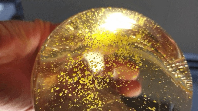
Regarding turbidity conditions, please find below a photo of microplasmodial liquid culture after a day of inoculation:
Obtaining a single Physarum tube:
The diverse morphology of Physarum network, even within a set of networks obtained from the same batch of microplasmodia, allows for finding particularly long tubes with connections on both or even just one end. Once identified, the tube can be isolated by pressing on it vertically using a flat-edge spatula and scraping away the rest of the network.
- Alim, K(2022). Experiments. Bio-protocol Preprint. bio-protocol.org/prep1579.
- Fleig, P., Kramar, M., Wilczek, M. and Alim, K.(2022). Emergence of behaviour in a self-organized living matter network. eLife. DOI: 10.7554/eLife.62863
Category
Do you have any questions about this protocol?
Post your question to gather feedback from the community. We will also invite the authors of this article to respond.