
- Home
- Protocols

-

Quantification of lysosome number and volume
Last updated date: Aug 9, 2021 Views: 830 Forks: 0
Fluorescence images of C. elegans adults at different ages (days 1, 3, 5, 9) expressing NUC-1::CHERRY in 10–15 z-series (0.5 µm/section) were captured by spinning-disk microscopy. Serial optical sections were analyzed, and the volume and number of NUC-1::CHERRY-positive vesicular lysosomes per unit area (31 X 43 µm2) was quantified by Volocity software (PerkinElmer). At least eight animals were quantified in each strain at each stage. The total volume of vesicular and tubular lysosomes was quantified by Volocity. At least 10 worms were quantified in each strain at each day.
Quantification of the number and volume of vesicular lysosomes:
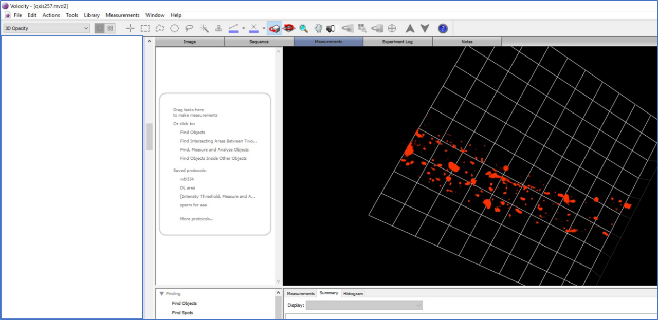
- We open the file in Volocity, the image is first displayed in 2D, it is cropped into the same size: 31 X 43 µm2.
- Now, the new image is displayed as “3D Opacity” option. Then, it goes to “Measurements” section to quantify.
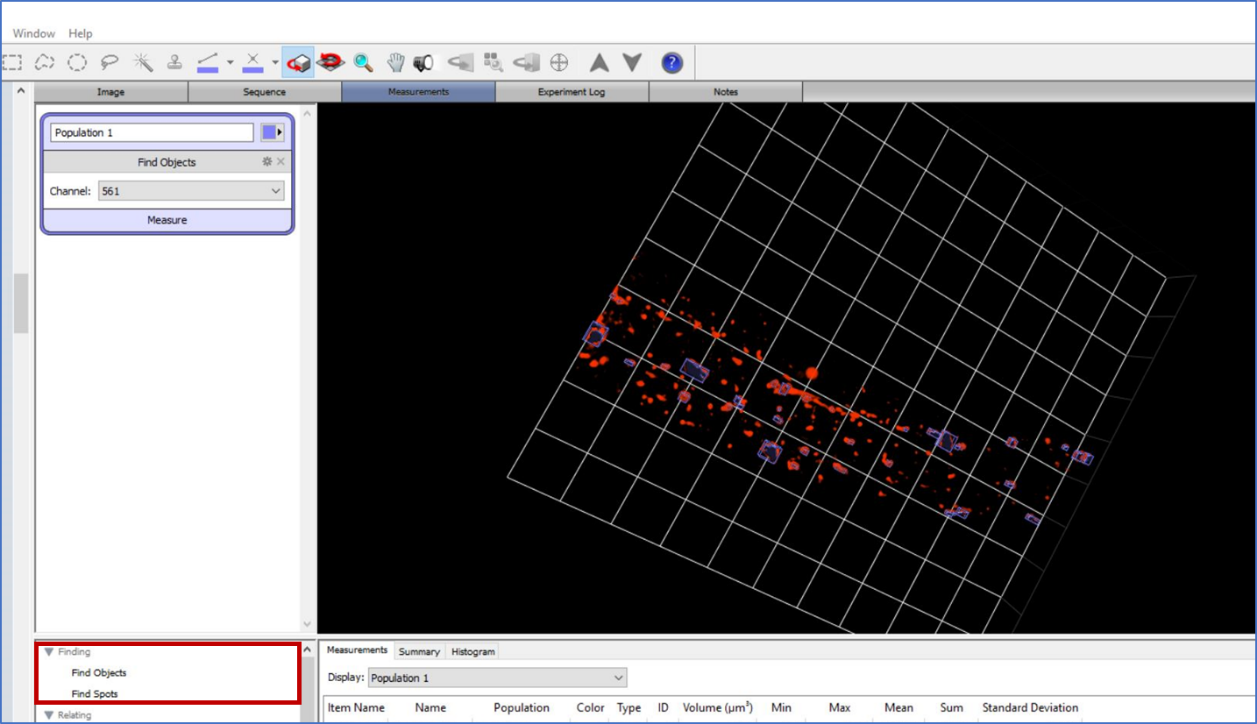
- In the “Measurements” section, select and double-click “Find Objects” to open the window. Volocity automatically counts every single fluorescence object and measures 3D volume including vesicular and tubular lysosomes. Note: at day 5 and day 9, there are many tubular lysosomes formed in wild-type animals. So, we manually pick the vesicular lysosomes as many as possible in Volocity. The individual value of each selected object will be grouped together to calculate the total and average volume of vesicular lysosomes.
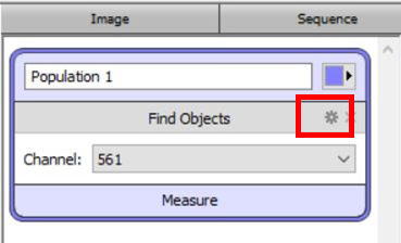
- To cover more objects during measurement, we change the settings by clicking button shown in the find objects window.
- Change “minimum object” size to 0.05 μM3, change fluorescence intensity level by adjusting the offset threshold. Then click OK.
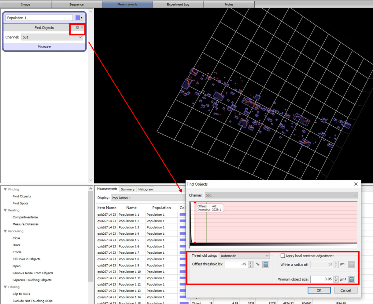
- Go to “Summary” page. The “count” of objects is the total number of vesicular lysosomes. On the “Volume” column, “mean” volume is the average size of vesicular lysosome,
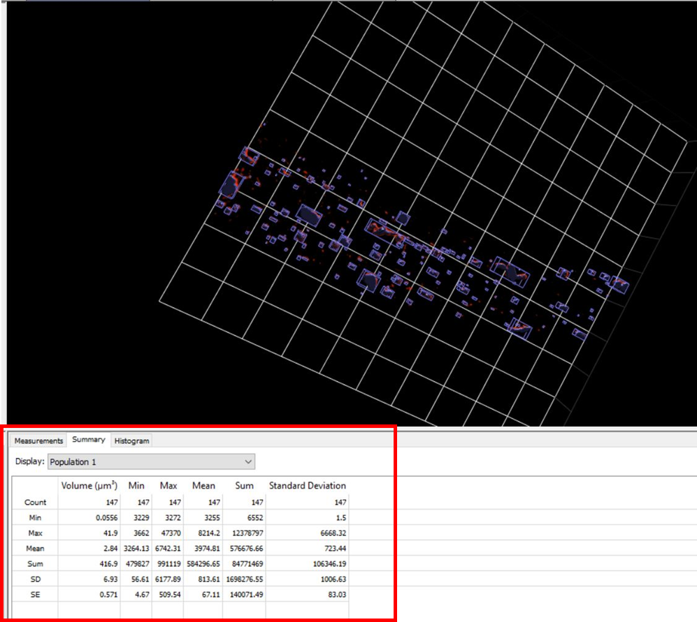
For the total volume of lysosomes:
- In the “Measurements” section, select and double-click “Find Objects” to open the window. Let the software counts and quantifies every single object, which includes vesicular and tubular lysosomes. Finally go to “Summary” page, “sum” of volume indicates the total volume of all lysosomes.
- Li, X, Sun, Y, Li, L and Wang, X(2021). Quantification of lysosome number and volume. Bio-protocol Preprint. bio-protocol.org/prep1335.
- Sun, Y., Li, M., Zhao, D., Li, X., Yang, C. and Wang, X.(2020). Lysosome activity is modulated by multiple longevity pathways and is important for lifespan extension in C. elegans. eLife. DOI: 10.7554/eLife.55745
Do you have any questions about this protocol?
Post your question to gather feedback from the community. We will also invite the authors of this article to respond.