
- Home
- Protocols

-

Oxygen measurements
Published: Aug 2, 2021 Views: 695
Original research article
The authors used this protocol in:
Jun 19, 2018
We measured oxygen concentration in vivo using a hydrogel oxygen microsensor coupled with an optical reader.
The microsensors work based on the principle of phosphorescence quenching of metalloporphyrins, a well-established technique with excellent sensitivity and specificity to physiologic oxygen. They are composed of a biocompatible hydrogel, poly (2-hydroxyethyl methacrylate) (pHEMA) and a near infrared (NIR) oxygen-sensitive palladium-benzoporphyrin molecule (Pd-MABP) (for more details see Montero-Baker et al., 2015; Wisniewski et al., 2017; Chien et al., 2017). The Pd-MABP molecules are covalently attached to the pHEMA hydrogel, ensuring that the sensing chemistry is retained in the hydro-gel structure. The sensors were manufactured by Profusa Inc. (https://profusa.com/) and cut to a miniature size (0.75 x 0.75 x 2.5 mm). For each measurement, a sensor was placed via rectal insertion in the distal large intestine (~1 cm from anus) of mice under isofluorane anesthesia.
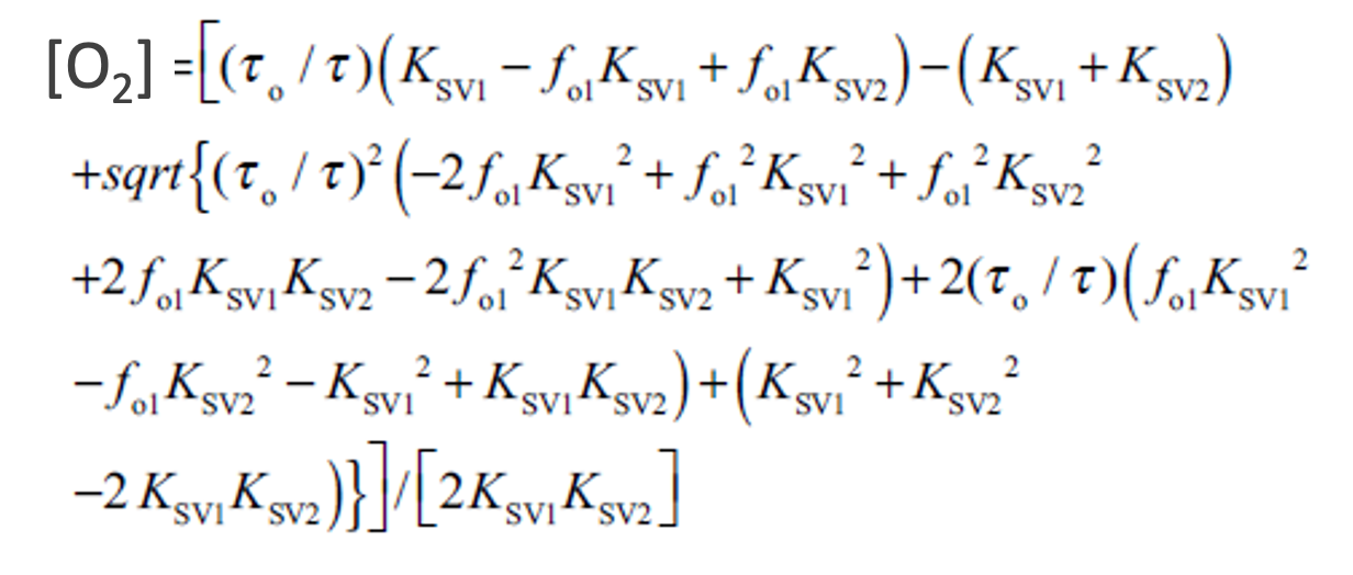
The non-invasive optical reader (also manufactured by Profusa) was manually positioned over the sensor and a LED pulsed illumination light into the skin above the sensor. A photodetector collected emission light emanating from the sensor for two minutes. The phosphorescent lifetime, a property of the oxygen sensitive dye, was measured and averaged across the two minutes before conversion to oxygen concentration. The temperature was assumed to be at 37 ̊C to convert the lifetime measurement (τ) to oxygen concentration with the following equation:
Upon reawakening from anesthesia, the sensor was passed naturally from the mouse via peristalsis and disposed of.
Do you have any questions about this protocol?
Post your question to gather feedback from the community. We will also invite the authors of this article to respond.