
- Protocols

- About
-


Optimized ex vivo preservation and FACS isolation of Drosophila ovarian germline and follicle cells for cell-type-specific gene-expression analysis

Last updated date: Sep 18, 2026 Views: 31 Forks: 0
 Download PDF
 Ask a
question
Ask a
question
 How to
cite
How to
cite
 Favorite


Optimized ex vivo preservation and FACS isolation of Drosophila ovarian germline and follicle cells for cell-type-specific gene-expression analysis
Rounab Sarkar¹, Sautan Show¹, Anjali Mohan¹, Upendra Nongthomba¹*
1 Department of Developmental Biology and Genetics; Indian Institute of Science; Bengaluru – 560012, India.
* Corresponding author: Prof. Upendra Nongthomba, upendra@iisc.ac.in
Abstract:
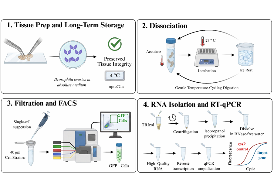
Cell-type-specific molecular profiling of the Drosophila melanogaster ovary is important for understanding germline development, somatic–germline interactions, and the molecular regulation of oogenesis. However, obtaining sufficient numbers of viable, cell-type-specific populations for downstream RNA analysis remains technically challenging because ovarian tissues are sensitive to prolonged ex vivo manipulation and enzymatic dissociation. Here, we describe an optimized workflow for the ex vivo preservation, dissociation, fluorescence-activated cell sorting (FACS), and gene-expression analysis of Drosophila ovarian cells. The protocol incorporates an insulin- and serum-supplemented Schneider's medium for ex vivo tissue maintenance, controlled Accutase-mediated enzymatic dissociation combined with gentle mechanical disruption, filtration of the resulting single-cell suspension, and FACS-based isolation of GFP-labeled germline cell and somatic follicle cell populations. Under the conditions tested, dissected ovaries could be maintained at 4 °C for up to 72 h before processing, allowing ovarian tissue to be accumulated over multiple dissection sessions. Following dissociation and FACS, total RNA was successfully recovered from the sorted populations and used for cDNA synthesis, reverse-transcription PCR (RT-PCR) and reverse-transcription quantitative PCR (RT-qPCR). Analysis of cell-type-associated transcripts by RT-PCR/RT-qPCR enabled molecular assessment of the enriched germline and somatic follicle populations and demonstrated the suitability of the isolated RNA for downstream quantitative gene-expression analysis. Overall, this workflow provides a practical and adaptable approach for isolating defined ovarian cell populations from Drosophila and facilitates targeted gene-expression analysis.
Keywords: Drosophila melanogaster, Ovarian cell isolation, Cell sorting, RNA extraction
Drosophila ovarian organization and oogenesis
An adult female Drosophila melanogaster has two ovaries, each containing approximately 16–20 egg-producing ovarioles, depending on the genotype and physiological condition. Each ovariole consists of a linear series of progressively developing egg chambers that originate from the germarium. The germarium is located at the anterior tip of each ovariole and contains both germline and somatic stem-cell populations. During early oogenesis, a germline stem cell gives rise to a cystoblast that undergoes four rounds of incomplete mitotic division to generate a 16-cell germline cyst. One of these cells adopts the oocyte fate, whereas the remaining 15 differentiate into nurse cells [Vallés AM & Huynh JR., 2020]. The resulting 16-cell germline cyst, comprising one oocyte and 15 nurse cells, becomes surrounded by a monolayer of somatic follicle cells to form an egg chamber. As oogenesis progresses, successive egg chambers develop along the anterior–posterior axis of the ovariole and undergo a series of morphological and physiological changes before mature eggs are released. Drosophila oogenesis is conventionally divided into 14 developmental stages. Early stages involve germline cyst formation, egg-chamber organization, growth, and patterning, whereas later stages are characterized by vitellogenesis, oocyte maturation, and preparation for ovulation. [Spradling, 1993; Bastock & St Johnston, 2008; Jouandin et al., 2022].
Functional roles of germline and follicle cells
During oogenesis, germline and somatic follicle cells undergo extensive structural and functional changes essential for proper oocyte development. The germline comprises an oocyte and nurse cells, which support oocyte growth and maturation. Nurse cells are highly polyploid and synthesize RNAs, proteins, and other cytoplasmic components that are subsequently transported to the growing oocyte before they undergo programmed cell death [Becalska, A.N. & Gavis, E.R., 2009; Kugler, J.M. & Lasko, P., 2009]. Moreover, the germ cells maintain close contacts with follicle cells through gap junctions, allowing coordinated intercellular signaling throughout egg-chamber development. Follicle cells form a somatic epithelium around the germline and provide both structural and signaling functions. They contribute to eggshell formation [Spradling & Mahowald, 1980; Waring, 2000], vitellogenesis [Brennan et al., 1982], embryonic axis patterning [Schüpbach, T., 1987; Neuman-Silberberg & Schüpbach, 1993; González-Reyes et al., 1995], and transport of yolk proteins [Schonbaum et al., 1995; Schonbaum et al., 2000]. Thus, interactions between germline and follicle cells are essential for normal oogenesis.
Technical challenges in ovarian cell isolation
Given the biological importance of germline and follicle cells in the Drosophila ovary, characterizing their molecular states at different stages of oogenesis is essential for understanding fundamental processes in cell and developmental biology. Reverse-genetic approaches, particularly RNA interference (RNAi) combined with the GAL4-UAS system, are widely used to achieve spatiotemporal depletion of target transcripts in defined cell populations. However, validating RNAi-mediated knockdown in specific ovarian cell types can be challenging because only limited numbers of target cells can be recovered. RT-qPCR provides a sensitive method for assessing transcript-level changes but requires enough high-quality RNA [Mainland et al., 2017; Vallés & Huynh, 2020].
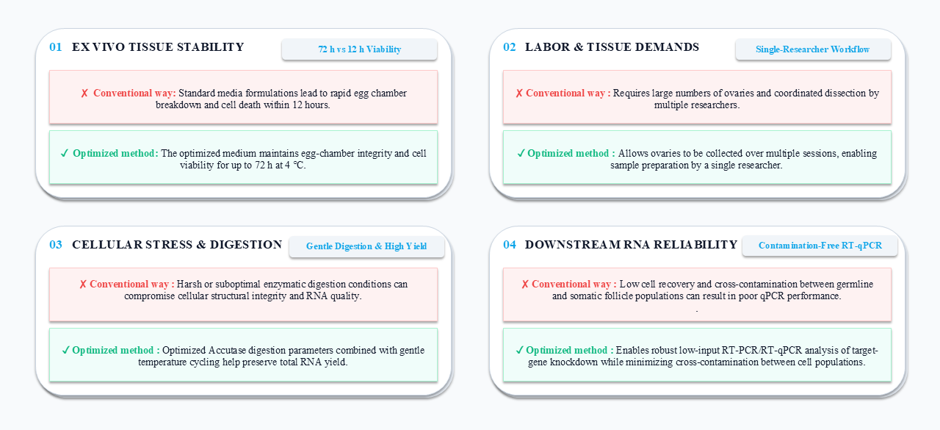
Isolation of ovarian cells is particularly challenging because the tissue is sensitive to prolonged ex vivo manipulation, which can affect cell viability and RNA quality. During Drosophila oogenesis, the developing egg chamber comprises exactly 16 germline cells (15 nurse cells and one oocyte) encased within a simple somatic epithelial monolayer. This follicular epithelium proliferates through active mitotic divisions during the early stages of development, reaching a final pool of approximately 650 to 1,000 follicle cells by the end of Stage 6 [Margolis & Spradling, 1995; Bilder & Haigo, 2012], at which point mitotic divisions cease and the cells transition to endocycling. Consequently, a substantial number of ovaries must often be dissected and processed within a limited time to obtain sufficient material for downstream RNA analysis (Fig. 2). The challenges associated with the convention are as follows:
Limited ex vivo survival: Prolonged ex vivo maintenance can lead to deterioration of egg chambers, restricting the time available for tissue collection, processing, and cell isolation (Fig. 2).
High starting-material and labor requirements: Obtaining enough target cells may require the dissection of large numbers of ovaries within a limited period, increasing the time and labor required for sample preparation (Fig. 2).
Low RNA recovery and quality: Enzymatic and mechanical dissociation can stress or damage ovarian cells, potentially reducing cell recovery and compromising the quantity and quality of RNA available for downstream analysis (Fig. 2).
Key methodological advantages
This protocol addresses key limitations of conventional Drosophila ovarian cell-isolation methods through several methodological improvements. Our optimized ex vivo culture formulation maintains ovarian tissue integrity and GFP signal for up to 72 h, whereas standard media cause rapid egg-chamber deterioration within 12 h. Consequently, a single researcher could do the whole harvesting and isolation procedure without relying on a coordinated dissection team. Controlled enzymatic digestion combined with gentle mechanical dissociation minimizes cellular damage while preserving RNA integrity during single-cell preparation. Overall, this simple and cost-effective pipeline yields high-quality RNA suitable for downstream RT-qPCR analysis. The method overcomes low-input limitations and provides a reliable way to assess the effectiveness of GAL4-UAS/RNAi knockdown in specific cell types, with a clear distinction between germline and somatic follicle cells(Fig. 2).
Methodological basis and adaptations
The cell isolation and profiling protocol described in this study represents a consolidated, optimized workflow developed by adapting and modifying key methodologies from several established methods in Drosophila reproductive biology and flow cytometry:
ex vivo culture and tissue stabilization
Reference Followed: Peters, N. C., & Berg, C. A. (2016). in vitro culturing and live imaging of Drosophila egg chambers: A history and adaptable method.
Adaptation:We adapted the ex vivo culture principles described by Peters and Berg (2016) to establish an insulin-supplemented cold-storage (4 °C) medium that maintains cell viability and follicle and germline marker integrity for up to 72 h, allowing ovaries to be collected over several days prior to sorting.
Enzymatic Dissociation and FACS Gating
Reference Followed: Vallés, A. M., & Huynh, J. R. (2020). Isolation of stage-specific germ cells using FACS in Drosophila germarium.
Adaptation: We adapted the cell dissociation and flow cytometry gating strategies described by Vallés and Huynh (2020) for sorting germline cells. We modified the enzymatic digestion profile by implementing an intermittent, temperature-cycling Accutase treatment (alternating digestion at 25 °C–27 °C with brief incubation on ice) to facilitate tissue dissociation while limiting prolonged exposure to enzymatic and mechanical stress..
Follicle Cell Handling and Marker Validation
Reference Followed: Prasad, M., & Montell, D. J.(2007). Cellular and molecular mechanisms of border cell migration analyzed using time-lapse live-cell imaging and Prasad, M. (2015). Border cell migration: A model system for live imaging and genetic analysis of collective cell movement.
Adaptation: Dissection and somatic cell-handling procedures were adapted from the methods described by Prasad (2015) for the analysis of follicle-cell and border-cell behavior. Specifically, gentle mechanical handling of follicle cell layers (including migrating border cell clusters marked by the BS-6458 reporter line) was optimized to prevent epithelial disruption, ensuring clean cell separation and minimal somatic-to-germline cross-contamination during FACS.
Methods
Reagents for media preparation
1. Basal medium: Drosophila Schneider’s Medium (Sigma-Aldrich, Cat. No. S0146 or equivalent).
2. Fetal bovine serum (FBS): Heat-inactivated FBS (Sigma-Aldrich, Cat. No. F2442 or equivalent).
3. Bovine insulin: Lyophilized powder (Sigma-Aldrich, Cat. No. I5500 or equivalent).
4. Antibiotic cocktail: Penicillin-Streptomycin solution containing 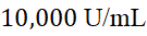 Penicillin G-sodium and
 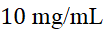 Streptomycin sulfate in
  saline (HIMEDIA, Cat. No. A002A) or equivalent.
Media preparation
Drosophila Schneider’s medium was supplemented with 20% (v/v) heat-inactivated fetal bovine serum (FBS; comprising 10% filtered and 10% unfiltered FBS) Note1and 1% penicillin-streptomycin. A stock solution of bovine insulin ( ) was prepared by dissolving insulin in acidified ultrapure water (
 concentrated
  in
in  Milli-Q
  ) and added to the medium to achieve a final working concentration of
 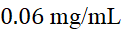 Note2. The final pH of the formulation was adjusted to 6.85-6.95. This complete formulation is designated as "absolute medium." All preparation steps were conducted under strict aseptic conditions in a laminar flow hood.
*Note 1 (FBS fractionation): To achieve the best nutritional support, a mixture of 10% membrane-filtered (0.22 µm PVDF syringe filter (Whatman™ Uniflo™, Cat. No. 9913-2502)) and 10% untreated, heat-inactivated FBS was used. This is because too much sterile filtration can remove important trace growth factors and lipid-bound components from serum [Freshney, R. I., (2010); van der Valk et al., 2018].
*Note 2 (Insulin optimization): The final insulin concentration was set to 0.06 mg/mL to provide metabolic support while minimizing potential alterations in ovarian tissue development.
Fly stocks
1. w¹¹¹⁸ (Negative Control): A non-GFP control used to establish baseline background autofluorescence and define GFP-negative gating thresholds for FACS analysis and confocal imaging.
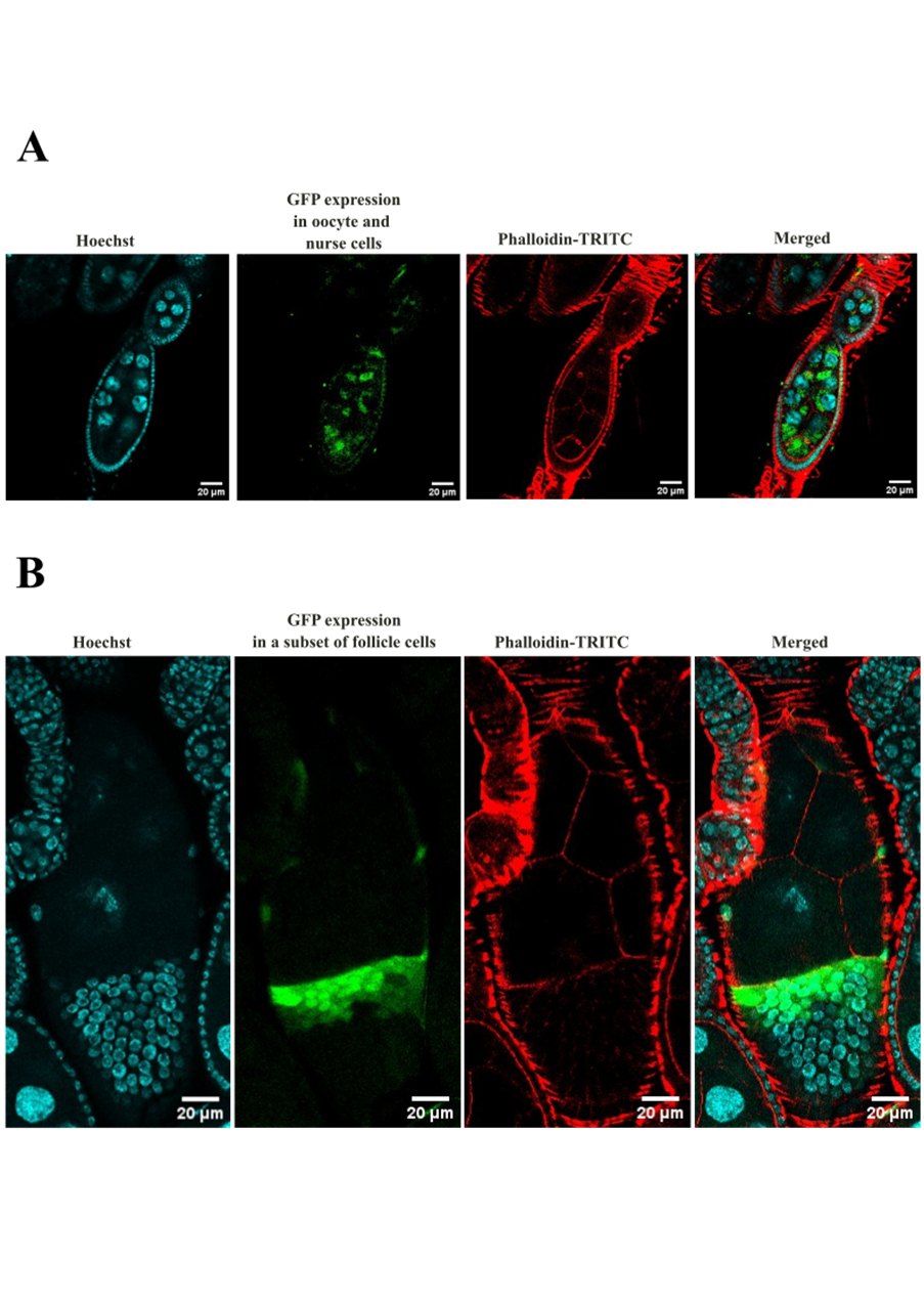
2. nos-GAL4; UAS-GFP-NLS (Germline Reporter): Drives GFP expression in the germline and was used for germline-specific visualization and staging (Fig. 3-A).
3. BS 6458 (Somatic Follicle Reporter): Drives targeted GFP expression in migrating border cell clusters and subsets of follicle cells to monitor somatic follicle dynamics (Fig. 3-B).
Fly husbandry
Fly pad and etherizer
Fly incubators (29 °C and 25 °C)
Dissecting microscope (Model-Olympus SZ51) with dark stage and side illumination
Fly food vials/bottles
Nunc 35 mm Dish IVF Certified Dishes (Cat. No. 150255)
Fly preparation
Females, along with males (2:1 ratio), are maintained on fresh vials supplemented with dry yeast for at least 2 days before dissection to promote active oogenesis before dissection. Two-day-old mated females were used for ovarian dissection. Selecting young females enriched with Stage 6 to Stage 10 egg chambers ensures a high density of highly viable somatic and germline cells with minimal background debris.
Ovary dissection
Anesthetize female flies using an etherizer and transfer them to a fly pad.
Transfer one anesthetized female fly to a glass slide with absolute medium placed under a dissecting microscope.
Using a pair of forceps, gently hold the thorax with one pair and open the posterior abdomen with the other to release the ovaries (white and opaque) into the medium. Gently separate the ovaries from the surrounding tissues of the fly body and keep them submerged in medium.
Repeat the process for remaining female flies and collect the dissected ovaries in absolute medium on a glass slide.
After dissection, transfer cleaned ovary pairs (free of gut/Malpighian tubule contamination) into Nunc 35 mm Dish IVF Certified Dishes.
Ex vivo Ovary Collection and Short-Term Storage
To achieve optimal downstream yields, a minimum threshold of  pairs of ovaries are required. Freshly dissected ovaries were maintained ex vivo in 35 mm culture dishes containing sufficient absolute medium until the required number were collected. Ovaries were stored under sterile conditions at 4 °C for up to 72 h Note3 with absolute medium replacement every 12 h.
Once the required number of ovaries was harvested, samples were processed immediately for FACS.
*Note 3 (Temperature and Storage Optimization): Proper temperature control during storage is crucial to preserve tissue viability and the GFP signal. Dissected ovaries ex vivo can be stored in absolute media at 4 °C for up to 72 h or at 25 °C (room temperature) for up to 48 h without loss of GFP expression. The ovaries may be pooled directly in a sterile sample collection plate during dissection. However, the incubation medium has to be changed every 12 h to ensure the best viability and minimize metabolic deterioration.
Reagents and consumables for FACS:
1. Enzymatic and Dissociation Reagents
Accutase® Cell Dissociation Reagent (Sigma-Aldrich, Cat. No. A6964)
Usage: Used for gentle tissue dissociation to yield highly-viable single cells.
2. Filtration and Cell Isolation
40 μm Cell Strainer (NEST Biotechnology, Cat. No. 258366)
Usage: Essential for filtering out tissue debris and clumps to prevent FACS nozzle clogging.
3. Plasticware and Tubes
1.5 mL Microcentrifuge Tubes (Eppendorf)
Usage: Used for tissue collection, enzymatic digestion, centrifugation, and cell recovery.
5 mL Round-Bottom FACS Tubes (Polypropylene)
Brand: Falcon® (Corning, Cat. No. 352063)
Usage: Standard tubes for loading samples directly onto the flow cytometer.
Tissue Digestion and Single Cell Isolation
1. Tissue Collection & Initial Pelleting
- Step 1.1: Using a wide-bore pipette tip (cut the end of a sterile 200 µL or 1000 µL tip to widen the opening), carefully transfer the dissected ovaries (minimum 200 pairs) in dissecting medium into a 1.5 mL microcentrifuge tube.
- Step 1.2: Centrifuge at 300 × g (approx. 1,200–1,500 RPM, depending on your rotor radius) for 3 min at 4 °C to pellet the intact tissue.
- Step 1.3: Carefully discard the supernatant without disturbing the tissue pellet.
2. Enzymatic digestion & dissociation
Step 2.1: Add 150 µL of Accutase (pre-warmed) Note4.
Step 2.2: Dissociate the tissue by pipetting up and down gently but firmly using a standard 200 µL pipette. Avoid introducing air bubbles, as the air-water interface is highly damaging to fragile ovarian cells and causes massive cell lysis.
Step 2.3: Incubate the tube at 25 °C (room temperature) or in a 27 °C dry bath for 15–20 min total. To facilitate complete tissue breakdown without overheating the cells, perform the following temperature-cycling steps: Every 3 to 5 min during the incubation, remove the tube and gently pipette the sample 10–15 times. Immediately after pipetting, transfer the tube to ice for 1–2 min to rest and stabilize the fragile cells before returning it to the dry bath/room temperature.
3. Quenching & pelleting single cells
Step 3.1: Immediately stop the enzymatic reaction by adding 300 µL of absolute medium.
Step 3.2: Centrifuge at 400 × g for 5 min at 4 °C to pellet the single cells.
Step 3.3: Carefully discard the supernatant and gently resuspend the cell pellet in 300 µL of cold, fresh absolute medium.
4. Filtration & FACS preparation
Step 4.1: Place a 40 µm cell strainer on top of a fresh 5 mL polypropylene FACS tube.
Step 4.2: Wet the strainer membrane with 50 µL of cold absolute medium first, then load your resuspended cell sample onto the strainer to filter out mature eggs, cellular aggregates, and undigested tissue debris.
Step 4.3: Rinse the strainer once with an additional 100 µL of cold absolute medium to maximize cell recovery.
Step 4.4: Keep the filtered single-cell suspension on ice and proceed immediately to the FACS facility.
5. FACS collection
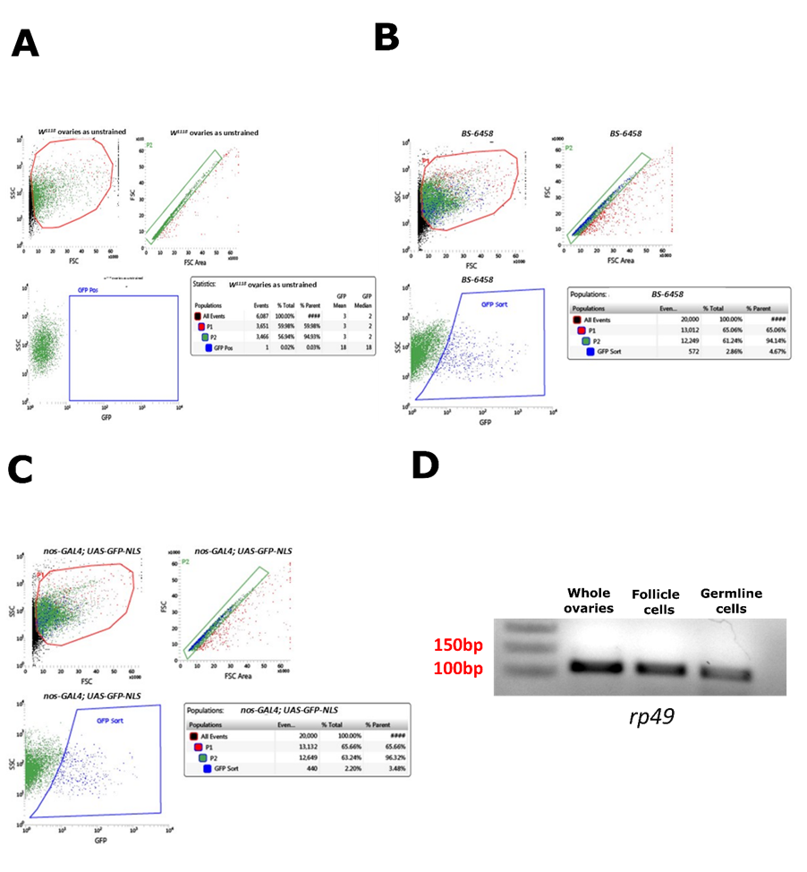
Step 5.1: Set up your collection FACS tube by adding 300 µL of absolute medium to the bottom of the tube. This helps preserve GFP-positive cell viability upon collection into the tube. (Fig. 4-A, B, C).
*Note 4: For highly efficient digestion, pre-warm the Accutase alone in a 37 °C dry bath for 5 min before adding it to the tissue. Once added to the ovaries, immediately bring the temperature down to the 25° C–27 °C range as outlined in step 2.3. To accommodate a larger sample volume, the quantity of Accutase can be increased up to 300 µL.
6. Post-sorting harvest and RNA preservation
Step 6.1: After the FACS run, keep the collection tube on ice. Gently pipette the sorted GFP-positive cells to ensure they are thoroughly resuspended in the medium.
Step 6.2: Transfer the entire volume of the medium containing the sorted cells into a sterile, RNase-free 1.5 mL microcentrifuge tube.
Step 6.3: Centrifuge the tube at 5,000 RPM (approximately 2,000 × g) for 10 min at 4 °C to pellet the sorted cells Note5.
Step 6.4: Carefully aspirate and discard the supernatant medium. Take extreme care during this step, as the pellet of sorted cells will be very small and fragile.
Step 6.5: Add 200 µL of TRIzol™ Reagent directly to the cell pellet. Homogenize and resuspend the pellet thoroughly by pipetting up and down until the sample is completely clear and no visible cell clumps remain.
Step 6.6: Immediately transfer the homogenized lysate to a -80 °C to preserve RNA integrity for subsequent RNA isolation and RT-PCR/qPCR.
*Note 5: If the cell pellet is not clearly visible after centrifugation (common with low-yield FACS-sorted cells), increase the centrifugation speed to 6,000–8,000 RPM (approximately 3,000–5,000 g) and extend the run time to 12–15 min at 4 °C.
RNA extraction, cDNA synthesis, and RT-qPCR analysis
Total RNA was isolated from sorted Drosophila ovarian cells using the standard TRIzol™ Reagent (Thermo Fisher Scientific) protocol. The concentration and purity of the extracted RNA were assessed using a spectrophotometer.
For first-strand cDNA synthesis, total RNA was reverse transcribed using the iScript™ Reverse Transcription Supermix (Bio-Rad, Cat. No. 1708841) according to the manufacturer’s instructions. The reaction was performed in a thermal cycler using the following program: priming at 25 °C for 5 min, reverse transcription at 46 °C for 20 min, and RT inactivation at 95 °C for 1 min.
RT-PCR (Fig. 4-D) and RT-qPCR were subsequently performed using cDNA templates, gene-specific primers. Ribosomal Protein 49 (rp49) was utilized as the internal reference/housekeeping gene for normalization.
Key technical considerations
Efficient cell recovery and preservation of sample quality depend on three key methodological parameters. Below, we outline these procedural parameters alongside empirical recommendations derived from our optimization pipeline.
1. Standardization of fly age and stage selection
To maximize single-cell yields and minimize debris during sorting, it is critical to select fly ovaries of the correct developmental stage. The use of aged female flies (e.g., >3 days post-eclosion) is strongly discouraged, as their ovaries accumulate fully mature, vitellogenic oocytes (Stage 14) that resist gentle enzymatic digestion. These mature egg chambers can release yolk-rich material during dissociation, increasing debris and background fluorescence, which clogs the 40 µm cell strainer and disrupts FACS gating. We strongly recommend using young, well-fed mated females (2–3 days post-eclosion) conditioned on fresh yeast paste, as their ovaries are highly enriched in Stage 6 to Stage 10 egg chambers, which dissociate cleanly into a high-density single-cell suspension (Huynh & St Johnston, 2004; Bastock & St Johnston, 2008).
2. Somatic driver selection and target cell yield
In our validation, the somatic follicle cell reporter line utilized (BS-6458) exhibits restricted GFP expression in a subset of the follicular epithelium rather than the entire follicle population. While this is ideal for analyzing specific sub-lineages, it naturally limits the total sorted cell count. For downstream applications requiring maximum cell yield or broad somatic profiling, we recommend substituting restricted reporters with pan-follicle cell drivers (e.g., c204-GAL4) to drive GFP expression across the entire follicular epithelium, thereby potentially increasing the sorting output (Vallés & Huynh, 2020).
3. Tissue Scaling for Rare Germline Populations
Germline-specific sorting can require substantial starting material because each developing egg chamber contains a limited number of germline cells relative to the surrounding somatic population. In our optimization trials, the dissection of approximately 200 ovaries (from 100 female flies) yielded approximately 48,000 GFP-positive germline cells (Fig. 4-C). Therefore, for robust, high-integrity downstream molecular analyses (such as low-input RNA-seq or RT-qPCR), we strongly advise scaling starting material to a minimum of 200–300 pair of ovaries. Importantly, the 72 h ex vivo stability enabled by our absolute medium formulation makes this scale entirely feasible for a single researcher working independently.
Summary of the Protocol
This protocol provides an optimized workflow for isolating GFP-labeled germline and somatic follicle cells from Drosophila ovaries. The “absolute medium” supports ex vivo ovarian storage at 4 °C for up to 72 h, enabling tissue collection across multiple dissection sessions. Combined with controlled dissociation and FACS, the workflow yields cell-type-specific populations suitable for downstream RNA isolation and gene-expression analysis.
Author Contribution: R.S. and S.S. optimized the protocol and performed the experiments. R.S., S.S., A.M., and U.N. drafted the manuscript. U.N. supervised the project and reviewed the final manuscript draft. All authors have read and approved the final manuscript.
Acknowledgment: The authors would like to acknowledge BioRender (BioRender.com) for assistance in generating the graphical abstract (Fig. 1). The authors also acknowledge Grammarly (Grammarly, Inc.) for proofreading, grammatical corrections, and writing/language improvements during the preparation and refinement of this manuscript. The authors also thank Bilal Jamadar for technical assistance with FACS.
Conflict of interest: The authors declare no conflict of interest.
References
Bastock, R., & St Johnston, D. (2008). Drosophila oogenesis. Current Biology, 18, R1082–R1087. https://doi.org/10.1016/j.cub.2008.09.011
Becalska, A. N., & Gavis, E. R. (2009). Lighting up mRNA localization in Drosophila oogenesis. Development, 136, 2493–2503. https://doi.org/10.1242/dev.032391
Bilder, D., & Haigo, S. L. (2012). Expanding the morphogenetic repertoire: perspectives from the Drosophila egg. Developmental Cell, 22(1), 12–23. https://doi.org/10.1016/j.devcel.2011.12.003
Brennan, M. D., Weiner, A. J., Goralski, T. J., & Mahowald, A. P. (1982). The follicle cells are a major site of vitellogenin synthesis in Drosophila melanogaster. Developmental Biology, 89, 225–236. https://doi.org/10.1016/0012-1606(82)90310-X
Freshney, R. I. (2010). Culture of animal cells: a manual of basic technique and applications (6th ed.). John Wiley & Sons. ISBN: 978-0470528129.
González-Reyes, A., Elliott, H., & St Johnston, D. (1995). Polarization of both major body axes in Drosophila by gurken-torpedo signalling. Nature, 375, 654–658. https://doi.org/10.1038/375654a0
Huynh, J. R., & St Johnston, D. (2004a). The origin of asymmetry: Early polarization of the Drosophila germline cyst and oocyte. Current Biology, 14, R438–R449. https://doi.org/10.1016/j.cub.2004.05.040
Huynh, J. R., & St Johnston, D. (2004b). The origin of asymmetry in Drosophila oogenesis. Seminars in Cell & Developmental Biology, 15, 89–98. https://doi.org/10.1016/j.semcdb.2003.09.012
Jevitt, A., Ghosh, S., & Lehmann, R. (2020). Single-cell RNA-seq reveals developmental and cell-type-specific signatures in the Drosophila ovary. Cell Reports, 32, 108155. https://doi.org/10.1016/j.celrep.2020.108155
Jouandin, P., Ghiglione, C., & Noselli, S. (2022). Ex vivo culture and live imaging of Drosophila egg chambers. Journal of Visualized Experiments, 181, e63529. https://doi.org/10.3791/63529
Kugler, J. M., & Lasko, P. (2009). Localization, anchoring, and translational control of oskar, gurken, bicoid and nanos mRNA during Drosophila oogenesis. Fly, 3, 15–28. https://doi.org/10.4161/fly.3.1.15
Mainland, J. D., Li, Y. R., Zhou, T., Liu, H. L., & Matsunami, H. (2017). Optimization of low-input RNA-seq and qPCR workflows for FACS-sorted cell populations. BMC Genomics, 18, 412. https://doi.org/10.1186/s12864-017-3801-4
Mainland, R. L., Lyons, T. A., Ruth, M. M., & Kramer, J. M. (2017). Optimal RNA isolation method and primer design to detect gene knockdown by qPCR when validating Drosophila transgenic RNAi lines. BMC Research Notes, 10, 596. https://doi.org/10.1186/s13104-017-2918-z
Margolis, J., & Spradling, A. C. (1995). Identification and behavior of epithelial stem cells in the Drosophila ovary. Development, 121(11), 3797–3807. [PMID: 8521831]
Neuman-Silberberg, F. S., & Schüpbach, T. (1993). The Drosophila dorsoventral patterning gene Gurken produces a dorsally localized RNA and encodes a TGFα-like protein. Cell, 75, 165–174. https://doi.org/10.1016/0092-8674(93)80075-8
Peters, N. C., & Berg, C. A. (2016). In vitro culturing and live imaging of Drosophila egg chambers: A history and adaptable method. Methods, 98, 70–81. https://doi.org/10.1016/j.ymeth.2015.11.020
Prasad, M. (2015). Border cell migration: A model system for live imaging and genetic analysis of collective cell movement. Methods in Molecular Biology, 370, 177–186. https://doi.org/10.1007/978-1-59745-388-214
Prasad, M., & Montell, D. J. (2007). Cellular and molecular mechanisms of border cell migration analyzed using time-lapse live-cell imaging. Developmental Cell, 997–1005. https://doi.org/10.1016/j.devcel.2007.03.021
Rust, K., Byrnes, L. E., Yu, K. S., Park, J. S., Sneddon, J. B., Tward, A. D., & Nystul, T. G. (2020). A single-cell atlas and lineage analysis of the Drosophila ovary. Nature Communications, 11, 5628. https://doi.org/10.1038/s41467-020-19361-0
Schonbaum, C. P., Lee, S., & Mahowald, A. P. (1995). The Drosophila yolkless gene encodes a vitellogenin receptor belonging to the low density lipoprotein receptor superfamily. Proceedings of the National Academy of Sciences, 92, 1485–1489. https://doi.org/10.1073/pnas.92.5.1485
Schonbaum, C. P., Perrino, J. J., & Mahowald, A. P. (2000). Regulation of the vitellogenin receptor during Drosophila melanogaster oogenesis. Molecular Biology of the Cell, 11, 511–521. https://doi.org/10.1091/mbc.11.2.511
Schüpbach, T. (1987). Germ line and soma cooperate during oogenesis to establish the dorsoventral pattern of egg shell and embryo in Drosophila melanogaster. Cell, 49, 699–707. https://doi.org/10.1016/0092-8674(87)90537-7
Slaidina, M., Banisch, T. U., Gupta, S., & Lehmann, R. (2020). A single-cell atlas of the Drosophila ovary identifies a niche-supporting precursor population. Genes & Development, 34, 239–253. https://doi.org/10.1101/gad.334417.119
Spradling, A. C. (1993). Developmental genetics of oogenesis. In M. Bate & A. Martinez Arias (Eds.), The Development of Drosophila melanogaster. Cold Spring Harbor Laboratory Press. ISBN: 978-0879694234.
Spradling, A. C., & Mahowald, A. P. (1980). Amplification of genes for chorion proteins during oogenesis in Drosophila melanogaster. Proceedings of the National Academy of Sciences, 77, 1096–1100. https://doi.org/10.1073/pnas.77.2.1096
Vallés, A. M., & Huynh, J. R. (2020). Isolation of stage-specific germ cells using FACS in Drosophila germarium. Methods in Cell Biology, 158, 11–24. https://doi.org/10.1016/bs.mcb.2020.02.003
van der Valk, J., Bieback, K., Buta, C., Cochrane, B., Dirks, W. G., Fu, J., Gstraunthaler, G., Hokao, R., Ivanov, C., Keat, A. L., Kolf, A. M., Luzyanina, N., Millitre, D., Mulry, S., Peasley, M., & Weinshilboum, R. (2018). Fetal bovine serum (FBS): Past – Present – Future. ALTEX - Alternatives to Animal Experimentation, 35(1), 99–118. https://doi.org/10.14573/altex.1705101
Waring, G. L. (2000). Morphogenesis of the eggshell in Drosophila. International Review of Cytology, 198, 67–108. https://doi.org/10.1016/s0074-7696(05)19802-8


- SARKAR, R, Show, S, Mohan, A and Nongthomba, U(2026). Optimized ex vivo preservation and FACS isolation of Drosophila ovarian germline and follicle cells for cell-type-specific gene-expression analysis. Bio-protocol Preprint. bio-protocol.org/prep3000.
Do you have any questions about this protocol?
Post your question to gather feedback from the community. We will also invite the authors of this article to respond.
 Post a Question


