
- Home
- Protocols

-

Generation of single cell suspensions of Mtb
Last updated date: Sep 25, 2023 DOI: 10.21769/p2428 Views: 352 Forks: 0
Generation of single cell suspensions of Mycobacteria tuberculosis
Ekansh Mittal* and Jennifer A. Philips*
Division of Infectious Diseases, Department of Medicine, Department of Molecular Microbiology, Washington University School of Medicine, St. Louis, MO, USA
*Corresponding (philips.j.a@wustl.edu, emittal@wustl.edu)
Abstract
Mycobacteria tuberculosis (Mtb) is the causative agent of tuberculosis (TB), one of the leading causes of death worldwide. M. tuberculosis has growth characteristics that make it a challenge to work with experimentally, including extremely slow and clumpy growth. Investigators have devised a variety of approaches to disperse M. tuberculosis, but there is little consistency in the approaches or attention to how these methods impact experimental findings. Our recent publication in eLife highlights the potential experimental artifacts introduced by widely used methodologies in Mtb-host interaction studies and their outcomes in models of TB infection1. To assist in bringing consistency to these endeavors, we have outlined detailed specific methods for generating single cell suspensions of Mtb in this protocol.
Note: Work with M. tuberculosis must be performed in a Biosafety Level 3 (BSL3) facility by individuals trained and approved for the work. Safety precaution for work with BSL3 agents are not described as part of this protocol. All the work should be done in compliance with local standards of practice.
Materials and Reagents
- Middlebrook 7H9 broth base powder (Sigma-Aldrich, catalog number: M0178)
- Middlebrook OADC growth supplement (Sigma-Aldrich, catalog number: M0678)
- Tyloxapol (Sigma-Aldrich, catalog number: T8761)
- Glycerol (Sigma-Aldrich catalog number: G7893)
- 7H9 liquid medium (see Recipes)
- Phosphate-Buffered Saline (PBS) (Gibco, catalog number: J61196.AP)
- Bacterial strain: Mtb strains H37Rv and Erdman were used in our study.
- DMEM (Gibco, catalog number: 11995-065)
- Fetal bovine serum (FBS) (Gibco, catalog number: A5256701)
- L-Glutamine (Gibco, catalog number: 25030-081)
- Pyruvate (Corning, catalog number: 25-000-cl)
Equipment
- Appropriate biocontainment facilities, biosafety cabinets, disinfectants, and personal protective equipment.
- P20 Pipetman (Gilson, catalog number: F123600)
- P200 Pipetman (Gilson, catalog number: F123601)
- P1000 Pipetman (Gilson, catalog number: F123602)
- 37 °C incubator
- Filtered pipet tips {DNase and RNase free, sterilized microfilter, Crystalgen, Catalog number: SR-5107 (1000 ml), SR-5106 (200 ml), SR-5009 (10 ml)}
- Media bottles {DNase and RNase free, sterile, square, Crystalgen, Catalog number: 24-30 (30 ml), 24-125 (125 ml), 24-250 (250 ml)}
- Serological plastic pipets {DNase and RNase free, sterile, Nunc serological pipet, Catalog number: 170356 (10 ml) and 170357N (25 ml)}
- 0.22 mm Filter unit (GenClone Cell Culture, catalog number: 25-225)
- 10 ml Syringe (BD Bioscience, catalog number: 302995)
- Acrodisc PES syringe filters with supor membrane, sterile - 5 mm, 32 mm (Pall Life Science, catalog number: 4650)
- Conical centrifuge tubes {MIDSCI, sterile, catalog number: C50B (50 ml) and C15B (15 ml)}
- Water bath sonicator (Branson Ultrasonics Corporation, Digital Sonifier 450)
Spectrophotometer (Ultrospec 10, Amersham Biosciences)
Procedure
A. Bacterial Culture
- Prepare 7H9 liquid medium as described below.
- Thaw 1 frozen vial of 1 ml Mtb (stored in 20% glycerol and 80% 7H9 liquid medium) and resuspend in 10 ml 7H9 liquid medium in a 30 ml bottle.
- Grow bacterial culture in an incubator at 37°C with gentle agitation (120 rpm) for one week.
- Transfer 1-2 ml Mtb culture from the growing culture and add to 30-50 ml 7H9 liquid medium in a 125 or 250 ml media bottle.
- Measure the Optical Density (OD)600 every 2 days until the OD600 reaches 0.50-0.80 (mid-log phase).
- Pellet the bacteria by centrifugation at room temperature, 3,000 x g for 5 min.
- Resuspend bacterial pellet in 25 ml PBS to remove detergent.
Repeat step 6 and resuspend bacterial pellet in 25 ml media that will be used for subsequent analysis. For macrophage infections, the media will depend upon the type of macrophages used. For bone marrow-derived macrophages (BMDM), we use 10% BMO (see details in recipe section).
- Dived the bacterial suspension in three different aliquots (8 ml each) in 15 ml falcon tubes and proceed with single cell suspension.
B. Preparing single cell suspensions
Single cell suspensions of Mtb were generated using one or a combination of the following methods:
a. Low-speed spin (sp)
1. Centrifuge 8 ml of bacteria culture from above at 206 x g (1000 rpm) for 10 min.
2. Collect the supernatant in fresh tube and centrifuge at 132 x g (800 rpm) for 8 min.
3. Collected the supernatant and measure the bacterial suspensions with OD600. It should be between 0.04 and 0.12. This value is used to calculate the concentration of bacteria 1 OD600=3 x 10⁸ bacteria per ml.
Note: if the OD600 is more than 0.12, repeat steps 2 and 3 again. If the OD600 is still more than 0.12, dilute the sample 1:1 with the same media and repeat steps 2 and 3 again. Repeat this up to three times as necessary to achieve an OD600 between 0.04 and 0.12 before using for infection. If the OD600 remains above 0.12 after three repeated attempts, there may be an issue with the culture. We recommend refraining from using this culture for further analysis.
b. Filter (5 μm filter; 5μmF)
4. Place the second 8 ml aliquot from above in a 10 mL syringe and then, with gentle pressure applied to the syringe plunger, passed through a 5 mm polyethersulfone (PES) filter (PALL Life Sciences; cat. 4650) as seen in Figure 1.
5. Collect the filtrate and measure the concentrations with OD600. In this preparation method, 1 OD600=2 x 10⁸ bacteria per ml (instead of 1 OD600=3 x 10⁸ bacteria per ml for the spin preparation)
Note: There is a loss of bacteria viability with this method. Hence, it is necessary to use 1.5-times more 5μmF bacilli (based upon OD600) to achieve the same number of viable bacteria. After filtration, we never find that the OD600 is more than 0.12, so we never repeat the filtration process.
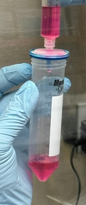
Figure 1: Passing the bacterial culture through 5 μm filter (5μmF).
Bacterial suspension was added to a 10 mL syringe and then, with gentle pressure applied to the syringe plunger, passed through a 5 μm polyethersulfone (PES) filter and collected in a 50 ml tube.
c. Sonication followed by low-speed spin (so/sp)
1. Place the 15 ml conical tube with the third 8 ml bacterial aliquot in a water bath sonicator (Branson Ultrasonics Corporation, Digital Sonifier 450), as seen in Figure 2, and sonicate with 3 pulses lasting 10 second each, with an amplitude of 70% and 5 s rests between each pulse.
Note: Use of a sonicator often produces noise. To mitigate potential hazards, we recommend appropriate sound protection during sonication.
2. Following sonication, perform the low-speed spin procedure as described above.
3. Collect the supernatant and measure the concentration. The OD600 should be between 0.04 and 0.12. The bacterial concentration is calculated as 1 OD600=3 x 10⁸ bacteria per ml.
Note: If the OD600 is more than 0.12, repeat the procedure as mentioned in low- speed spin procedure.
Figure 2: Sonicate the bacteria in water bath ultrasonic sonifier.
Bacterial suspension in a 15 ml conical tube were placed in a water bath ultrasonic sonifier and sonicated with three pulses lasting 10 second each, with an amplitude of 70% and 5 s rests between each pulse.
Recipes
7H9 liquid medium: Dissolve 4.7 g of 7H9 powder in 900 ml dH2O. Add 4 ml 50% glycerol (filter sterilized, final conc. 0.20%), 2.5 ml 20% Tyloxapol (filter sterilized, final conc. 0.05%), and 10% Middlebrook OADC (oleic acid, albumin, dextrose, catalase) supplement, mix well, filter through 0.22 μm filter. Store medium at 4 °C and use within several weeks.
Note: Instead of filtering, the media can be autoclaved and cooled before the adding 10% OADC supplement. Store medium at room temperature in the dark. We recommend against using media that has been stored for longer than a month. Prior to use, add 10% OADC supplement (filter sterilized). Media to which the supplement has been added should be stored at 4 °C and used within several weeks.
10% BMO: Dulbecco's Modified Eagle Medium (DMEM) with 10% FBS, 2 mM L-glutamine, and 1 mM pyruvate (DMEM complete). DMEM complete media was supplemented with 10% L929 cell supernatant [as a source of macrophage colony stimulating factor (M-CSF), 100 U/mL final concentration].
Note: This is the media used for BMDMs at the time of infection. So, bacteria are resuspend in this media when generating single cell suspensions.
Acknowledgments
Funding for this study came from NIAID/NIH (R01 AI087682 and AI30454) to JAP. The authors have no conflicts of interest. This protocol was used for studies reported in Mittal et al. (2023).
References
1. Mittal, E. et al. Single cell prepara2ons of Mycobacterium tuberculosis damage the mycobacterial envelope and disrupt macrophage interac2ons. Elife 12 (2023). https://doi.org/10.7554/eLife.85416
- Mittal, E and Philips, J(2023). Generation of single cell suspensions of Mtb. Bio-protocol Preprint. 10.21769/p2428.
- Mittal, E., Roth, A. T., Seth, A., Singamaneni, S., Beatty, W. and Philips, J. A.(2023). Single cell preparations of Mycobacterium tuberculosis damage the mycobacterial envelope and disrupt macrophage interactions. eLife. DOI: 10.7554/eLife.85416
Do you have any questions about this protocol?
Post your question to gather feedback from the community. We will also invite the authors of this article to respond.