

Pancreatic Acinar Cell 3-Dimensional Culture
胰腺腺泡细胞的三维培养

发布: 2013年10月05日第3卷第19期 DOI: 10.21769/BioProtoc.930 浏览次数: 15115
评审: Lin FangFanglian HeAnonymous reviewer(s)
参见作者原研究论文
The authors used this protocol in:
Apr 2013


Abstract
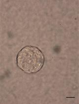
Normal pancreatic acinar cells are difficult to maintain on traditional plastic culture surfaces due to their physical properties of housing large quantities of digestive enzymes and the formation of intercellular tight junctions and gap junctions (Apte and Wilson 2005; Rukstalis et al., 2003). However, placing primary acinar cells within a 3-dimensional matrix (3D-culture) maintains the cells for sufficient time so that they can be monitored for physiological changes to different stimuli. We have used a modified collagen 3D-culture system that has been adapted from Means et al. (2005) to model the very early events associated with pancreatic cancer development. In this model, KrasG12D-expressing pancreatic acinar cells, or wildtype acinar cells treated with EGFR-dependent growth factors (i.e., TGFα), convert to ductal cysts that mimic the acinar-to-ductal metaplasia (ADM) stage that precedes formation of Pancreatic Intraepithelial Neoplasia (PanIN) and Pancreatic Ductal Adenocarcinoma (PDAC) (Means et al., 2005; Shi et al., 2013).
Keywords: Tissue culture (组织文化)Materials and Reagents
- Rat Tail Collagen I (Life Technologies, Invitrogen™, catalog number: A10483 )
- Fetal Bovine Serum (FBS)
- Pen-Strep
- Hanks Balanced Salt Solution (HBSS) (Life Technologies, Gibco®, catalog number: 14065 )
- E.Z.N.Z Total RNA Kit I (Omega Bio-Tek, catalog number: R6834-01 )
- BME-phenol blue
- Xylene
- 10x RPMI 1640 medium (Life Technologies, Gibco®, catalog number: 31800-022 ) (see Recipes)
- 4.2% NaHCO3 (see Recipes)
- Collagenase P (Roche, catalog number: 11213857001 ) (see Recipes)
- 100x Soybean Trypsin Inhibitor (STI) (Sigma-Aldrich, catalog number: T6522 ) (see Recipes)
- 2,000x Dexamethasone (Dex) (Sigma-Aldrich, catalog number: D4902 ) (see Recipes)
- 3D-culture medium (see Recipes)
Equipment
- Centrifuge (Beckman Coulter, model: GS-15R or equivalent, rotor S4180)
- Standard 5% CO2 tissue culture incubator
- 24-well tissue culture plates
- 37 °C shaker
- 6 cm culture dish
- Dissection scissors
- 15 ml plastic screw cap tube
- Laminar flow hood
- Rocking platform
- Tissue processor
- Polypropylene Mesh (105 μm & 500 μm sizes) (Spectra/Mesh) (Fisher Scientific, catalog number: 146436 and 146418 )
Note: Cut meshes into 10 x 10 cm squares and sterilize by autoclaving.
- Homogenizer spin column (Omega Bio-Tek, catalog number: HCR003 )
- Tissue-Teck Biopsy Uni-cassette (Sakura, catalog number: 4087)
- Falcon tube (15 ml and 50 ml)
- Nanodrop spectrophotometer
Procedure
文章信息
版权信息
© 2013 The Authors; exclusive licensee Bio-protocol LLC.
如何引用
Qu, C. and Konieczny, S. F. (2013). Pancreatic Acinar Cell 3-Dimensional Culture. Bio-protocol 3(19): e930. DOI: 10.21769/BioProtoc.930.
分类
癌症生物学 > 通用技术 > 细胞生物学试验 > 细胞分离和培养
细胞生物学 > 细胞分离和培养 > 3D细胞培养
细胞生物学 > 组织分析 > 组织分离
您对这篇实验方法有问题吗?
在此处发布您的问题,我们将邀请本文作者来回答。同时,我们会将您的问题发布到Bio-protocol Exchange,以便寻求社区成员的帮助。