

Transmission Electron Microscopy (TEM) Protocol: Observation Details within Cells
透射电子显微镜(TEM)法观察细胞内的细胞器结构
发布: 2013年07月05日第3卷第13期 DOI: 10.21769/BioProtoc.816 浏览次数: 20243
参见作者原研究论文
The authors used this protocol in:
Sep 2012
Advertisement


Abstract
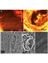
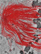
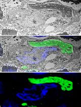
Transmission electron microscopy is a technique for observing the fine details of organelles in cells or tissues. This protocol is to be used to exam the membrane structure in cells with or without virus infection. Modifications should be made if users want to get images from tissues.
Keywords: Transmission Electron Microscopy (透射电子显微镜)Materials and Reagents
- Trypsin: working solution is 0.5% trypsin-0.2% EDTA (Life Technologies, Gibco®, catalog number: 15400-054 )
- NaH2PO4 (Kanto, catalog number: 37239-01 )
- Na2HPO4 (Chemical, catalog number: 37240-01)
- Alcohol
- Poly/Bed 812 (Electron microscopy sciences, catalog number: 14900 )
- Araldite 502 (Electron microscopy sciences, catalog number: 10900 )
- Dodecenylsuccinic anhydride (DDSA) (TAAB, catalog number: D025 )
- DMP-30 (Electron microscopy sciences, catalog number: 13600 )
- Propylene oxide (Merk, catalog number: S4912527-745 )
Note: Toxic, need to handle with care and collect after use.
- Syringe
- Dropper
- Grid (200 mesh) (TAAB)
- 0.1 M Phosphate buffer (PB) (see Recipes)
- Paraformaldehyde (4% in PB) (Bionovas, catalog number: AP0130-0500 ) (see Recipes)
- Osimium tetraoxide (1% in PB) (Electron microscopy sciences, catalog number: 51007 ) (see Recipes)
Note: Toxic, need to handle with care and collect after use.
- Epon (see Recipes)
- Toluidune Blue (Sigma-Aldrich, catalog number: NO202-146-2) (see Recipes)
- Uranyl acetate (Art, catalog number: 8473) (see Recipes)
- Lead citrate (TAAB, catalog number: L003 ) (see Recipes)
Equipment
- Eppendorf tubes
- Centrifuge
- Vacuum drying oven
- Diamond knife (Diatome)
- Ultracut (Leica)
- TEM (HITACHI, model: H-7100 )
Procedure
文章信息
版权信息
© 2013 The Authors; exclusive licensee Bio-protocol LLC.
如何引用
Readers should cite both the Bio-protocol article and the original research article where this protocol was used:
- Peng, W., Lu, K., Lai, S., Shy, H. and Kung, H. (2013). Transmission Electron Microscopy (TEM) Protocol: Observation Details within Cells. Bio-protocol 3(13): e816. DOI: 10.21769/BioProtoc.816.
- Lee, C. P., Liu, P. T., Kung, H. N., Su, M. T., Chua, H. H., Chang, Y. H., Chang, C. W., Tsai, C. H., Liu, F. T. and Chen, M. R. (2012). The ESCRT machinery is recruited by the viral BFRF1 protein to the nucleus-associated membrane for the maturation of Epstein-Barr Virus. PLoS Pathog 8(9): e1002904.
分类
细胞生物学 > 细胞成像 > 电子显微镜
您对这篇实验方法有问题吗?
在此处发布您的问题,我们将邀请本文作者来回答。同时,我们会将您的问题发布到Bio-protocol Exchange,以便寻求社区成员的帮助。