

MACS Isolation and Culture of Mouse Liver Mesothelial Cells
小鼠肝间皮细胞的MACS分离和培养

发布: 2013年07月05日第3卷第13期 DOI: 10.21769/BioProtoc.815 浏览次数: 11671
评审: Lin Fang
参见作者原研究论文
The authors used this protocol in:
Feb 2013


Abstract
Mesothelial cells (MCs) form a single squamous epithelial cell layer and cover the surfaces of the internal organs, as well as the walls of cavities. The isolation of MCs is of great importance to study their function and characteristics for the understanding of physiology and pathophysiology of the liver. Glycoprotein M6a (GPM6A) was originally identified as a cell surface protein expressed in neurons and recently its expression was reported in epicardium and liver MCs (Wu et al., 2001; Bochmann et al., 2010; Li et al., 2012). Here we describe a method to isolate MCs from the adult mouse liver with anti-GPM6A antibodies. Under the low glucose and serum concentration, primary MCs grow and form epithelial colonies (Figure 1).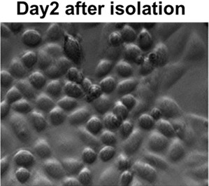
Figure 1. Liver MCs 2 days in culture (20x objective)
Materials and Reagents
- Dulbecco’s Modified Eagle’s Medium (DMEM) Low glucose with stable L-glutamine (Thermo Fisher Scientific, catalog number: SH30021.01 )
- DMEM/F-12 (Thermo Fisher Scientific, catalog number: SH30023.FS )
- Fetal Bovine Serum (FBS) (Sigma-Aldrich, catalog number: F9665 )
- PBS, pH 7.4 (Sigma-Aldrich, catalog number: P3813-10PAK )
- Bovine Collagen Solution, Type I (Advanced BioMatrix, catalog number: 5005-B )
- Pronase (Roche, catalog number: 11459643001 )
- Antibiotic-Antimycotic (100x) (Life Technologies, catalog number: 15240-062 )
- BD Falcon polypropylene conical tube 50 ml (BD Biosciences, catalog number: 352070 )
- BD Falcon cell strainer with 70 μm nylon mesh (BD Biosciences, catalog number: 352350 )
- Rat anti-mouse glycoprotein M6a (GPM6A) antibodies (MBL International, catalog number: D0553 )
- Goat anti-rat IgG microbeads (Miltenyl Biotec, catalog number: 130-048-501 )
- Hydrocortisone solution (Sigma-Aldrich, catalog number: H6909-10ML )
- Insulin-Transferrin-Selenium-X (Life Technologies, catalog number: 41400-045 )
- Ketamine (Clipper Distributing Company, catalog number: NDC57319-542-02 )
- 5% low glucose DMEM medium (see Recipes)
- MC medium (see Recipes)
- Ketamine solution (see Recipes)
Equipment
- 37 °C shaker
- Surgery Tools: Forceps, scissors and glass petri dish
- 37 °C incubator
- Centrifuge
- 24-well plate (VWR, catalog number: 29442-044 )
- G24 Environmental Incubator Shaker (New Brunswick Scientific)
- Miltenyi AutoMacs machine
Procedure
文章信息
版权信息
© 2013 The Authors; exclusive licensee Bio-protocol LLC.
如何引用
Li, Y., Lua, I. and Asahina, K. (2013). MACS Isolation and Culture of Mouse Liver Mesothelial Cells. Bio-protocol 3(13): e815. DOI: 10.21769/BioProtoc.815.
分类
细胞生物学 > 细胞分离和培养 > 细胞分离
您对这篇实验方法有问题吗?
在此处发布您的问题,我们将邀请本文作者来回答。同时,我们会将您的问题发布到Bio-protocol Exchange,以便寻求社区成员的帮助。