- EN - English
- CN - 中文
Subcellular Fractionation of Cultured Human Cell Lines
人源细胞系的亚细胞分离
发布: 2013年05月05日第3卷第9期 DOI: 10.21769/BioProtoc.754 浏览次数: 50641

相关实验方案

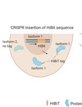
通过CRISPR-Cas9介导的HiBiT标签对高度同源蛋白水平进行异构体特异性半定量测定
Kristina Seiler [...] Mario P. Tschan
2023年07月20日 2505 阅读
Abstract
Subcellular localization is crucial for the proper functioning of a protein. Deregulation of subcellular localization may lead to pathological consequences and result in diseases like cancer. Immuno-fluorescent staining and subcellular fractionation can be used to determine localization of a protein. Here we discuss a protocol to separate the nuclear, cytosolic, and membrane fractions of cultured human cell lines using a centrifuge and ultracentrifuge. The membrane fraction contains plasma membranes and ER-golgi membranes, but no mitochondria or nuclear structures. The fractions can be further analyzed using Western blotting. This protocol is based on that from Dr. Richard Patten at Abcam, and was modified and utilized in a publication by Huang et al. (2012).
Keywords: Nuclear (细胞核)Materials and Reagents
- Sucrose
- HEPES
- Potassium chloride (KCl)
- Magnesium chloride (MgCl2)
- Ethylene diamine tetraacetic acid (EDTA)
- Ethylene glycol tetraacetic acid (EGTA)
- Dithiothreitol (DTT)
- Tris (Affymetrix-USB, catalog number: 75825 )
- Sodium chloride (NaCl) (Sigma-Aldrich, catalog number: 13565 )
- Nonidet P40 substitute (NP40)
- Sodium deoxycholate
- Glycerol
- Sodium dodecyl sulfate (SDS)
- Protease inhibitor (PI) cocktails (F. Hoffmann-La Roche, catalog number: 11836145001 )
- Methanol
- Acetic acid
- Brilliant Blue R (Affymetrix, catalog number: 32826 )
- Phosphate buffered saline (PBS)
- Histone H3 antibody (Cell Signaling Technology, catalog number: 9715 )
- Alpha-tubulin antibody (GeneTex, catalog number: GTX108784 )
- Cell scraper (BD Biosciences, Falcon®, catalog number: 353086 )
- Subcellular fractionation buffer (SF buffer) (see Recipes)
- Nuclear Lysis buffer (NL buffer) (see Recipes)
- Brilliant Blue R staining solution and destaining solution (see Recipes)
Equipment
- 4 °C Microcentrifuge (Eppendorf, catalog number: 5415R );
- Ultracentrifuge (Beckman Coulter, model number: Optima TLX )
- Optional: Sonicator (Sonics, model number: VC505 )
- Whatman filter paper
- 37 °C incubator
- 1.5 ml Eppendorf microtubes
- Tube roller (Maplelab-scientific, model number: MTR-1D )
Procedure
文章信息
版权信息
© 2013 The Authors; exclusive licensee Bio-protocol LLC.
如何引用
Yu, Z., Huang, Z. and Lung, M. L. (2013). Subcellular Fractionation of Cultured Human Cell Lines. Bio-protocol 3(9): e754. DOI: 10.21769/BioProtoc.754.
分类
癌症生物学 > 通用技术 > 生物化学试验 > 蛋白质分析
生物化学 > 蛋白质 > 分离和纯化
细胞生物学 > 细胞器分离 > 分级分离
您对这篇实验方法有问题吗?
在此处发布您的问题,我们将邀请本文作者来回答。同时,我们会将您的问题发布到Bio-protocol Exchange,以便寻求社区成员的帮助。
Share
Bluesky
X
Copy link


