


Capturing Proteins Co-Sorted into Budding Vesicles via an Integrated Proximity Labeling and In Vitro Vesicle Reconstitution Assay


发布: 2026年12月05日第16卷第23期 DOI: 10.21769/BioProtoc.5897 浏览次数: 34
评审: Anik TuladharDevika AndhareAnonymous reviewer(s)
 PDF
 Q&A






Abstract
Protein trafficking relies on the selective packaging of cargo into transport vesicles, yet the molecular machinery governing specific cargo sorting remains incompletely understood. Co-immunoprecipitation, although widely used, often fails to capture weak or transient interactions within dynamic vesicle populations. Conventional proximity labeling methods also fail to distinguish bona fide vesicle-associated interactors from proteins merely near a cargo. Here, we describe a stepwise protocol that integrates proximity biotinylation with an in vitro vesicle reconstitution assay to selectively capture proteins that are co-packaged with a cargo of interest into budding vesicles. The cargo is fused to ascorbate peroxidase 2 (APEX2), and the resulting cargo–APEX2 fusion mediates proximity biotinylation of nearby proteins in live cells. The cells are then subjected to a cell-free vesicle formation reaction, followed by affinity capture of biotinylated proteins that are specifically incorporated into vesicles. This strategy reduces background contributions. Validated here with ShhN-APEX2 in HEK293T cells, this assay is expected to be adaptable to other secretory cargoes and provides a platform for systematic identification of trafficking regulators in development and disease.
Key features
• This protocol integrates proximity biotinylation with an in vitro vesicle formation assay to capture proteins co-sorted into budding vesicles with a specific cargo protein.
• This method minimizes background noise by isolating only biotinylated proteins that are specifically co-incorporated into reconstituted transport vesicles.
• This method was validated using Sonic hedgehog (Shh), applicable to diverse secretory cargoes.
Keywords: Proximity biotinylation APEX2
Vesicle reconstitution
Cargo sorting
Protein trafficking
Graphical overview
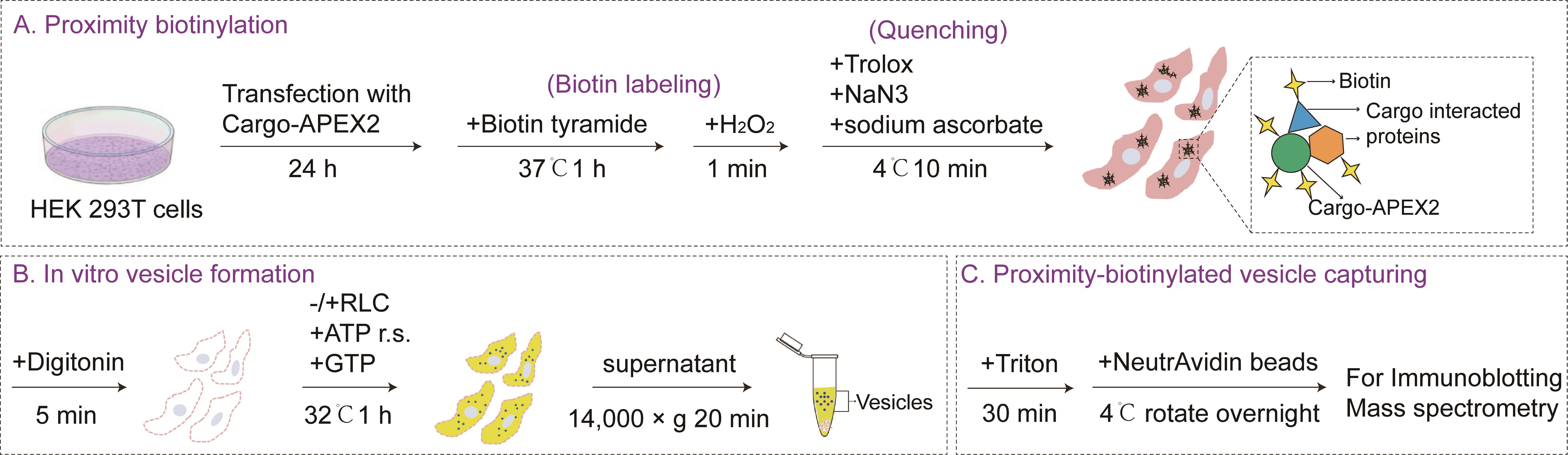
Schematic overview of the combined proximity biotinylation and in vitro vesicle reconstitution assay. Methods for proximity biotinylation, in vitro vesicle formation, and proximity-biotinylated vesicle capture are described in sections A, B, and C, respectively, of the Procedure. Efficiency of packaging of cargos into vesicles and the precipitation of biotinylated proteins were measured by immunoblotting, as described in steps C5–6 of the Procedure. RLC, rat liver cytosol. This figure is adapted from Song et al. [1].
Background
Intracellular protein trafficking relies on the selective packaging of cargo proteins into transport vesicles, a process fundamental to cellular organization, signaling, and development. However, the molecular mechanisms underlying cargo-specific vesicle sorting remain largely unclear. A critical challenge is to identify the protein mediators that govern cargo recognition, vesicle budding, and cargo delivery [2]. Conventional approaches for identifying trafficking regulators include co-immunoprecipitation (Co-IP) and proximity biotinylation [3,4]. Co-IP often fails to capture weak or transient interactions within dynamic vesicle populations. Proximity biotinylation labels proteins in the spatial vicinity of a bait but cannot discriminate between bona fide vesicle-associated interactors and abundant residents of donor compartments, leading to high background and frequent false positives. Both methods provide information about protein proximity but cannot directly distinguish vesicle-associated candidates from background proteins.
To overcome these limitations, we developed a protocol that integrates proximity biotinylation and an in vitro vesicle reconstitution assay. In this approach, the cargo of interest is fused to the engineered ascorbate peroxidase 2 (APEX2) and biotinylated in live cells. The cells are then permeabilized and subjected to a cell-free vesicle budding reaction [5,6]. Briefly, digitonin permeabilizes the plasma membrane, allowing cytosolic proteins to be removed by washing while intracellular membranes remain intact. The addition of rat liver cytosol (RLC), the ATP regeneration system (ATP r.s.), and GTP then supports vesicle budding, and the released vesicles are separated from the donor membranes by centrifugation. Biotinylated proteins that are co-packaged with the cargo into the released vesicles are subsequently enriched by affinity capture using NeutrAvidin agarose, effectively reducing background proteins from static organelles or non-vesicular compartments. Notably, proteins recovered by this approach represent candidates that are co-sorted or co-packaged with the cargo into vesicle-containing fractions. This enrichment does not necessarily indicate a direct physical interaction with the cargo, and such interactions require further validation.
This protocol has the potential to be adapted to map vesicle-associated interactomes of a wide range of secretory cargoes, including morphogens, growth factors, and disease-relevant cargoes. It may also be modified to study trafficking in different cell types or at distinct steps of the secretory pathway. Furthermore, the platform could be combined with CRISPR-based screening for high-throughput screening [7]. Overall, this protocol will provide a flexible tool for discovering trafficking regulators across diverse biological contexts.
Materials and reagents
Biological materials
1. HEK 293T cells; kindly provided by the University of California–Berkeley Cell Culture Facility, confirmed by short tandem repeat profiling and tested negative for Mycoplasma contamination
2. ShhN-HA-APEX2 plasmid; the ShhN-HA-APEX2 construct encodes the N-terminal fragment of mouse Shh (ShhN, aa 25–198) fused to a C-terminal 3×HA tag and APEX2; generated by BGI (Beijing Genomics Institute)
Note: The position of APEX2 should be considered during construct design, as it determines the subcellular scope of proximity labeling. For soluble cargoes such as ShhN, APEX2 labeling is restricted to the lumen side and is unlikely to reach cytosolic proteins involved in vesicle budding across an intact membrane. For transmembrane cargoes, the accessible protein populations depend on the topology of APEX2. If APEX2 is fused to a luminal domain, only luminal proteins are labeled. If it is fused to a cytosolic domain, cytosolic proteins can be labeled prior to permeabilization. Furthermore, when APEX2 is positioned on the cytosolic side, the order of the assay can be adjusted according to the experimental purpose. The vesicle budding reaction can be performed first, followed by biotin labeling, to capture cytosolic factors supplied by the RLC.
Reagents
1. Dulbecco's modified Eagle medium (DMEM) (Thermo Fisher Scientific, catalog number: 12800082)
2. Opti-MEM (Gibco, catalog number: 31985070)
3. Fetal bovine serum (FBS) (Thermo Fisher Scientific, catalog number: 10270106)
4. Penicillin-streptomycin (Thermo Fisher Scientific, catalog number: 15140122)
5. Polyethylenimine (Polysciences, catalog number: 23966-1)
6. 0.25% trypsin-EDTA (Thermo Fisher Scientific, catalog number: 25200056)
7. Sodium chloride (NaCl) (Sigma-Aldrich, catalog number: S5886)
8. Potassium chloride (KCl) (VWR Chemicals BDH®, catalog number: 2576)
9. Di-sodium hydrogen phosphate dihydrate (Na2HPO4·2H2O) (Scharlab, catalog number: SO03390500)
10. Potassium dihydrogen phosphate (KH2PO4) (Thermo Fisher Scientific, catalog number: A12142.36)
11. Biotin tyramide (Sigma-Aldrich, catalog number: SML2135)
12. Dimethyl sulfoxide (DMSO) (Sigma-Aldrich, catalog number: D8418)
13. Hydrogen peroxide solution (Millipore, catalog number: 1.07298)
14. Sodium ascorbate (Supelco, catalog number: PHR1279)
15. Trolox (MedChemExpress, catalog number: HY-101445)
16. Sodium azide (NaN3) (Sigma-Aldrich, catalog number: S8032)
17. HEPES (Acros Organics, catalog number: 172571000)
18. Acetic acid, potassium salt (KOAc) (Fisher Scientific, catalog number: 127082)
19. Magnesium acetate tetrahydrate [Mg(OAc)2] (Sigma-Aldrich, catalog number: M0631)
20. Digitonin (Sigma-Aldrich, catalog number: D141)
21. Creatine phosphate (Roche, catalog number: 10621722001)
22. Creatine kinase (Roche, catalog number: 10736988001)
23. Adenosine 5'-triphosphate (ATP) (Sigma-Aldrich, catalog number: 2383)
24. Guanosine 5'-triphosphate disodium salt (GTP) (FUJIFILM Wako Pure, catalog number: 073-03113)
25. Triton® X-100 (Sigma-Aldrich, catalog number: X100)
26. Protease inhibitor cocktail tablets (Roche, catalog number: 05056489001)
27. DL-dithiothreitol (DTT) (Sigma-Aldrich, catalog number: D9779)
28. PierceTM NeutrAvidinTM agarose (Thermo Fisher Scientific, catalog number: 29201)
29. Sodium dodecyl sulfate (SDS) (Sigma-Aldrich, catalog number: 151213)
30. Bromophenol blue (Sigma-Aldrich, catalog number: 114391)
31. Tris (Affymetrix, catalog number: 75825)
32. Glycerol (Sigma-Aldrich, catalog number: G7757)
33. 2-Mercaptoethanol (β-ME) (Sigma-Aldrich, catalog number: M6250)
34. Antibodies
a. Rabbit anti-HA (Cell Signaling Technology, catalog number: 3724), 1:2,000
b. Rabbit anti-calnexin (Abcam, catalog number: ab22595), 1:4,000
c. Mouse anti-SURF4 antibodies; kindly provided by Prof. Xiaowei Chen (Peking University, China), 1:2,000; generated by the Proteintech Group (www.ptglab.com) against the C-terminal epitope of SURF4 (N-glllvvalpgpgvsmedkekkw-C) [8]
d. Horseradish peroxidase (HRP)-coupled goat antibodies against rabbit IgG (Beyotime, catalog number: A0208), 1:4,000
e. HRP-coupled goat antibodies against mouse IgG (Beyotime, catalog number: A0216), 1:4,000
Solutions
A. Buffer solutions (see Recipes)
1. 1× PBS
2. 1× KHM buffer
3. Lysis buffer
4. Labeling medium
5. Quenching solution
6. High-salt KHM buffer
B. Stock solutions (see Recipes)
1. 10× PBS, pH 7.4
2. 1 M KOAc
3. 1 M HEPES-KOH, pH 7.2
4. 1 M Mg(OAc)2
5. 50 mM biotin tyramide
6. 1 M H2O2
7. 0.5 M Trolox
8. 3% NaN3
9. 0.25 M sodium ascorbate
10. 40 mg/mL digitonin
11. ATP regeneration system (ATP r.s.)
12. 10 mM GTP
13. 20 mg/mL rat liver cytosol (RLC)
14. 10% Triton® X-100
15. 100× protease inhibitors
16. 500 mM DTT
17. 1 mg/mL polyethylenimine
18. 1 M Tris-HCl, pH 6.8
19. 5× SDS-PAGE protein loading buffer
Recipes
A. Buffer solutions
1. 1× PBS (1 L)
Add 100 mL of 10× PBS to 900 mL of ddH2O.
2. 1× KHM buffer (1 L)
a. Add 5 mL of 1 M Mg(OAc)2, 20 mL of 1 M HEPES-KOH, pH 7.2, and 110 mL of 1 M KOAc to 0.5 L of ddH2O.
b. Adjust volume to 1 L.
c. Filter the buffer.
d. Store at 4 °C.
3. Lysis buffer (1 mL)
Add 0.05 mL of 10% Triton® X-100, 10 μL of 100× protease inhibitors, and 2 μL of 500 mM DTT in 938 μL of KHM buffer.
4. Labeling medium (10 mL)
Add 100 μL of 50 mM biotin tyramide to 10 mL of cell culture (DMEM containing 10% FBS, 10 milliunits/mL of penicillin, and 0.1 mg/mL of streptomycin).
5. Quenching solution (10 mL)
Add 0.4 mL of 0.25 M sodium ascorbate, 0.1 mL of 0.5 M Trolox, and 0.217 mL of 3% NaN3 in 10 mL of 1× PBS.
6. High-salt KHM buffer (1 L)
a. Dissolve 98.14 g of potassium acetate in 0.5 L of ddH2O.
b. Add 5 mL of 1 M Mg(OAc)2 and 20 mL of 1 M HEPES-KOH, pH 7.2.
b. Adjust volume to 1 L.
c. Filter the buffer.
d. Store at 4 °C.
B. Stock solutions
1. 10× PBS, pH 7.4 (2 L)
a. Dissolve 160 g of NaCl, 4 g of KCl, 28.8 g of Na2HPO4, and 4.8 g of KH2PO4 in 1.9 L of ddH2O.
b. Adjust pH to 7.4 with NaOH.
c. Add ddH2O to 2 L.
d. Sterilize by autoclaving and store at room temperature (RT).
2. 1 M KOAc (1 L)
a. Dissolve 98.14 g of potassium acetate in 1 L of ddH2O.
b. Sterilize by autoclaving and store at RT.
3. 1 M HEPES-KOH, pH 7.2 (1 L)
a. Dissolve 238.3 g of HEPES in 0.5 L of ddH2O.
b. Adjust pH to 7.2 with KOH
c. Make up to 1 L with ddH2O.
d. Store at RT.
4. 1 M Mg(OAc)2 (1 L)
a. Dissolve 155.41 g of Mg(OAc)2 in 1 L of ddH2O.
b. Sterilize by autoclaving and store at RT.
5. 50 mM biotin tyramide (1 mL)
a. Dissolve 18.17 mg of biotin tyramide in 1 mL of DMSO.
b. Store at -20 °C.
6. 1 M H2O2 (1 mL)
a. Add 102.1 μL of 30% H2O2 to 897.9 μL of ddH2O.
b. Prepare fresh and protect from light.
7. 0.5 M Trolox (1 mL)
a. Dissolve 0.125 g of Trolox in 1 mL of DMSO.
b. Store at -20 °C.
8. 3% NaN3 (50 mL)
a. Dissolve 1.5 g of NaN3 in 50 mL of ddH2O.
b. Store at RT.
9. 0.25 M sodium ascorbate (1 mL)
a. Dissolve 49.5 mg of sodium ascorbate in 1 mL of ddH2O.
b. Store at 4 °C.
10. 40 mg/mL digitonin (10 mL)
a. Dissolve 400 mg of digitonin in 10 mL of ddH2O.
b. Aliquot in volumes of 30 μL and store at -20 °C.
11. ATP regeneration system (20 mL)
a. Dissolve 2.04 g of creatine phosphate, 40 mg of creatine kinase, and 101.44 mg of ATP in 20 mL of 1× KHM.
b. Aliquot in small volumes and store at -80 °C.
12. 10 mM GTP (20 mL)
a. Dissolve 113.43 mg of GTP in 20 mL of 1× KHM.
b. Aliquot in small volumes and store at -80 °C.
13. 20 mg/mL RLC
RLC was prepared as described previously [9]. Briefly, livers from Sprague–Dawley rats were homogenized and subjected to sequential centrifugation at 1,000× g for 10 min, 18,459× g for 20 min, and 125,171× g for 1 h at 4 °C. The deep-red cytosolic fraction was collected, and the high-speed spin was repeated 2–3 times. The protein concentration was determined by the Bradford assay, and aliquots were snap-frozen in liquid nitrogen and stored at -80 °C. Avoid repeated freeze/thaw cycles.
14. 10% Triton® X-100 (50 mL)
a. Dissolve 5 mL of Triton® X-100 in 45 mL of ddH2O.
b. Store at 4 °C.
15. 100× protease inhibitors (1 mL)
a. Dissolve two protease inhibitor cocktail tablets in 1 mL of ddH2O.
b. Aliquot into volumes of 30 μL and store at -20 °C.
16. 500 mM DTT (1 mL)
a. Dissolve 0.077 g of DL-dithiothreitol in 1 mL of ddH2O.
b. Aliquot in volumes of 30 μL and store at -20 °C.
17. 1 mg/mL polyethylenimine (100 mL)
a. Dissolve 0.1 g of polyethylenimine in 100 mL of ddH2O.
b. Filter using a 0.22 μm filter.
c. Aliquot in small volumes and store at -80 °C.
18. 1 M Tris-HCl, pH 6.8 (1 L)
a. Dissolve 121.1 g of Tris in 0.8 L of ddH2O.
b. Adjust pH to 6.8 with HCl.
c. Add ddH2O to 1 L.
d. Sterilize by autoclaving and store at RT.
19. 5× SDS-PAGE protein loading buffer
a. Dissolve 3.75 g of SDS and 93.75 mg of bromophenol blue in a small beaker with 12.5 mL of 1 M Tris, pH 6.8, with constant stirring.
b. Mix in 25 mL of glycerol.
c. Adjust volume to 37.5 mL with ddH2O.
d. Aliquot to 750 μL/tube and store at -20 °C.
e. Add 250 μL of β-ME to an aliquot fresh before use.
Laboratory supplies
1. 10 cm tissue culture dishes (Biofil, catalog number: TCD-010100)
2. 15 mL centrifuge tubes (Biofil, catalog number: CFT-011150)
3. Falcon® 50 mL centrifuge tubes (Corning, catalog number: 352070)
4. 1,000 μL tips (USA Scientific, catalog number: 11112021)
5. 200 μL tips (Axygen, catalog number: T200-Y)
6. 10 μL tips (Axygen, catalog number: T300)
7. 1.5 mL microtubes (Axygen, catalog number: 20220415)
Equipment
1. 100 mL, 500 mL, 1 L, and 5 L beakers
2. 4 °C, -20 °C, and -80 °C freezers
3. Water bath
4. Eppendorf® 5418R centrifuge, refrigerated, with rotor FA-45-18-11 and rotor lid (Eppendorf, model: 5418R)
5. Light microscope with a 10× or 20× objective (any simple or compound light microscope is fine)
6. Barnstead Thermolyne (Thermo Fisher, model: DB16520-26)
7. Elite dry bath incubator (Major Science, model: EL-02)
8. Benchmark RotoMini rotator with tube holders (Benchmark, model: BMSR2020)
9. Thermo Scientific FormaTM Series 3 Water Jacketed CO2 Incubator (Thermo Fisher, model: 4120)
Software and datasets
1. ImageLab software v6.1
2. Adobe Photoshop
3. Adobe Illustrator
4. Proteome Discoverer
5. Perseus software (v1.6.6.0)
6. R packages: Vennerable (v3.1.0.9), clusterProfiler (v4.12.6), and ggplot2 (v3.5.12)
7. Alternatively, other image analysis programs and graphics software can be used, such as Fiji.
Procedure
登录 / 注册后阅读全文
文章信息
稿件历史记录
提交日期: Aug 24, 2026
接收日期: Oct 28, 2026
在线发布日期: Nov 17, 2026
出版日期: Dec 5, 2026
版权信息
© 2026 The Author(s); This is an open access article under the CC BY-NC license (https://creativecommons.org/licenses/by-nc/4.0/).
如何引用
Readers should cite both the Bio-protocol article and the original research article where this protocol was used:
- Zhao, X., Song, Z., Guo, Y. and Tang, X. (2026). Capturing Proteins Co-Sorted into Budding Vesicles via an Integrated Proximity Labeling and In Vitro Vesicle Reconstitution Assay. Bio-protocol 16(23): e5897. DOI: 10.21769/BioProtoc.5897.
- Song, Z., Chen, Z., Tian, Y., Wang, Q., Zhao, X., Guo, Y. and Tang, X. (2026). An integrated proximity labeling and vesicle reconstitution assay identifies novel regulators of Sonic hedgehog secretion. Mol Biol Cell. 37(6): ee25–12–0627. https://doi.org/10.1091/mbc.e25-12-0627

 Download Citation in RIS Format
您对这篇实验方案有问题吗?
在此处发布您的问题,我们将邀请本文作者来回答。同时,我们会将您的问题发布到Bio-protocol Exchange,以便寻求社区成员的帮助。
 发布问题


