


Generation, Cryopreservation, and Drug Assessment of 3D Tumor Spheroids Using the Spheromatrix Platform

(*contributed equally to this work) 发布: 2026年12月05日第16卷第23期 DOI: 10.21769/BioProtoc.5896 浏览次数: 37
评审: Tanxi CaiAnonymous reviewer(s)
 PDF
 Q&A







Abstract
Three-dimensional (3D) tumor spheroids provide tissue-like models for preclinical oncology, yet their scalable production, long-term preservation, and on-demand use remain technically demanding. This protocol describes the Spheromatrix, a paper-based platform combining wax-patterned hydrophilic U-wells with a custom 3D-printed cell loading device (Spherobox). The protocol covers Spherobox and U-well fabrication, uniform Spherobox-assisted cell seeding into the U-wells, spheroid culture, controlled-rate freezing, liquid nitrogen storage, rapid post-thaw recovery, and drug response assessment. The demonstrated 14 × 16 format provides 224 spatially separated U-wells for parallel spheroid generation and can be scaled by expanding the wax-patterned paper array. The platform was validated for spheroid generation using six cancer cell lines (MCF7, PC3, HeLa, SKBR3, HCT15, and U87) and the noncancerous MRC5 fibroblast cell line. U87 glioblastoma spheroids served as a proof-of-concept model for the complete cryopreservation-to-drug response assessment workflow and, following recovery, retained their morphology and metabolic activity and remained responsive to cisplatin and temozolomide. The compact, stackable format supports space-efficient generation, storage, and retrieval of ready-to-use 3D tumor models for drug screening and spheroid biobanking.
Key features
• Supports uniform Spherobox-assisted cell loading and 3D cell culture, demonstrated with six cancer cell lines and noncancerous fibroblasts, and adaptable to additional cell lines.
• The 14 × 16 format provides 224 spatially separated U-wells for parallel spheroid generation and can be scaled for application-specific needs.
• Compact, stackable paper chips support space-efficient spheroid banking and reduce medium and cryoprotectant volumes, offering potential savings in consumables, cryogenic capacity, and storage-related energy.
• Supports cryopreservation, post-thaw recovery, and drug response testing in cryopreserved and non-cryopreserved spheroids, demonstrated with cisplatin and temozolomide, and extendable to other candidate therapeutics.
Keywords: Spheromatrix Paper-based platform
3D cell culture
Tumor spheroids
Cryopreservation
Drug screening
Spheroid banking
Glioblastoma
Graphical overview
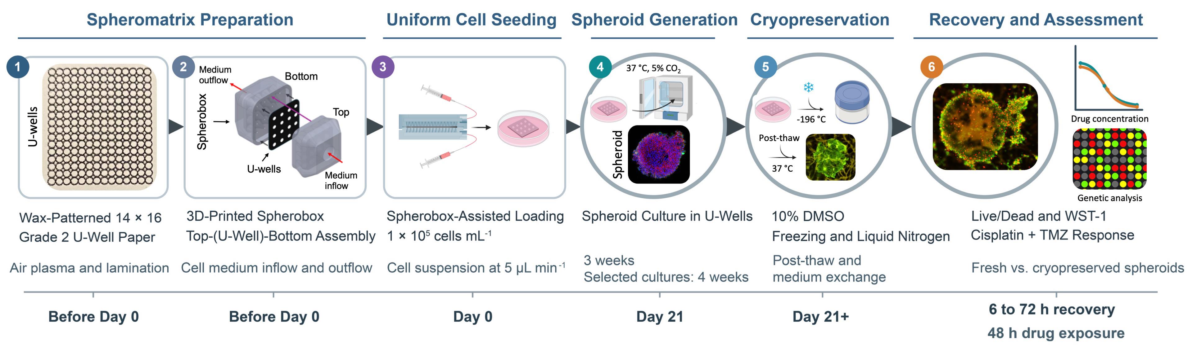
Spheromatrix workflow for scalable spheroid generation, cryopreservation, recovery, and drug response assessment. (1) Wax printing and thermal diffusion generate a 14 × 16 array of hydrophilic U-wells in Whatman Grade 2 filter paper, followed by air plasma treatment to improve liquid filling and lamination to enhance chip stability. (2) The 3D-printed Spherobox is assembled around the U-well chip to provide controlled cell medium inflow and outflow. (3) Cells are loaded through the Spherobox at 1 × 10⁵ cells/mL and a flow rate of 5 μL/min. (4) Spheroids are cultured at 37 °C and 5% CO2 for 3 weeks. Where indicated, selected cultures are maintained for 4 weeks. (5) Spheroids are cryopreserved in complete medium containing 10% (v/v) DMSO using controlled-rate freezing, stored in liquid nitrogen, rapidly thawed, and subjected to medium exchange. (6) Recovered spheroids and non-cryopreserved controls are evaluated using Live/Dead imaging, WST-1 metabolic activity measurements, cisplatin and temozolomide drug response assays, and, optionally, qPCR-based transcriptional integrity assessment. Device schematics and representative experimental images are adapted from Glia et al. [1].
Background
Three-dimensional (3D) in vitro tumor models have greater physiological relevance for preclinical oncology studies than conventional 2D cell cultures [2]. Tumor spheroids reproduce important features of solid tumors, including cell–cell and cell–matrix interactions, spatial organization, and gradients in oxygen, nutrients, metabolites, and waste [3]. These properties influence cell proliferation, drug penetration, and treatment response [4]. Common spheroid generation approaches include hanging-drop culture, low-attachment plates, scaffold-based systems, microwell arrays, and microfluidic devices [5]. Despite continued advances, these approaches involve trade-offs related to spheroid uniformity, matrix dependence, cost, operational complexity, standardization, scalability, and compatibility with downstream analysis.
Paper-based platforms offer porous, readily patterned, and comparatively inexpensive substrates for 3D cell culture [6]. Their interconnected fibrous networks provide structural support and capillary transport, while their planar geometry facilitates patterning, stacking, sectioning, imaging, and compact storage. Recent paper-based systems extended beyond basic scaffold-supported cell culture to quantitative cytotoxicity testing [7], tumor-stroma co-culture [8], rapid spheroid formation and retrieval [9], and multilayer models of immune cell migration into tumor spheroids [10]. Collectively, these studies demonstrate the versatility of paper for constructing spatially organized tumor models and integrating culture with functional assessment. However, these capabilities are generally distributed across separate platforms, and a unified workflow combining array-scale spheroid generation, uniform cell loading, mechanically robust extended culture, cryogenic storage, and post-thaw functional testing remains uncommon.
Our earlier studies established paper-based mammalian cell cryopreservation and demonstrated that the fibrous paper microenvironment could support the formation and cryopreservation of 3D tumor cultures [11,12]. This work was subsequently extended to cryopreservable arrays of paper-based 3D tumor models for drug screening [13]. Together, these studies established paper as both a culture scaffold and a compact storage substrate, while also highlighting the need for improved control over cell localization, spheroid uniformity, chip stability, and array-scale cell loading. The Spheromatrix addresses these technical needs by combining wax-patterned U-wells, lamination, and a 3D-printed Spherobox for controlled cell distribution across the array [1]. Wax patterning confines cells within defined hydrophilic U-wells, air plasma treatment improves wetting and liquid filling, and lamination enhances chip stability during extended culture and cryopreservation.
This protocol provides a step-by-step implementation of the Spheromatrix method reported by Glia et al. [1]. It expands the methodological description with explicit reagent recipes, assembly and alignment checks, handling precautions, assay guidance, and troubleshooting to support reproducibility. The platform was demonstrated for spheroid generation with six cancer cell lines (MCF7, PC3, HeLa, SKBR3, HCT15, and U87) and noncancerous MRC5 fibroblasts. U87 glioblastoma spheroids served as the proof-of-concept model for the complete cryopreservation-to-drug assessment workflow.
Several limitations should be considered when implementing this protocol. Although spheroid formation was demonstrated with six cancer cell lines and MRC5 fibroblasts, the complete cryopreservation-to-drug response assessment workflow was evaluated using U87 spheroids and two chemotherapeutic drugs. Performance with other cell types, treatments, and storage durations requires separate validation. MRC5 fibroblasts showed weaker aggregation and lower viability, indicating cell-type-dependent performance. Changes in array geometry require reassessment of cell loading and culture conditions. Paper autofluorescence, staining penetration, and aggregate recovery can also affect downstream measurements; metabolic activity should therefore be interpreted alongside morphology and an independent viability readout.
Materials and reagents
Note: The materials and reagents below support the validated cell lines described in this protocol. Use appropriate complete culture medium listed in the Procedure and Recipes sections.
Biological materials
Cell lines
1. MCF7 human breast cancer cells (ATCC, catalog number: HTB-22)
2. PC3 human prostate cancer cells (ATCC, catalog number: CRL-1435)
3. HeLa human cervical cancer cells (ATCC, catalog number: CCL-2)
4. SKBR3 human breast cancer cells (AddexBio, catalog number: C0006007)
5. MRC5 human noncancerous lung fibroblasts (AddexBio, catalog number: T0016002)
6. HCT15 human colorectal cancer cells (AddexBio, catalog number: C0009010)
7. U87 human glioblastoma cells (AddexBio, catalog number: C0005002)
8. MDA-MB-231 human breast cancer cells (ATCC, catalog number: HTB-26)
Reagents
Cell culture and cryopreservation
1. Dulbecco’s phosphate-buffered saline (DPBS) (10× stock solution) (Sigma-Aldrich, catalog number: 59331C)
2. Dulbecco’s modified Eagle’s medium (DMEM) (Gibco, catalog number: 11965092)
3. Roswell Park Memorial Institute Medium (RPMI) (Gibco, catalog number: 11875093)
4. Eagle’s minimum essential medium (EMEM) (Gibco, catalog number: 11095080)
5. Fetal bovine serum (FBS) (Sigma-Aldrich, catalog number: F7524)
6. Penicillin-streptomycin solution (Pen-Strep) (Sigma-Aldrich, catalog number: P4333)
7. TrypLE Express enzyme (Gibco, catalog number: 12604021)
8. Dimethyl sulfoxide (DMSO) (Sigma-Aldrich, catalog number: D2650)
9. Isopropyl alcohol (2-propanol), ≥99.9% (VWR Chemicals, catalog number: 84881.290)
10. Ethanol, ≥70% (v/v), TechniSolv® (VWR Chemicals, catalog number: 83801.290)
Viability and metabolic activity assessment
11. LIVE/DEAD Viability/Cytotoxicity kit for mammalian cells (Thermo Fisher, catalog number: L3224)
12. Cell proliferation reagent WST-1 (Sigma-Aldrich, catalog number: 11644807001)
13. Trypan Blue solution (Sigma-Aldrich, catalog number: T6146)
Drug response assessment
14. Cisplatin, cis-diamminedichloroplatinum(II) (Sigma-Aldrich, catalog number: P4394)
15. Temozolomide (TMZ) (Sigma-Aldrich, catalog number: T2577)
Spherobox fabrication
16. PlasCLEAR photopolymer resin (Asiga, catalog number: PN/00930)
Immunostaining (Optional)
17. Paraformaldehyde (Sigma-Aldrich, catalog number: 158127)
18. Triton X-100 (Sigma-Aldrich, catalog number: T8787)
19. Bovine serum albumin (BSA) (Sigma-Aldrich, catalog number: A9418)
20. 4′,6-diamidino-2-phenylindole (DAPI) (Invitrogen, catalog number: 62248)
21. PE mouse anti-human cytokeratin antibody (BD Biosciences, catalog number: 347204)
22. FITC Anti-CD326/EpCAM mouse monoclonal antibody (Clone 9C4) (BioLegend, catalog number: 324204)
Transcriptional integrity assessment (Optional)
23. Cells-to-CT kits (Thermo Fisher, catalog number: A25600)
24. Primers for ACTB, GAPDH, and 18S rRNA (Integrated DNA Technologies)
Solutions
1. Complete culture medium (see Recipes)
2. Freezing medium containing 10% DMSO (see Recipes)
3. LIVE/DEAD assay (see Recipes)
4. WST-1 assay (see Recipes)
5. DPBS working solution (see Recipes)
6. Cisplatin stock solution (see Recipes)
7. TMZ stock solution (see Recipes)
8. Combination (Cisplatin + TMZ) solution (see Recipes)
9. Immunostaining solutions (see Recipes)
Recipes
1. Complete culture medium
Prepare 100 mL using 89 mL of the appropriate basal medium, 10 mL of FBS, and 1 mL of Pen-Strep:
• DMEM: HeLa, MCF7, MDA-MB-231, and SKBR3
• RPMI: PC3 and HCT15
• EMEM: MRC5 and U87
Store at 4 °C. Prewarm only the required volume and avoid repeatedly warming the complete stock.
2. Freezing medium containing 10% DMSO
Prepare 10 mL immediately before use by mixing 9 mL of the appropriate complete culture medium with 1 mL of DMSO. Add DMSO slowly and mix thoroughly.
3. LIVE/DEAD assay
For 5 mL of working solution, add 2.5 μL of 4 mM calcein-AM stock and 10 μL of 2 mM ethidium homodimer-1 stock to 4,987.5 μL of sterile 1× DPBS (see Recipe 5). The final concentrations are 2 μM calcein-AM and 4 μM ethidium homodimer-1. Allow the kit components to reach room temperature before opening, mix gently, and prepare the working solution immediately before use. Protect it from light.
Note: The added stock volumes correspond to 0.05% (v/v) calcein-AM stock and 0.2% (v/v) ethidium homodimer-1 stock in the final solution. These percentages refer to stock solution volume fractions of the dyes.
4. WST-1 assay
Add WST-1 reagent to complete culture medium at a 1:10 ratio. Prepare immediately before use and protect from light. Incubate for 2–4 h at 37 °C and 5% CO2.
5. DPBS working solution
Prepare 1 L by mixing 100 mL of 10× DPBS stock solution with 900 mL of sterile deionized water. Mix thoroughly and store at room temperature.
6. Cisplatin stock solution
Prepare a 1 mM cisplatin stock in sterile DPBS (0.300 mg/mL). To prepare 10 mL of 100 μM cisplatin containing 1% (v/v) DMSO, combine 1000 μL of the stock, 100 μL of DMSO, and 8.9 mL of complete culture medium.
Note: Store cisplatin powder at room temperature, as used in this workflow. Prepare the 1 mM stock in sterile DPBS and the working solutions fresh on the day of treatment; protect solutions from light. The protocol does not use stored drug stocks, and no frozen stock shelf life is specified.
7. TMZ stock solution
Prepare a 100 mM TMZ stock in DMSO (19.4 mg/mL). To prepare 10 mL of 800 μM TMZ containing 1% (v/v) total DMSO, combine 80 μL of the stock, 20 μL of DMSO, and 9.9 mL of complete culture medium.
Note: Store TMZ powder at room temperature, as used in this workflow. Prepare the 100 mM stock in DMSO and the aqueous working solutions fresh on the day of treatment; protect solutions from light. The protocol does not use stored drug stocks, and no frozen stock shelf life is specified.
Note: Using the highest working concentration for each single-agent chemotherapeutic drug solution, prepare the lower concentrations by dilution with complete culture medium containing 1% (v/v) DMSO. From 100 μM cisplatin, prepare 10, 1, and 0.1 μM solutions. From 800 μM TMZ, prepare 400, 200, and 100 μM solutions. Prepare a final volume of 10 mL for each concentration. Count the DMSO contributed by the TMZ stock toward the final 1% DMSO concentration. Protect drug solutions from light and prepare them immediately before treatment.
8. Combination (cisplatin + TMZ) solution
Maintain 1:100 cisplatin:TMZ molar concentration ratio. For 10 mL working solutions containing 1% (v/v) total DMSO, prepare:
• 4 μM cisplatin + 400 μM TMZ: Combine 40 μL of 1 mM cisplatin stock, 40 μL of 100 mM TMZ stock, 60 μL of DMSO, and 9.860 mL of complete culture medium.
• 8 μM cisplatin + 800 μM TMZ: Combine 80 μL of 1 mM cisplatin stock, 80 μL of 100 mM TMZ stock, 20 μL of DMSO, and 9.82 mL of complete culture medium.
Critical: Include separate untreated complete medium and 1% (v/v) DMSO controls. Use the 1% DMSO control for all cisplatin, TMZ, and combination conditions.
9. Immunostaining solutions
• Fixation solution: 4% paraformaldehyde in DPBS
• Permeabilization solution: 0.1% Triton X-100 in DPBS
• Blocking solution: 5% BSA in DPBS
Prepare the solutions for fixation, permeabilization, and blocking, and store at 4 °C.
Laboratory supplies
1. Sterile 10 mL serological pipettes (Corning Costar Stripette, catalog number: 4488)
2. Sterile 15 mL centrifuge tubes (Thermo Fisher, catalog number: 339650)
3. Sterile 1–20, 20–200, and 100–1,000 μL pipette tips (Thermo Fisher, catalog numbers: 10380792, 10619331, and 10390792, respectively)
4. Sterile T75 tissue culture flasks (Thermo Fisher, catalog number: 156499)
5. Sterile 35 mm × 10 mm Petri dishes (Corning, catalog number: 430165)
6. Sterile 60 mm × 10 mm Petri dishes (Falcon, catalog number: 351007)
7. Whatman Grade 2 filter paper, 185 mm diameter (Cytiva, catalog number: 1002-185)
8. 96-well plates compatible with absorbance measurements (Thermo Fisher, catalog number: N8010560)
9. Cell counting chamber slides (Thermo Fisher, catalog number: C10228)
10. Sterile syringes (CETONI, capacity and catalog number as specified for the neMESYS syringe pump), tubing (Cole-Parmer Microbore, sized for the syringe pump and Spherobox inlet/outlet), and connectors (CETONI, compatible with the syringe and tubing)
Equipment
Spheromatrix fabrication
1. Stereolithography 3D printer (27 μm pixel resolution) (Asiga, model: MAX X27 UV)
2. Wax printer (Xerox, model: ColorQube 8570/DN)
Note: These printer models were used in the validation study. Equivalent instruments may be used if they reproduce the Spherobox geometry and continuous wax barriers at the specified dimensions.
3. Digital cutting machine (Cricut, model: Explore Air 2)
4. Autoclave (Runyes, model: SEA 18L-DIG-USB)
5. Analytical balance (Mettler Toledo, model: ME54)
6. High-precision tweezers (Dumont, catalog number: 5627)
7. Timer (Sunnex, catalog number: 360594)
8. Air plasma cleaner (Harrick Plasma, PDC series), connected to a PlasmaFlo gas flow controller (Harrick Plasma, model: PDC-FMG)
9. Digital hot plate (Thermo Fisher SuperInnova+, catalog number: HP88857195)
10. UV sterilizer cabinet (StateRiver)
11. Polyethylene terephthalate (PET) lamination film (Maxi A4)
12. Thermal pouch laminator (Monolith)
13. Microbalance (Mettler Toledo)
Cell culture, loading, and cryopreservation
14. Syringe pump (CETONI, model: neMESYS)
Note: An equivalent pump may be used provided that it is capable of stable delivery at 5 μL/min.
15. Centrifuge (Eppendorf, model: 5810)
16. Laboratory water bath (Witeg, catalog number: WITEG 20002)
17. Humidified CO2 incubator (New Brunswick, model: Galaxy 48R)
18. Water purification system (Millipore, model: Milli-Q)
19. Class II biological safety cabinet (NuAire, model: NU-437S)
20. Controlled-rate freezing container (Mr. Frosty, Thermo Fisher, catalog number: 5100-0001)
21. Custom 3D-printed cryogenic storage vessels (Cryobox; 1.5 cm × 1.5 cm × 2.5 cm; fabricated in-house from the same
resin and stereolithography approach as the Spherobox; up to 50 chips per vessel, fully immersed in 1 mL of freezing medium containing 10% DMSO; up to 18 vessels per Mr. Frosty container)
22. Freezer (-80 °C) (Haier, model: DW-86L100J)
23. Liquid nitrogen storage tank (–196 °C) (Air Liquide, model: Arpege 40, catalog number: 082102B)
Assessment and imaging
24. Multimode microplate reader (BioTek, model: Synergy H1)
25. Inverted fluorescence microscope (Nikon, model: Eclipse Ti2), equipped with a DS-Qi2 monochrome camera and FITC (MBE44727, 450–490 nm) and TRITC (MBE45607, 530–550 nm) filter cubes
26. Inverted confocal microscope (Olympus, model: FV1000/FV1000MPE FluoView)
Note: Equivalent microscopes and plate readers may be used if they provide the required optical filters, excitation/emission ranges, objectives, and absorbance wavelengths.
Optional
27. Automated cell counter (Thermo Fisher, catalog number: AMQAF1000)
28. qPCR instrument (Applied Biosystems, model: QS5)
Software and datasets
1. Fiji/ImageJ (NIH) for fluorescence image analysis
2. GraphPad Prism (Dotmatics) for graph preparation
3. OriginPro (OriginLab) for U-well size, profile, symmetry analysis
4. SPSS Statistics (IBM) for statistical analysis
5. Imaris (Oxford Instruments/Bitplane) for optional 3D confocal image reconstruction
Procedure
登录 / 注册后阅读全文
文章信息
稿件历史记录
提交日期: Sep 5, 2026
接收日期: Oct 26, 2026
在线发布日期: Nov 17, 2026
出版日期: Dec 5, 2026
版权信息
© 2026 The Author(s); This is an open access article under the CC BY-NC license (https://creativecommons.org/licenses/by-nc/4.0/).
如何引用
Deliorman, M., Sukumar, P., Glia, A. and Qasaimeh, M. A. (2026). Generation, Cryopreservation, and Drug Assessment of 3D Tumor Spheroids Using the Spheromatrix Platform. Bio-protocol 16(23): e5896. DOI: 10.21769/BioProtoc.5896.

 Download Citation in RIS Format
您对这篇实验方案有问题吗?
在此处发布您的问题,我们将邀请本文作者来回答。同时,我们会将您的问题发布到Bio-protocol Exchange,以便寻求社区成员的帮助。
 发布问题


