


Modeling Tumor Cell Transendothelial Migration In Vitro: A Functional Assay for Metastatic Potential
体外模拟肿瘤细胞跨内皮迁移:评估转移潜能的功能性实验

(*contributed equally to this work) 发布: 2026年11月20日第16卷第22期 DOI: 10.21769/BioProtoc.5895 浏览次数: 54
 PDF
 Q&A






Abstract
Metastasis of breast cancer (BC) cells remains one of the leading causes of cancer-related mortality among women worldwide and is therefore a major focus in molecular oncology research. Before disseminating and affecting vital organs, tumor cell intravasation into the circulatory system represents a key step in the complex metastatic process. To address this, we developed a robust and reproducible in vitro model to investigate the transmigration of BC cells across the endothelial barrier. The assay is based on a modified Boyden chamber assay, in which human umbilical vein endothelial cells (HUVECs) model the vascular wall. By optimizing cultivation conditions for endothelial cells and migration conditions for tumor cells, we established a stringent assay that allows the isolation of tumor cell subpopulations with more invasive characteristics for further use or analysis. The assay was established for estrogen receptor-positive (ER+) BC cells and their hormone therapy-resistant counterparts and can potentially be adapted to other tumor entities upon optimization, following the exemplary procedure for MCF7 and MCF7-TR cells demonstrated here.
Key features
• Adaptable assay parameters enable optimization for diverse cancer cell types and experimental needs, including FBS concentration, seeding timing, coating, pore size, and migration duration.
• Validated for endocrine therapy-resistant ER+ BC, this model captures the enhanced migratory behavior in resistant subpopulations, reflecting clinical metastatic progression.
• Enables isolation of migratory and non-migratory cells for downstream functional, molecular, or phenotypic analysis.
• Uses primary HUVECs to provide a physiologically relevant barrier, improving translational relevance over generic cell lines.
Keywords: Transendothelial tumor cell migration (肿瘤细胞跨内皮迁移) Intravasation (血管内渗)
Transwell assay (Transwell小室实验)
Therapy-resistant ER+ breast cancer (治疗抵抗性雌激素受体阳性乳腺癌)
GFP labeling (绿色荧光蛋白标记)
Graphical overview
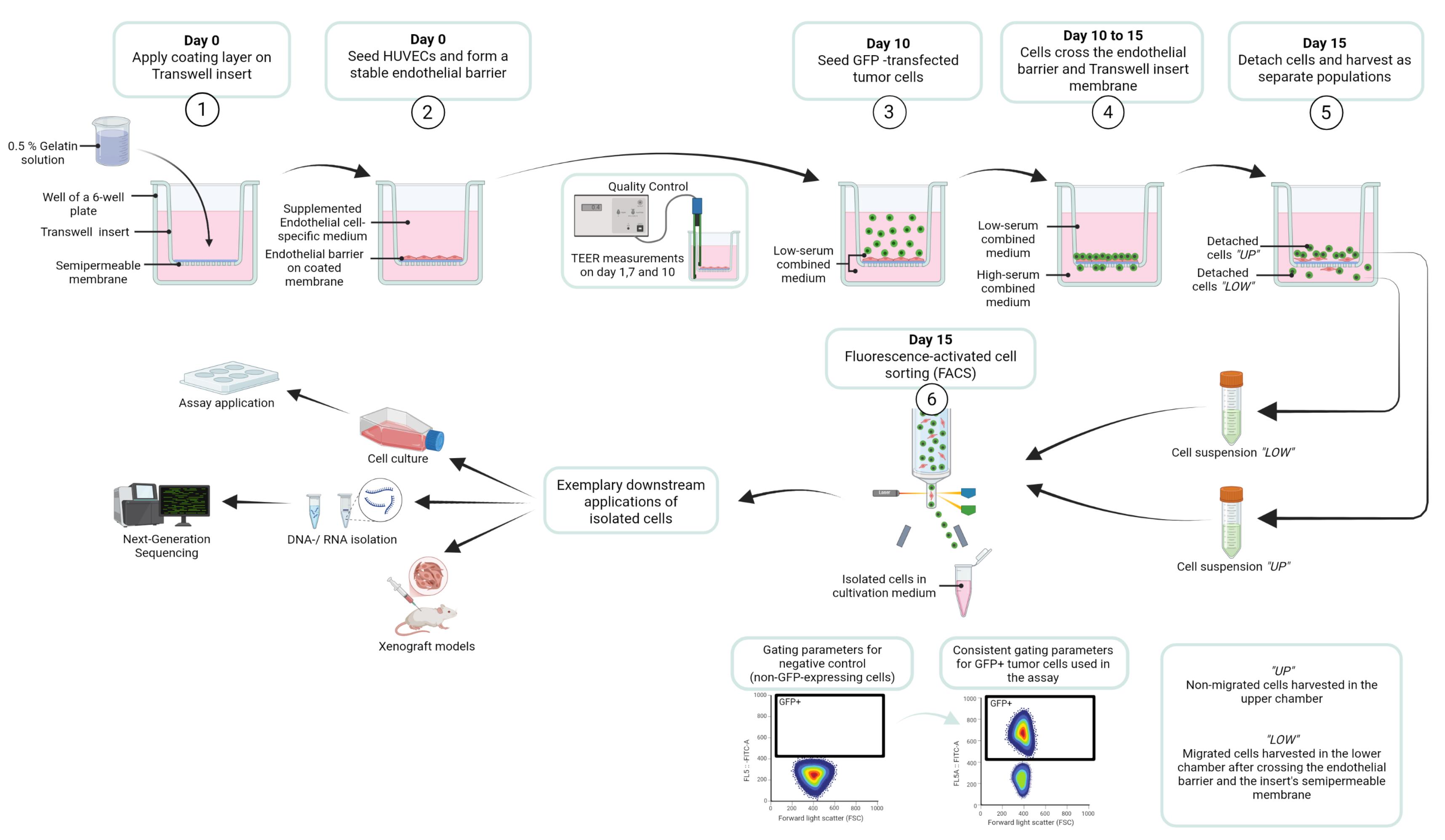
Workflow for the transendothelial migration assay designed to identify and isolate tumor cell subpopulations in MCF7 GFP and MCF7-TR GFP cells, based on their invasive capacity. Created in BioRender [1].
Background
Globally, approximately 1 in 20 women will be diagnosed with breast cancer (BC) during their lifetime, and 1 in 70 will die from the disease, though these rates vary significantly across countries and continents [2]. Estrogen receptor-positive (ER+) tumors account for approximately 70% of cases [3], underscoring the clinical and molecular significance of this subtype in both research and clinical settings.
Initially, ER+ BC typically exhibits sensitivity to selective estrogen receptor modulators (SERMs), including tamoxifen, which blocks estrogen-mediated proliferative signaling. However, acquired resistance during the therapeutic process remains a major clinical challenge, as it severely limits treatment efficacy [4].
During BC progression, formation of distant metastasis marks a critical turning point, transforming a potentially curable condition into a life-threatening disease [3]. As with most solid tumors, hematogenous dissemination is the predominant route of metastatic spread in breast cancer [5]. The metastatic cascade—driven by dynamic interactions between tumor cells and the tumor microenvironment—relies on intravasation into the bloodstream as a key, rate-limiting step. Once tumor cells breach the endothelial barrier and enter the bloodstream, disseminated tumor cells can seed distant organs, leading to metastatic colonization, poor prognosis, and increased metastasis-related mortality [6]. Understanding the mechanisms of intravasation, particularly across endothelial barriers, is therefore crucial for developing strategies to prevent metastatic spread.
To model tumor cell intravasation in vitro, we developed a physiologically relevant assay in which human endothelial cells are seeded on the apical side of porous tissue culture inserts to form a confluent, functional monolayer mimicking the vascular endothelium. For this purpose, HUVECs were chosen as an endothelial model due to their availability as a readily accessible and cost-effective human endothelial cell source with well-characterized responses to canonical stimuli and suitability for a wide range of standard in vitro vascular assays. FBS concentrations in the culture medium were optimized to enhance endothelial barrier stability on the one hand and to modulate migratory stimuli on the other hand, enabling controlled assessment of tumor cell migration and transendothelial invasion.
In order to facilitate the isolation and analysis of migratory versus non-migratory cell populations, BC cells were stably transfected with a GFP expression cassette using Sleeping Beauty transposon-mediated genome integration prior to seeding. After a predefined migration period, cells that had crossed the endothelial barrier were harvested and purified by fluorescence-activated cell sorting (FACS) for downstream analysis.
To compare migratory behavior, a 4-hydroxytamoxifen (4OHT)-resistant cell line established through chronic exposure to escalating 4OHT concentrations over several months was used [7]. This model enables the characterization of molecular adaptations associated with enhanced metastatic potential in therapy-resistant breast cancer.
Overall, this Transwell-based migration assay provides a robust platform for identifying key genes and signaling pathways driving metastasis, offering promising targets for future therapeutic strategies aimed at improving long-term patient outcomes.
Materials and reagents
Biological materials
1. HUVECs, isolated from human umbilical veins at the Department of Clinical Chemistry and the Department of Gynecology and Obstetrics, UMG Göttingen, following an in-house protocol as described in Willer et al. [8]; utilized in experiments at passages 3–5
2. MCF7 cells (ATCC, HTB-22); utilized in experiments up to passage 33
3. MCF7 GFP cells (ATCC, HTB-22, stably expressing a GFP reporter gene; see Section A); utilized in experiments up to passage 40
4. MCF7-TR cells (ATCC, HTB-22 variant with 4-OHT resistance developed by [7]); utilized in experiments up to passage 42
5. MCF7-TR GFP cells (ATCC, HTB-22 variant with 4-OHT resistance developed by [7], stably expressing a GFP reporter gene; see Section A); utilized in experiments up to passage 38
6. MG-63 cells (ATCC, CRL-1427); utilized in experiments up to passage 35
Reagents
A. Antibodies and staining solutions
1. APC anti-human CD31 antibody (BioLegend, catalog number: 303116)
2. DAPI (Novus Biologicals, catalog number: NBP2-31156), 1:1,000
3. Phalloidin 633 (Abcam, catalog number: ab176758), 1:400
4. Phalloidin 488 (Biolegend, catalog number: 424201), 1:400
5. Primary CD31 monoclonal antibody (mAb), mouse anti-human (DAKO, catalog number: M0823), 1:100
6. Primary ZO-1 (D7D12) mAb, rabbit anti-human (Cell Signaling Technology, catalog number: 8193), 1:100
7. Secondary antibody donkey anti-mouse IgG, Alexa Fluor 488 (Thermo Scientific, catalog number: A-21202), 1:400
8. Secondary antibody donkey anti-rabbit IgG, Alexa Fluor 555 (Thermo Scientific, catalog number: A-31572), 1:400
B. Cell culture reagents
1. Collagen I (BD Biosciences, catalog number: 354231)
2. DNAse I (NEB, catalog number: M0303S)
3. Fetal bovine serum (FBS) (Capricorn, catalog number: FBS-11A)
4. GFP Plasmid (Vector Builder: pSB[Exp]-CMV>EGFP, VB010000-9778kgx)
5. Insulin (Huminsulin 100, Eli Lilly, catalog number: HI0210)
6. Lipofectamine 2000 (Invitrogen, catalog number: 11668027)
7. Lipofectamine LTX reagent (Invitrogen, catalog number: 15338500)
8. Penicillin/streptomycin (Biowest, catalog number: L0022-100)
9. Paraformaldehyde (PFA) 4.5% (Chemsolute, catalog number: 2135.5000)
10. Poly-L-Lysine hydrochloride (Sigma-Aldrich, catalog number: P2658-25MG)
11. ROTI cell gelatin solution 2% (Roth, catalog number: 0646.1)
12. ROTI cell water (Roth, catalog number: 9186.1)
13. Transposase plasmid (Vector Builder, pRP[Exp]-CMV>T7/SB100X, VB010000-9366jzq)
14. Trypsin 0.05% EDTA (Pan-Biotech, catalog number: p10-027100)
15. Trypsin 0.25% EDTA (Pan-Biotech, catalog number: p10-020100)
16. TurboFect transfection reagent (Thermo Scientific, catalog number: R0531)
C. Cell culture media
1. Dulbecco's modified Eagle medium (DMEM) (Gibco, catalog number: 41966-029)
2. Dulbecco’s phosphate-buffered saline (DPBS) (Pan-Biotech, catalog number: P04-36500)
3. Endothelial cell growth medium 2 (EGCM2) (PromoCell, catalog number: C-22111)
4. Endopan 3 basal medium for endothelial cells (Pan-Biotech, catalog number: P04-0010K)
5. Medium 199 (Pan Biotech, catalog number: P04-07050)
6. Minimum essential medium (MEM) (Pan-Biotech, catalog number: P04-08500)
7. Opti-MEM I reduced serum medium (Gibco, catalog number: 51985026)
D. Other reagents
1. Bovine serum albumin (BSA) (Roth, catalog number: 3737.3)
2. Ethanol (99.9%) (Chemsolute, catalog number: 2212.5000)
3. Glycerol (Sigma-Aldrich, catalog number: 49767)
4. HCl (Merck, catalog number: 109057)
5. Mowiol 488 (Calbiochem, catalog number: 475904)
6. Sterile water (from Arium Mini Plus)
7. Tris hydrochloride (Roth, catalog number: 9090.3)
8. Triton X-100 (Merck, catalog number: 1086031000)
9. Tween-20 (ITW Reagents, catalog number: A4974)
Solutions
1. BSA (0.5% in DPBS) (see Recipes)
2. Collagen solution (0.1%) (see Recipes)
3. Combined medium (1.75% FBS) (see Recipes)
4. Combined medium (20% FBS) (see Recipes)
5. DMEM medium (see Recipes)
6. ECGM2 medium (see Recipes)
7. Endopan 3 medium (see Recipes)
8. Endopan 10 medium (see Recipes)
9. Endopan 15 medium (see Recipes)
10. Endopan 20 medium (see Recipes)
11. Ethanol 70% (see Recipes)
12. FACS buffer (see Recipes)
13. Gelatin solution (0.1%) (see Recipes)
14. Gelatin solution (0.2%) (see Recipes)
15. Gelatin solution (0.3%) (see Recipes)
16. Gelatin solution (0.4%) (see Recipes)
17. Gelatin solution (0.5%) (see Recipes)
18. Gelatin solution (0.6%) (see Recipes)
19. M199 medium (see Recipes)
20. MEM medium (see Recipes)
21. MEM medium (0.5% FBS) (see Recipes)
22. MEM medium (15% FBS) (see Recipes)
23. Mixed medium for endothelial cells (see Recipes)
24. Mowiol mounting medium (see Recipes)
25. DPBS-T (0.1% Tween-20) (see Recipes)
26. PFA (0.04%) (see Recipes)
27. Poly-L-Lysine solution (20 mg/mL) (see Recipes)
28. Poly-L-Lysine solution (0.1%) (see Recipes)
29. Staining solution with DAPI and Phalloidin (see Recipes)
30. Staining solution with primary antibodies (see Recipes)
31. Staining solution with secondary antibodies and Phalloidin, DAPI (see Recipes)
32. Tris-HCl (0.2 M, pH 8.5) (see Recipes)
33. Triton X-100 (1%) (see Recipes)
Recipes
1. BSA (0.5% in DPBS)
| Reagent | Final concentration | Volume for 50 mL |
|---|---|---|
| DPBS | n/a | 50 mL |
| BSA | 0.5% | 250 mg |
Stir for 1 h at room temperature. Aliquot and use or store at -20 °C.
2. Collagen solution (0.1%)
| Reagent | Final concentration | Volume for 1 mL |
|---|---|---|
| ROTI cell water | n/a | 966.7 μL |
| Collagen (30 mg/mL) | 0.1% | 33.3 μL |
3. Combined medium (1.75% FBS)
| Reagent | Final concentration | Volume for 500 mL |
|---|---|---|
| MEM medium (0.5% FBS) | 50% | 250 mL |
| Endopan 3 medium | 50% | 250 mL |
4. Combined medium (20% FBS)
| Reagent | Final concentration | Volume for 500 mL |
|---|---|---|
| MEM medium (10% FBS) | n/a | 200 mL |
| Endopan 3 medium | n/a | 200 mL |
| FBS | 20% | 67.5 mL |
5. DMEM medium
| Reagent | Final concentration | Volume for one bottle |
|---|---|---|
| DMEM | n/a | 500 mL |
| FBS | 20% | 126.6 mL |
| Penicillin/streptomycin | 1% | 6.4 mL |
6. ECGM2 medium
| Reagent | Final concentration | Volume for one bottle |
|---|---|---|
| ECGM2 | n/a | 500 mL |
| 7 supplements from kit | 1× | 700 μL |
| FBS from kit | 1× | 15 mL |
Follow the manufacturer’s recommendations.
7. Endopan 3 medium
| Reagent | Final concentration | Volume for one bottle |
|---|---|---|
| Endopan 3 | n/a | 500 mL |
| 8 supplements from kit | 1× | 800 μL |
| FBS from kit | 1× | 15 mL |
Follow the manufacturer’s recommendations.
8. Endopan 10 medium
| Reagent | Final concentration | Volume for one bottle |
|---|---|---|
| Endopan 3 | n/a | 500 mL |
| 8 supplements from kit | 1× | 800 μL |
| FBS from kit | 1× | 15 mL |
| Supplemented FBS | 10% | 40 mL |
9. Endopan 15 medium
| Reagent | Final concentration | Volume for one bottle |
|---|---|---|
| Endopan 3 | n/a | 500 mL |
| 8 supplements from kit | 1× | 800 μL |
| FBS from kit | 1× | 15 mL |
| Supplemented FBS | 15% | 73 mL |
10. Endopan 20 medium
| Reagent | Final concentration | Volume for 500 mL |
|---|---|---|
| Endopan 3 | n/a | 500 mL |
| 8 supplements from kit | 1× | 800 μL |
| FBS from kit | 1× | 15 mL |
| Supplemented FBS | 20% | 110 mL |
11. Ethanol 70%
| Reagent | Final concentration | Volume for 50 mL |
|---|---|---|
| Sterile water | n/a | 15 mL |
| Ethanol (99.9%) | 70% | 35 mL |
12. FACS buffer
| Reagent | Final concentration | Volume for 50 mL |
|---|---|---|
| DPBS | n/a | 45 mL |
| FBS | 10% | 5 mL |
13. Gelatin solution (0.1%)
| Reagent | Final concentration | Volume for 1 mL |
|---|---|---|
| ROTI cell water | n/a | 950 μL |
| ROTI cell gelatin solution (2%) | 0.1% | 50 μL |
14. Gelatin solution (0.2%)
| Reagent | Final concentration | Volume for 1 mL |
|---|---|---|
| ROTI cell water | n/a | 900 μL |
| ROTI cell gelatin solution (2%) | 0.2% | 100 μL |
15. Gelatin solution (0.3%)
| Reagent | Final concentration | Volume for 1 mL |
|---|---|---|
| ROTI cell water | n/a | 850 μL |
| ROTI cell gelatin solution (2%) | 0.3% | 150 μL |
16. Gelatin solution (0.4%)
| Reagent | Final concentration | Volume for 1 mL |
|---|---|---|
| ROTI cell water | n/a | 800 μL |
| ROTI cell gelatin solution (2%) | 0.4% | 200 μL |
17. Gelatin solution (0.5%)
| Reagent | Final concentration | Volume for 1 mL |
|---|---|---|
| ROTI cell water | n/a | 750 μL |
| ROTI cell gelatin solution (2%) | 0.5% | 250 μL |
18. Gelatin solution (0.6%)
| Reagent | Final concentration | Volume for 1 mL |
|---|---|---|
| ROTI cell water | n/a | 700 μL |
| ROTI cell gelatin solution (2%) | 0.6% | 300 μL |
19. M199 medium
| Reagent | Final concentration | Volume for one bottle |
|---|---|---|
| Medium 199 | n/a | 500 mL |
| FBS | 20% | 126.6 mL |
| Penicillin/streptomycin | 1% | 6.4 mL |
20. MEM medium
| Reagent | Final concentration | Volume for one bottle |
|---|---|---|
| MEM | n/a | 500 mL |
| FBS | 10% | 56 mL |
| Penicillin/streptomycin | 1% | 5.6 mL |
| Insulin 100 I.E./mL | 0.3% | 1.6 mL |
21. MEM medium (0.5% FBS)
| Reagent | Final concentration | Volume for one bottle |
|---|---|---|
| MEM | n/a | 500 mL |
| FBS | 0.5% | 2.6 mL |
| Penicillin/streptomycin | 1% | 5.1 mL |
| Insulin 100 I.E/mL | 0.3% | 1.5 mL |
22. MEM medium (15% FBS)
| Reagent | Final concentration | Volume for one bottle |
|---|---|---|
| MEM | n/a | 500 mL |
| FBS | 15% | 89 mL |
| Penicillin/streptomycin | 1% | 6 mL |
| Insulin 100 I.E./ mL | 0.3% | 1.8 mL |
23. Mixed medium for endothelial cells
| Reagent | Final concentration | Volume for one bottle |
|---|---|---|
| ECGM2 medium | 50% | 250 mL |
| M199 medium | 50% | 250 mL |
24. Mowiol mounting medium
| Reagent | Final concentration | Mass or volume for 50 mL |
|---|---|---|
| Mowiol 488 | 9.6% | 4.8 g |
| Glycerol | 24% | 12 g |
| Sterile water | n/a | 12 mL |
| Tris-HCl (0.2 M, pH 8.5) | 0.1 M | 24 mL |
Mix Mowiol 488 and glycerol with frequent agitation for 1 h at room temperature. Add sterile water and use a stir bar and magnetic stirrer to mix for 1 h at room temperature. Add Tris-HCl and incubate for 2 h at 50 °C with occasional stirring (e.g., stir for 2 min every 20 min). Centrifuge at 5,000× g for 15 min. Aliquot the supernatant and store at -20 °C.
25. DPBS-T (0.1% Tween-20)
| Reagent | Final concentration | Volume for 50 mL |
|---|---|---|
| DPBS | n/a | 50 mL |
| Tween | 0.1% | 50 μL |
26. PFA (0.04%)
| Reagent | Final concentration | Volume for 50 mL |
|---|---|---|
| DPBS | n/a | 49.6 mL |
| PFA (4.5%) | 0.04% | 440 μL |
27. Poly-L-lysine solution (20 mg/mL)
| Reagent | Final concentration | Volume for 10 mL |
|---|---|---|
| DPBS | n/a | 10 mL |
| Poly-L-lysine hydrochloride | 2% | 200 mg |
Mix thoroughly, aliquot, and either use immediately or store at -20 °C.
28. Poly-L-lysine solution (0.1%)
| Reagent | Final concentration | Volume for 1 mL |
|---|---|---|
| ROTI cell water | n/a | 950 μL |
| Poly-L-lysine (20 mg/mL) | 0.1% | 50 μL |
Use immediately.
29. Staining solution with DAPI and Phalloidin
| Reagent | Final concentration | Volume for 2.5 mL |
|---|---|---|
| DPBS | n/a | 2.47 mL |
| DAPI (1 mg/mL) | 1:1,000 | 2.5 μL |
| Phalloidin 488 (1 mg/mL) | 1:400 | 25 μL |
30. Staining solution with primary antibodies
| Reagent | Final concentration | Volume for 1.5 mL |
|---|---|---|
| BSA (0.5% in DPBS) | n/a | 1.47 mL |
| Primary CD31 mAb, mouse anti-human | (1:100) | 15 μL |
| Primary ZO-1 mAb, rabbit anti-human | (1:100) | 15 μL |
31. Staining solution with secondary antibodies and Phalloidin, DAPI
| Reagent | Final concentration | Volume for 1.5 mL |
|---|---|---|
| BSA (0.5% in DPBS) | n/a | 1.49 mL |
| Secondary antibody donkey anti-mouse IgG | (1:400) | 3.75 μL |
| Secondary antibody donkey anti-rabbit IgG | (1:400) | 3.75 μL |
| Phalloidin 633 (1 mg/mL) | (1:400) | 3.75 μL |
| DAPI (1 mg/mL) | (1:1,000) | 1.5 μL |
32. Tris-HCl (0.2 M, pH 8.5)
| Reagent | Final concentration | Volume for 1 L |
|---|---|---|
| Sterile water | n/a | 1 L |
| Tris hydrochloride | 0.2 M | 24.2 g |
| HCl | 1× | Dropwise to pH 8.5 |
Dissolve Tris hydrochloride in 800 mL of sterile water. While stirring with a stir bar on a magnetic stirrer, add HCl dropwise to adjust the pH to 8.5. Then, add sterile water to a final volume of 1 L. Store at room temperature.
33. Triton X-100 (1%)
| Reagent | Final concentration | Volume for 50 mL |
|---|---|---|
| DPBS | n/a | 49.5 mL |
| Triton-X 100 | 1% | 0.5 mL |
Mix and store at room temperature.
Laboratory supplies
1. Aluminum foil (Labsolute, catalog number: 7696857)
2. Conical tube sterile 15 mL (Greiner Bio-One, catalog number: 188271-N)
3. Conical tube sterile 50 mL (Greiner Bio-One, catalog number: 227261)
4. Cotton buds 150 mm (Beese, catalog number: 1260)
5. Coverslips (Roth, catalog number: HKE5.1)
6. Disposable gloves (Nitril NextGen, catalog number: 1283)
7. Eppendorf tube 0.5 mL (Eppendorf, catalog number: 0030121023)
8. Eppendorf tube 1.5 mL (Eppendorf, catalog number: 0030120086)
9. FACS tubes (Sarstedt, catalog number: 55.526.006)
10. Glass slides (Marienfeld, catalog number: 08-104-01)
11. Handheld counter (Roth Selection, catalog number: EE53.1)
12. Monarch Spin RNA Isolation kit (Mini) (NEB, catalog number: T2110L)
13. Neubauer counting chamber (Blaubrand, catalog number: 717805)
14. Paper towels (Tapira, catalog number: 07730008)
15. Parafilm M 100 mm (Amcor, catalog number: PM999)
16. Pasteur pipettes (Labsolute, catalog number: 7691061)
17. Pipette tips 10 μL (Sarstedt, catalog number: 70.3010.100)
18. Pipette tips 200 μL (Sarstedt, catalog number: 70.3030.020)
19. Pipette tips 1,000 μL (Sarstedt, catalog number: 70.3050.200)
20. PluriStrainer filter mini 40 μm (PluriSelect, catalog number: 43-10040-50)
21. Pyrex beaker 100 mL (Brand, catalog number: BR91224)
22. Scalpel sterile (Swann-Morton, catalog number: 0507)
23. Serological pipette 2 mL (Sarstedt, catalog number: 86.1252.001)
24. Serological pipette 5 mL (Sarstedt, catalog number: 86.1253.001)
25. Serological pipette 10 mL (Sarstedt, catalog number: 86.1254.001)
26. Square Petri dish non-sterile (Greiner Bio-One, catalog number: 688102)
27. TC flask T25 (Sarstedt, catalog number: 83.3910.002)
28. TC flask T75 (Sarstedt, catalog number: 83.3911.002)
29. TC inserts 6-well 1 μm pores (Sarstedt, catalog number: 83.3930.100)
30. TC inserts 6-well 3 μm pores (Sarstedt, catalog number: 83.3930.300)
31. TC inserts 6-well 5 μm pores (Sarstedt, catalog number: 83.3930.500)
32. TC inserts 6-well 8 μm pores (Sarstedt, catalog number: 83.3930.800)
33. TC inserts 24-well 5 μm pores (Sarstedt, catalog number: 83.3932.500)
34. TC inserts 24-well 8 μm pores (Sarstedt, catalog number: 83.3932.800)
35. TC plate 6-well (Sarstedt, catalog number: 83.3920)
36. TC plate 24-well (Sarstedt, catalog number: 83.3922)
37. Tweezers, blunt and curved (Roth, catalog number: PX30.1)
Equipment
1. Arium Mini Plus (Sartorius, model: H2O-MA-UV-T)
2. Automated cell counter (DeNovix Cell Drop BF)
3. Bead bath (Thermo Fisher, model: Precision GP 10)
4. Benchtop centrifuge (Eppendorf, model: 5415D)
5. Centrifuge (Heraeus Instruments, model: Labofuge 400)
6. Chemical fume hood (Köttermann, model: 2-401-DAAAGY)
7. CO2 incubator (HERAcell 150i)
8. Digital CMOS camera (Hamamatsu Photonics, model: ORCA-spark)
9. FACS instrument (BD Biosciences, model: Aria II)
10. Flow cytometer (Beckman Coulter, model: Cytoflex S)
11. Fluorescent microscope (Olympus, model: IX 83)
12. Ice machine (Scotsman, model: AF80 XSafe)
13. Magnetic stir bars (Roth, catalog number: X171.1)
14. Magnetic stirrer (IKA RCT basic, catalog number: Z645060)
15. Microscope (Nikon, model: Eclipse TS100)
16. Mini centrifuge (Biozym, catalog number: 552032)
17. pH meter Lab 860 Set (Schott Instruments, catalog number: 102711330)
18. Pipettes 2.5 μL, 10 μL, 100 μL, 200 μL, 1,000 μL (Eppendorf, catalog number: 492940)
19. Pipette controller (Integra Biosciences, catalog number: 155016)
20. Sterile cell culture hood (Thermo Scientific, model: Herasafe 2025 1.2)
21. TEER electrode (Millicell ERS-2 Replacement Electrode)
22. TEER measurement device (Millicell ERS-2)
23. Vortex machine (Scientific Industries, model: Vortex-Genie 2)
Software and datasets
1. CellSens Dimension (Olympus, version 3.2) for acquiring images with the inverted fluorescence microscope Olympus IX83
2. CytExpert Software (Beckman Coulter, version 2.5) for flow cytometry
3. Excel (Microsoft Office 2019, version 16.0) for quantitative analyses
4. Fiji (open-source distribution of ImageJ, version 1.54) for visual and quantitative analyses
5. FlowJo software (BD Biosciences, version 10.0.1) for FACS data analyses
6. GraphPad Prism (version 8.0.1) for statistical analyses
Procedure
登录 / 注册后阅读全文
文章信息
稿件历史记录
提交日期: May 25, 2026
接收日期: Aug 10, 2026
在线发布日期: Nov 18, 2026
出版日期: Nov 20, 2026
版权信息
© 2026 The Author(s); This is an open access article under the CC BY license (https://creativecommons.org/licenses/by/4.0/).
如何引用
Koperski, A., Rieß, A. L., Schüürhuis, L., Andag, R., Fischer, A., Hasanov, M. F., Gallwas, J. and Wegwitz, F. (2026). Modeling Tumor Cell Transendothelial Migration In Vitro: A Functional Assay for Metastatic Potential. Bio-protocol 16(22): e5895. DOI: 10.21769/BioProtoc.5895.

 Download Citation in RIS Format
分类
癌症生物学 > 侵袭和转移 > 细胞生物学试验 > 细胞侵袭
细胞生物学 > 细胞运动 > 细胞迁移
您对这篇实验方案有问题吗?
在此处发布您的问题,我们将邀请本文作者来回答。同时,我们会将您的问题发布到Bio-protocol Exchange,以便寻求社区成员的帮助。
 发布问题


