


Flow Cytometry Assessment of T-Cell Exhaustion Markers and Cytokine Production in Cryopreserved Mouse Splenocytes

发布: 2026年12月05日第16卷第23期 DOI: 10.21769/BioProtoc.5894 浏览次数: 18
评审: Luis Alberto Sánchez VargasHepzibah Bacilio OcampoArundhati Mehta
 PDF
 Q&A







Abstract
Aging is associated with accumulating dysfunctional T cells, characterized by altered marker expression detectable by flow cytometry. However, since no individual marker reflects age-related processes specifically, assessing a combined set of markers is critical, yet often impractical on fresh cells when multiple panels or samples are involved. Here, we present a detailed protocol to comprehensively evaluate exhaustion markers and cytokine expression using cryopreserved mouse splenocytes and multi-parameter flow cytometry. The approach is validated by comparing cells isolated from young and aged mice.
Key features
• Protocol for cryopreservation, thawing, stimulation, and flow cytometry processing of mouse splenocytes.
• Cryopreservation enables separation of splenocyte isolation and staining into distinct procedures, enhancing experimental flexibility and scheduling.
• Multi-marker flow cytometry panels for parallel assessment of T-cell exhaustion markers and cytokines.
• Protocol validation in a relevant biological context (young and aged mice).
Keywords: Flow cytometry T-cell exhaustion
Immune suppression
Cytokines
Cryopreserved splenocytes
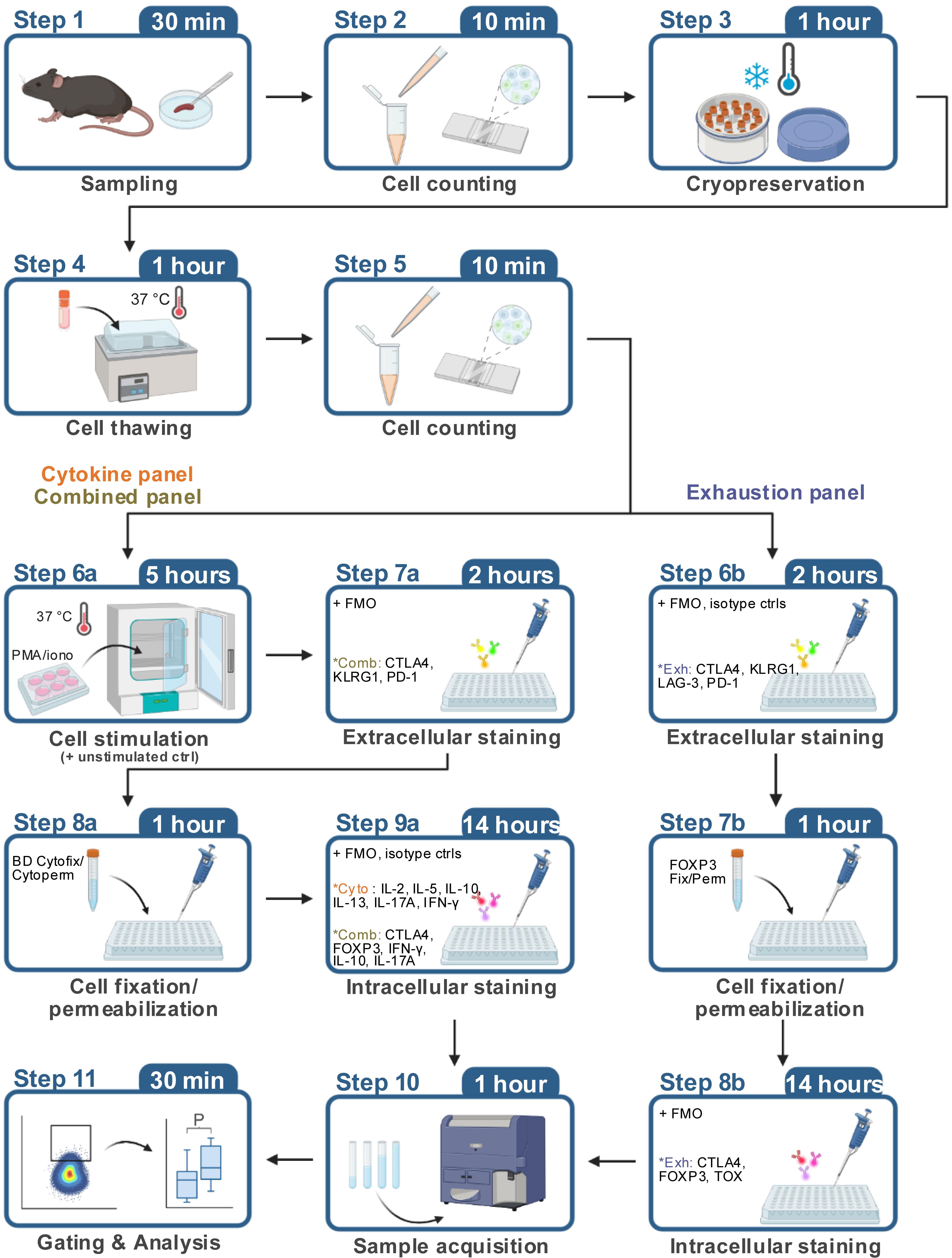
Graphical overview
Background
Aging is associated with gradual T-cell compositional and functional changes that may reflect altered activation and differentiation programs and effector competence. With aging, naïve T-cell frequency typically decreases, whereas differentiated effector memory and immunosuppressive regulatory FOXP3+ T cells increase [1]. Phenotypically, age-associated dysregulated T cells show increased expression of markers associated with immune exhaustion, senescence, or suppression. T-cell exhaustion and senescence-associated markers include inhibitory checkpoint molecules such as programmed cell death protein 1 (PD-1), cytotoxic T-lymphocyte antigen 4 (CTLA-4), T-cell immunoreceptor with Ig and ITIM domains (TIGIT), lymphocyte-activation gene 3 (LAG-3), and killer cell lectin-like receptor subfamily G member 1 (KLRG1); and transcription factors such as eomesodermin (EOMES) and thymocyte selection-associated high mobility group box (TOX) [2,3]. In addition, age-associated dysregulated T cells may increase production of effector cytokines [3].
However, none of the above markers is specific to age-associated T-cell dysregulation, as they can also be expressed in other states, such as during immune cell development, immune activation, or immune regulation. Furthermore, although T-cell exhaustion has been more extensively investigated in CD8+ T cells, increasing evidence suggests that CD4+ T cells may undergo a similar fate with age [4]. For these reasons, it is important to evaluate the expression of multiple markers simultaneously in well-defined experimental settings.
Flow cytometry can be used to quantify the expression of many of the molecules associated with T-cell dysregulation. This may be achieved through adapted antibody panels, for example targeting key surface markers or intracellular targets such as transcription factors and cytokines upon ex vivo stimulation. However, because different markers often require distinct strategies to process and stain samples, including fixation/permeabilization and stimulation conditions, their simultaneous assessment through multiple panels may be challenging in fresh cells, increasing the risk of technical variability. Cryopreservation of immune cells is an essential technique that allows for long-term storage of biological samples for subsequent analysis. While fresh samples are typically preferred for functional assays, cryopreserved specimens offer several key advantages, including the batch processing of samples collected at different points and the possibility to share samples with collaborators at different sites.
Here, we present an optimized protocol based on established procedures for immune cell preservation and flow cytometric analysis. The protocol includes a detailed description of the key steps for freezing, thawing, and subsequent flow cytometric analysis of mouse splenocytes, which provide a rich source of T cells. Using three distinct antibody panels (“exhaustion”, “cytokine”, and “combined”) on individual aliquots from the same animals, our protocol enables comprehensive phenotyping encompassing the assessment of inhibitory immune checkpoints (CTLA4, KLRG1, LAG-3, PD-1), transcription factors (FOXP3, TOX), and effector cytokines (IFN-γ, IL-2, IL-5, IL-10, IL-13, IL-17). The “exhaustion” panel is applied to thawed cells without a subsequent stimulation step, whereas the “cytokine” and “combined” panels require stimulation after thawing. The extracellular staining workflow is identical for the three panels, whereas the “exhaustion” panel uses a different fixation/permeabilization buffer from the “cytokine” and “combined” panels for the intracellular staining step. The overall staining steps are described once in the protocol below. Critically, the protocol recommends the use of controls for antibodies targeting rare populations, including fluorescence-minus-one (FMO) and isotype controls. Finally, our protocol is validated on cryopreserved splenocytes isolated from young (7–8 weeks) and aged (2 years) male C57BL/6J mice. Nonetheless, our protocol provides a versatile workflow that could be adapted to evaluate the same markers in other conditions associated with T-cell dysfunction, such as cancer, systemic inflammation, or metabolic disorders.
For a comprehensive analysis of T-cell activation, Treg functionality, and cytokine production in fresh mouse splenocytes, we recommend referring to the previously published protocol [5].
Materials and reagents
Biological materials
1. C57BL/6JRj mice (Janvier Labs, identifier: RRID: IMSR_RJ: C57BL-6JRJ)
2. C57BL/6JRj mice (aged; Janvier Labs, identifier: RRID: IMSR_RJ: AGED-C57BL-6JRJ)
Reagents
1. 10× PBS (Thermo Fisher Scientific, catalog number: BP399-1)
2. ACK lysis buffer (Gibco, catalog number: A10492-01)
3. Acridine orange/propidium iodide stain (Logos Biosystems, catalog number: F23001)
4. Benzonase (Sigma, catalog number: 70746-4)
5. Bovine serum albumin (BSA) (Sigma, catalog number: A7906)
6. Brefeldin A (Sigma, catalog number: B7651)
7. BV711 Streptavidin (BioLegend, catalog number: 405241)
8. Cytofix/CytopermTM Fixation/Permeabilization kit (BD, catalog number: 554714)
9. Dimethyl sulfoxide (DMSO) (Sigma, catalog number: D2650)
10. Ethanol 70%, filtered denatured (Contec, product code: FBC570DE)
11. Foxp3/Transcription Factor Staining Buffer Set (eBioscience, catalog number: 00-5523-00)
12. Fetal bovine serum (FBS) (Gibco, catalog number: 16140-071)
13. Horizon Brilliant Stain Buffer (BD, catalog number: 566349)
14. Ionomycin (Sigma, catalog number: I0634)
15. Isopropanol (2-propanol) for HPLC 99% (Sigma, catalog number: 34863-1L)
16. Ketamine 1000, 100 mg/mL (Virbac, GTIN: 03597132111010)
17. Live/DeadTM Fixable Blue Dead Cell Stain kit, for UV excitation (Thermo Fisher Scientific, catalog number: L23105)
18. Penicillin-streptomycin (Gibco, catalog number: 15140122)
19. Phorbol 12-myristate 13-acetate (PMA) (Sigma, catalog number: P8139)
20. Physiodose 5 mL (physiological serum) (Laboratoires Gilbert, reference: 3518646266638)
21. RPMI 1640 (Gibco, catalog number: 61870-010)
22. Xylasol 20 mg/mL (Bimeda, catalog number: 04042668304898)
23. Antibodies (Table 1)
Table 1. Antibodies used in the protocol. Antibody dilutions are reported in Table S1.
| Antibodies (clone) | Manufacturer | Catalog number; RRID |
|---|---|---|
| CD16/CD32 (93) | eBioscience | Catalog number: 14-0161-86; RRID:AB_467135 |
| CD19 BV650 (6D5) | BioLegend | Catalog number: 115541; RRID:AB_11204087 |
| CD3 APC-eF780 (17a2) | eBioscience | Catalog number: 47-0032-82; RRID:AB_1272181 |
| CD4 AF700 (RM4-5) | BD Biosciences | Catalog number: 557956; RRID:AB_396956 |
| CD45 PE-CF594 (30-F11) | BD Biosciences | Catalog number: 562420; RRID:AB_11154401 |
| CD8a BV786 (53-6.7) | BD Biosciences | Catalog number: 563332; RRID:AB_2721167 |
| CTLA4 APC (UC10-4B9) | BioLegend | Catalog number: 106310; RRID:AB_2087653 |
| CTLA4 BV605 (UC10-4B9) | BioLegend | Catalog number: 106323; RRID:AB_2566467 |
| EOMES PerCP-eF710 (Dan11mag) | eBioscience | Catalog number: 46-4875-82; RRID:AB_10597455 |
| FOXP3 AF488 (MF-14) | BioLegend | Catalog number: 126406; RRID:AB_1089113 |
| IFN-γ BV711 (XMG1.2) | BioLegend | Catalog number: 505835; RRID:AB_11219588 |
| IL-10 APC (JES5-16E3) | eBioscience | Catalog number: 17-7101-81; RRID:AB_469501 |
| IL-13 PE-Cy7 (eBio13A) | eBioscience | Catalog number: 25-7133-82; RRID:AB_2573530 |
| IL-17A AF488 (TC11-18H10) | BD Biosciences | Catalog number: 560220; RRID:AB_1645194 |
| IL-17A PE (TC11-18H10) | BD Biosciences | Catalog number: 559502; RRID:AB_397256 |
| IL-2 BV605 (JES6-5H4) | BioLegend | Catalog number: 503829; RRID:AB_11204084 |
| IL-4 PE (11B11) | BD Biosciences | Catalog number: 554435; RRID:AB_395391 |
| IL-5 BV421 (TRFK5) | BioLegend | Catalog number: 504311; RRID:AB_2563161 |
| KLRG1 PE-Cy7 (2F1) | BioLegend | Catalog number: 138416; RRID:AB_2561736 |
| LAG-3 Biotin (C9B7W) | BioLegend | Catalog number: 125206; RRID:AB_2133345 |
| Mouse IgG1, κ Isotype Ctrl BV605 (MOPC-21) | BioLegend | Catalog number: 400162; RRID:AB_11125373 |
| PD-1 eF450 (J43) | eBioscience | Catalog number: 48-9985-82; RRID:AB_2574139 |
| Rat IgG1, κ Isotype Ctrl BV421 (RTK2071) | BioLegend | Catalog number: 400429; RRID:AB_10900998 |
| Rat IgG1, κ Isotype Ctrl PE (R3-34) | BD Biosciences | Catalog number: 554685; RRID:AB_395140 |
| Rat IgG1, κ Isotype Ctrl PE-Cy7 (RTK2071) | BioLegend | Catalog number: 400416; RRID:AB_326522 |
| TIGIT BV605 (1G9) | BD Biosciences | Catalog number: 744212; RRID:AB_2742061 |
| TNF-α PerCP-Cy5.5 (MP6-XT22) | BioLegend | Catalog number: 506322; RRID:AB_961434 |
| TOX PE (TXRX10) | eBioscience | Catalog number: 12-6502-82; RRID:AB_10855034 |
Solutions
1. Acridine orange/propidium iodide staining (see Recipes)
2. Antibody cocktail (see Recipes)
3. Anesthesia (see Recipes)
4. BD Perm/Wash solution (see Recipes)
5. Cell culture medium (see Recipes)
6. Cell stimulation medium (see Recipes)
7. Dead-cell suspension (see Recipes)
8. FACS buffer (see Recipes)
9. FOXP3 Fix/Perm solution (see Recipes)
10. FOXP3 Fix/Perm wash buffer (see Recipes)
11. Freezing medium (see Recipes)
Recipes
1. Acridine orange/propidium iodide staining
Mix the cell suspension and acridine orange/propidium iodide stain thoroughly at a 9:1 ratio (18 μL of cell suspension + 2 μL of acridine orange/propidium iodide stain).
2. Antibody cocktail
Prepare the necessary volume of antibody cocktail by diluting the antibodies at the appropriate concentrations (Table S1) in Brilliant Stain Buffer. The antibody cocktail should be prepared fresh and used on the same day.
3. Anesthesia
For each 1 mL of final volume, add 250 μL of ketamine and 50 μL of xylazine and bring to the final volume with physiological serum.
4. BD Perm/Wash solution
Dilute one part of the 10× BD Perm/Wash solution concentrate stock with nine parts of distilled water (dilution 1:10). The 1× BD Perm/Wash solution can be kept at 4 °C until use.
5. Cell culture medium
Prepare complete cell culture medium under sterile conditions by supplementing RPMI 1640 base medium with 10% FBS, 100 μg/mL penicillin, and 100 μg/mL streptomycin.
6. Cell stimulation medium
Prepare cell culture medium supplemented with PMA (50 ng/mL), ionomycin (500 ng/mL), and 1× brefeldin A (10 μg/mL).
7. Dead-cell suspension
Place 1 × 10⁶ spleen cells in an Eppendorf tube and incubate them in a water bath pre-heated to 65 °C for 30 min to kill them. The obtained dead-cell suspension can be used on the same day or prepared in advance and kept at 4 °C for up to 2 weeks.
8. FACS buffer
Prepare FACS buffer under sterile conditions by dissolving 2.5 g of BSA and adding 2 mL of 0.5 M EDTA to 500 mL of PBS to achieve final concentrations of 0.5% BSA and 2 mM EDTA. Mix thoroughly until BSA is completely dissolved and store at 4 °C.
9. FOXP3 Fix/Perm solution
Dilute one part of the 4× FOXP3 Fix/Perm concentrate stock with three parts of the Fix/Perm diluent (dilution 1:4). The 1× FOXP3 Fix/Perm solution can be kept at 4 °C until use.
Notes: Prepare fresh before use. The solution contains formaldehyde, which is toxic and carcinogenic. Wear proper personal protective equipment and dispose of waste appropriately.
10. FOXP3 Fix/Perm wash buffer
Dilute one part of the 10× permeabilization buffer concentrate stock with nine parts of distilled water (dilution 1:10). The 1× FOXP3 Fix/Perm wash buffer can be kept at 4 °C until use.
Note: The 10× permeabilization buffer concentrate stock may precipitate. If precipitate forms, filter after diluting to 1×.
11. Freezing medium
On the day of the experiment, prepare fresh freezing medium under sterile conditions by mixing 90% FBS and 10% DMSO. If prepared in advance, keep the freezing medium at 4 °C and use it the same day.
Laboratory supplies
1. 6-well tissue culture plate, flat bottom (Corning, catalog number: 3516)
2. 96-well cell culture plate, round bottom (Dutscher Dominique SAS, catalog number: 353077)
3. Cell freezing container, Nalgene Mr. Frosty (Sigma, catalog number: 5100-0001)
4. Cell strainers 100 μm (Thermo Fisher Scientific, catalog number: 11517532)
5. Centrifuge tubes 15 mL (Dutscher Dominique SAS, catalog number: 352096)
6. Centrifuge tubes 50 mL (Dutscher Dominique SAS, catalog number: 352070)
7. Clearline 2 mL cryotube (Dutscher Dominique SAS, catalog number: 390708)
8. Falcon 5 mL polystyrene round-bottom tube with cell strainer cap (Dutscher Dominique SAS, catalog number: 352235)
9. Forceps, Dumont AA, standard tips (Fine Science Tools GMBH, catalog number: 11210-10)
10. Forceps, anatomical standard forceps, curved (Fine Science Tools GMBH, catalog number: 11001-12)
11. PhotonSlideTM ultra-low fluorescence cell counting slide (Logos Biosystems, catalog number: L12005)
12. Scissors, hardened fine, straight/sharp (Fine Science Tools GMBH, catalog number: 14090-09)
13. Syringe Plastipak 1 mL (Dutscher Dominique SAS, catalog number: 303172)
Equipment
1. BD LSRFortessa X-20 (BD), cytometer configuration is summarized in Table S2
2. Cell culture CO2 incubator, Heraeus Kendro HeraCell 150 (Thermo Fisher Scientific)
3. Centrifuge 5810/5810R (Eppendorf)
4. Centrifuge Sorvall ST 8R (Thermo Fisher Scientific)
5. LUNA-FLTM Dual Fluorescence Cell Counter (Logos Biosystems)
6. Microbiological safety cabinet BIOAIR Safeflow 1.2 (Bioair)
Software and datasets
1. Adobe Illustrator v30.5.1 (Adobe, identifier: RRID:SCR_010279)
2. BD FACSDiva Software v.9.2 (BD, identifier: RRID:SCR_001456)
3. FlowJo v.10.10.0 (FlowJo LLC, identifier: RRID:SCR_008520)
4. GraphPad Prism 11.0.0 (GraphPad Software, identifier: RRID:SCR_002798)
5. Real Statistics Resource Pack software (Release 9.9, copyright 2013 – 2026, Charles Zaiontz, https://real-statistics.com)
6. All data are in Table S3
Procedure
登录 / 注册后阅读全文
文章信息
稿件历史记录
提交日期: Jun 25, 2026
接收日期: Oct 19, 2026
在线发布日期: Nov 15, 2026
出版日期: Dec 5, 2026
版权信息
© 2026 The Author(s); This is an open access article under the CC BY license (https://creativecommons.org/licenses/by/4.0/).
如何引用
Song, M. H., Lafosse, I., Eberl, G., Petit, A. C., Lledo, P. M. and Suzzi, S. (2026). Flow Cytometry Assessment of T-Cell Exhaustion Markers and Cytokine Production in Cryopreserved Mouse Splenocytes. Bio-protocol 16(23): e5894. DOI: 10.21769/BioProtoc.5894.

 Download Citation in RIS Format
分类
免疫学 > 免疫细胞染色 > 流式细胞术
细胞生物学 > 细胞染色 > 全细胞
您对这篇实验方案有问题吗?
在此处发布您的问题,我们将邀请本文作者来回答。同时,我们会将您的问题发布到Bio-protocol Exchange,以便寻求社区成员的帮助。
 发布问题


