


Cy5-Fluorescent EMSA for Characterization of G-Quadruplex Structures and DNA–Protein Interactions Without Radioisotopes

(*contributed equally to this work) 发布: 2026年12月05日第16卷第23期 DOI: 10.21769/BioProtoc.5892 浏览次数: 16
评审: Jing LiRavi KumarAnonymous reviewer(s)
 PDF
 Q&A






Abstract
The characterization of DNA–protein interactions is fundamental to understanding the molecular mechanisms that regulate genome function. G-quadruplexes (G4s) are non-canonical nucleic acid secondary structures that influence diverse biological processes, including DNA replication, transcription, and DNA repair, primarily through their interactions with G-quadruplex-binding proteins (G4BPs). The electrophoretic mobility shift assay (EMSA) is one of the most widely used methods for studying DNA–protein interactions under native conditions. Conventional EMSA protocols commonly employ radioisotope-labeled oligonucleotides, which require specialized laboratory infrastructure, regulatory compliance, and radioactive waste disposal. Here, we describe a fluorescence-based EMSA protocol that uses commercially synthesized Cy5-labeled oligonucleotides for both the characterization of G4 structures and the analysis of DNA–protein interactions without the use of radioisotopes. G-rich oligonucleotides are folded under potassium-containing conditions and resolved by native polyacrylamide gel electrophoresis (PAGE), enabling discrimination between intramolecular and intermolecular G4 conformations based on their electrophoretic mobility. The folded G4 DNA can subsequently be incubated with purified proteins or cell lysates, and relative protein binding is detected by the reduced electrophoretic mobility of DNA–protein complexes. Fluorescent DNA and DNA–protein complexes are directly visualized using standard fluorescence gel imaging systems, eliminating the need for radioactive probe preparation while simplifying sample handling and data acquisition. This protocol provides a simple, sensitive, and reproducible approach for investigating G-quadruplex structures and their interactions with DNA-binding proteins and can be readily adapted to other structured or linear DNA substrates.
Key features
• Non-radioactive fluorescence-based EMSA using Cy5-labeled oligonucleotides for the analysis of DNA secondary structures and DNA-protein interactions.
• Eliminates the need for radioisotopes, reducing the regulatory burden while simplifying probe handling, storage, and waste disposal.
• Preserves native DNA conformation and DNA-protein complexes, enabling the analysis of non B-DNA structure formation and DNA-protein interaction assays.
• Direct visualization and quantitative analysis of fluorescently labeled DNA and DNA-protein complexes using standard fluorescence gel imaging systems.
Keywords: Non-radioactive EMSA G-quadruplex
Cy5
EMSA
DNA–protein interaction
G-quadruplex-binding proteins (G4BPs)
Relative binding
Fluorescent EMSA
Graphical overview
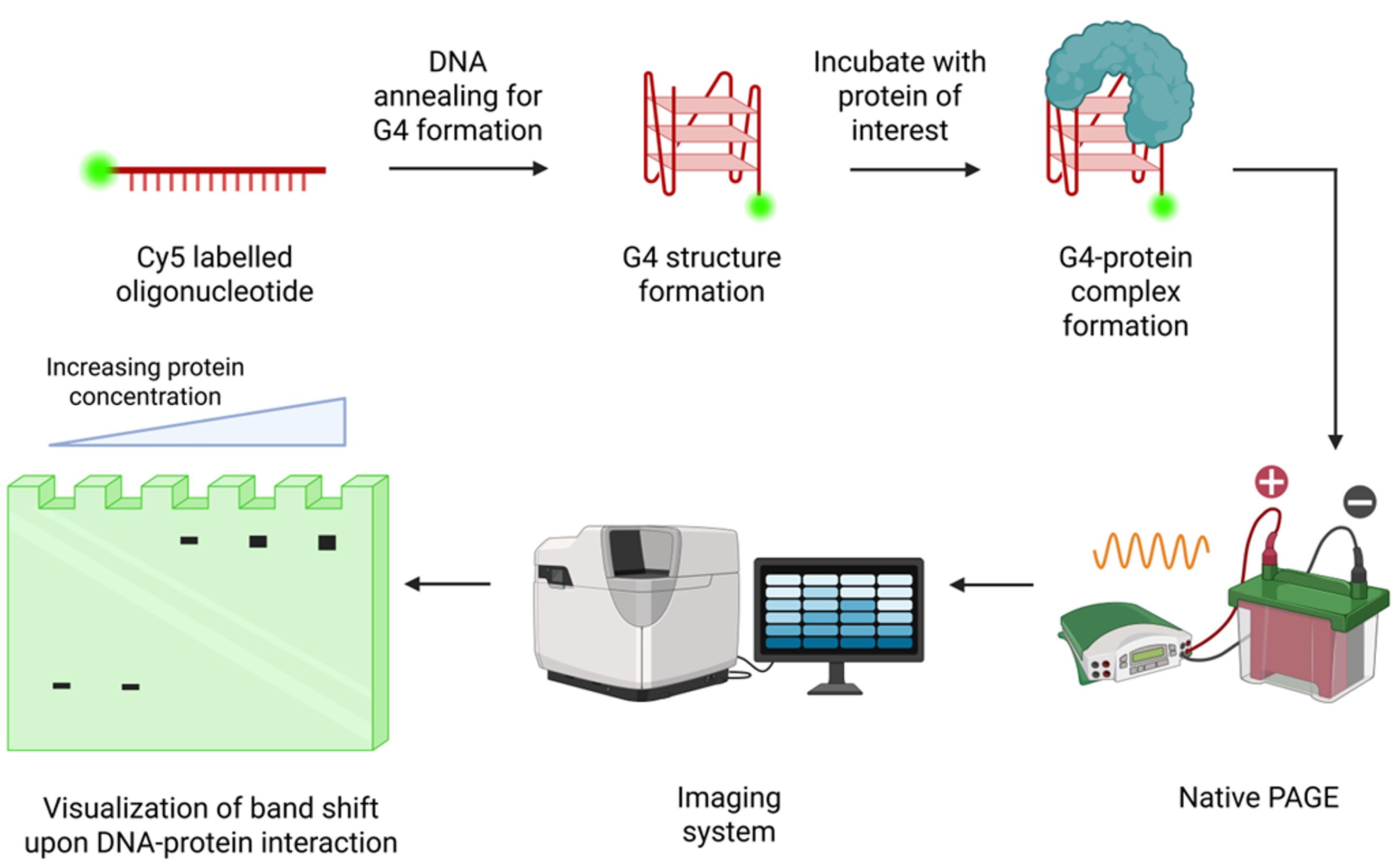
EMSA of Cy5-labeled G-quadruplex (G4) oligonucleotide with protein. The Cy5-labeled oligonucleotide sequences are annealed to form the G4 structure. This G4 form is incubated with the protein of interest or cell lysate and separated by polyacrylamide gel electrophoresis (PAGE) under low voltage at low temperature. The gel is imaged for Cy5 fluorescence using a fluorescence imager to detect mobility-shifted bands resulting from protein–DNA interactions. Created in BioRender, https://BioRender.com/4h275p6.
Background
G quadruplexes (G4s) are non-canonical nucleic acid secondary structures formed by guanine-rich DNA or RNA sequences through Hoogsteen hydrogen bonding. Four guanine bases associate to form a planar G-tetrad, and the stacking of two or more G-tetrads gives rise to a G4 structure [1]. These structures are dynamic and can adopt multiple topologies depending on their sequence composition, loop length, strand orientation, and ionic conditions. Monovalent cations, particularly potassium (K+), stabilize G4 structures by coordinating within the central channel formed by stacked G-tetrads [2].
Over the past two decades, G4s have emerged as important regulators of genome function. Genome-wide studies have demonstrated that G-rich sequences capable of forming G4s are enriched at promoters, telomeres, replication origins, untranslated regions, and other regulatory genomic elements [1,3–5]. G4 formation has been implicated in the regulation of DNA replication, transcription, translation, DNA repair, and epigenetic regulation [6]. Their involvement in these processes has generated considerable interest in exploiting G4 as potential therapeutic targets for cancer and neurodegenerative disorders [7,8].
Many of the biological functions of G4s are mediated through their interactions with G4BPs, which recognize, stabilize, remodel, or resolve these structures [9]. Characterizing these interactions is therefore essential for understanding how G4s influence genome regulation. A variety of biochemical and biophysical techniques have been developed to study DNA–protein interactions [10,11]. Among these, the electrophoretic mobility shift assay (EMSA) remains one of the most widely used methods because of its simplicity, versatility, and ability to directly visualize protein-bound DNA under native conditions. EMSA is routinely employed to evaluate the relative binding or concentration-dependent binding, perform competition assays, and investigate the impact of sequence mutations or DNA structural changes on protein binding [12,13]. Under native electrophoretic conditions, the assay can also resolve distinct DNA conformations, enabling the characterization of both intermolecular and intramolecular G4 structures based on their differential electrophoretic mobility.
Conventional EMSA protocols frequently employ radioactively labeled oligonucleotides generated using T4 polynucleotide kinase (T4 PNK) and γ-32P ATP because of their high sensitivity [14]. However, radioactive assays require specialized laboratory infrastructure, regulatory compliance, isotope handling expertise, and dedicated waste disposal procedures. Furthermore, exposure to radioactivity poses potential health and safety risks and requires stringent handling practices to minimize contamination and accidental exposure. In addition, the limited shelf life of radioisotopes necessitates repeated probe preparation. Fluorescence-based detection provides an effective alternative that eliminates these constraints while enabling direct visualization of DNA and DNA–protein complexes using standard fluorescence imaging systems.
Several fluorescent dyes, including 6-carboxyfluorescein (6-FAM) [15], tetramethylrhodamine (TAMRA) [16], cyanine 3 (Cy3) [17], cyanine 5 (Cy5) [18], and Alexa Fluor dyes (Alexa Fluor 488, 647) [19,20] have been successfully used for fluorescence-based EMSA. In this protocol, Cy5-labeled oligonucleotides are folded under defined ionic conditions, incubated with the protein of interest where required, and resolved by native polyacrylamide gel electrophoresis (PAGE). The assay enables direct visualization of free DNA, intramolecular and intermolecular G4 conformations, and DNA–protein complexes using standard fluorescence imaging systems (Graphical overview). By eliminating the need for radioisotopes, this protocol provides a simple, safe, and broadly accessible approach for investigating G-quadruplex structures and their interactions with DNA-binding proteins.
Materials and reagents
Biological materials
1. Purified protein of interest, protein extract, or cell lysate (as appropriate)
2. E. coli DH5α
3. E. coli Rosetta
Oligonucleotides
1. Cy5-labeled DNA oligonucleotides (commercially synthesized or labeled in-house)
Reagents
1. Tris base (Himedia, catalog number: MB029)
2. Boric acid (Himedia, catalog number: MB077)
3. EDTA (Himedia, catalog number: MB011)
4. Potassium chloride (KCl) (Himedia, catalog number: MB043)
5. Sodium chloride (NaCl) (Himedia, catalog number: GRM853)
6. Magnesium chloride hexahydrate (MgCl2·6H2O) (Himedia, catalog number: MBO40)
7. Dithiothreitol (DTT) (Himedia, catalog number: MB070)
8. Acrylamide (Himedia, catalog number: MB068)
9. N, N’-Methylenebisacrylamide (Himedia, catalog number: MB005)
10. Ammonium persulfate (APS) (Himedia, catalog number: MB003)
11. Glycerol (Himedia, catalog number: MB060)
12. Zinc chloride (ZnCl2) (Himedia, catalog number: TC184)
13. TEMED (Himedia, catalog number: MB026)
14. Purple loading dye (NEB, catalog number: B7025S)
15. Ultrapure water
16. Poly(deoxyinosic-deoxycytidylic) acid sodium salt (Sigma-Aldrich, catalog number: P4929)
Solutions
1. 5× Tris-borate EDTA (TBE) buffer (see Recipes)
2. 40% acrylamide/bis-acrylamide solution (29:1) (see Recipes)
3. 10% ammonium persulfate (APS) (see Recipes)
4. 3 M potassium chloride (KCl) (see Recipes)
5. Tris-EDTA (TE) buffer (see Recipes)
6. 10× protein binding buffer (see Recipes)
7. 50% glycerol (see Recipes)
8. 2 M Tris-HCl (pH 7.5) (see Recipes)
9. 5 M sodium chloride (NaCl) (see Recipes)
10. 0.5 M EDTA (pH 8.0) (see Recipes)
11. 0.1 M DTT (see Recipes)
12. 0.1 M ZnCl2 (see Recipes)
13. 1 M MgCl2 (see Recipes)
14. 1 M KCl (see Recipes)
15. Native polyacrylamide gel (8% or 15%) (see Recipes)
Recipes
1. 5× TBE buffer
| Reagent | Final concentration | Quantity or volume |
|---|---|---|
| Tris base | 445 mM | 54 g |
| Boric acid | 445 mM | 27.5 g |
| 0.5 M EDTA (pH 8.0) | 10 mM | 20 mL |
| Ultrapure water | ----- | To 1 L |
Dissolve Tris base and boric acid in approximately 800 mL of ultrapure water. Add EDTA, adjust the volume to 1 L, mix thoroughly, and store at 4 °C for up to 6 months.
2. 40% acrylamide/bis-acrylamide solution (29:1)
| Reagent | Quantity or volume |
|---|---|
| Acrylamide | 77.32 g |
| N, N’-Methylenebisacrylamide | 2.68 g |
| Ultrapure water | To 200 mL |
Dissolve completely, filter through a 0.45-μm filter, protect from light, and store at 4 °C for up to 1 month (or prepare freshly).
3. 10% APS
| Reagent | Final concentration | Quantity or volume |
|---|---|---|
| APS | 10% (w/v) | 1 g |
| Ultrapure water | ----- | To 10 mL |
Prepare fresh before use or aliquot and store at -20 °C for up to 1 month. Avoid repeated freeze/thaw cycles.
4. 3 M KCl
| Reagent | Final concentration | Quantity or volume |
|---|---|---|
| KCl | 3 M | 22.37 g |
| Ultrapure water | ---- | To 100 mL |
Store at room temperature for up to 6 months.
5. TE buffer
| Reagent | Final concentration | Quantity or volume |
|---|---|---|
| 2 M Tris-HCl (pH 7.5) | 100 mM | 0.5 mL |
| 0.5 M EDTA (pH 8.00) | 10 mM | 0.2 mL |
| Ultrapure water | ----- | To 10 mL |
Store at room temperature for up to 6 months.
6. 10× protein binding buffer
| Reagent | Final concentration | Quantity or volume |
|---|---|---|
| 2 M Tris-HCl (pH 7.5) | 250 mM | 0.125 mL |
| 5 M NaCl | 500 mM | 0.1 mL |
| 1 M MgCl2 | 10 mM | 0.01 mL |
| Glycerol (100%) | 50% (v/v) | 0.5 mL |
| ZnCl2 (0.1 M) | 0.1 mM | 1 μL |
| Ultrapure water | --- | To 1 mL |
Aliquot and store at -20 °C for 2 months.
7. 50% glycerol
| Reagent | Quantity or volume |
|---|---|
| Glycerol | 500 μL |
| Ultrapure water | 500 μL |
Mix thoroughly and store at room temperature for up to 6 months.
8. 2 M Tris-HCl (pH 7.5)
| Reagent | Final concentration | Quantity or volume |
|---|---|---|
| Tris base | 2 M | 12.12 g |
| Concentrated HCl | --- | Adjust to pH 7.5 |
| Ultrapure water | ---- | To 50 mL |
Adjust the pH to 7.5 using concentrated HCl and make up to the final volume. Store at room temperature for up to 6 months.
9. 5 M NaCl
| Reagent | Final concentration | Quantity or volume |
|---|---|---|
| NaCl | 5 M | 14.61 g |
| Ultrapure water | ---- | To 50 mL |
Store at room temperature for up to 6 months.
10. 0.5 M EDTA (pH 8.0)
| Reagent | Final concentration | Quantity or volume |
|---|---|---|
| EDTA disodium salt | 0.5 M | 9.31 g |
| NaOH | ---- | Adjust to pH 8.0 |
| Ultrapure water | --- | To 50 mL |
Adjust the pH to 8.0 with NaOH until EDTA dissolves completely. Bring to the final volume and store at room temperature for up to 6 months.
11. 0.1 M DTT
| Reagent | Final concentration | Quantity or volume |
|---|---|---|
| DTT | 0.1 M | 7.72 mg |
| Ultrapure water | --- | To 0.5 mL |
DTT is light-sensitive. Prepare small aliquots, store at -20 °C protected from light, and use within 1 month. Avoid repeated freeze/thaw cycles.
12. 0.1 M ZnCl2
| Reagent | Final concentration | Quantity or volume |
|---|---|---|
| Anhydrous ZnCl2 | 0.1 M | 136.4 mg |
| Ultrapure water | --- | To 10 mL |
Store at room temperature for up to 6 months.
13. 1 M MgCl2
| Reagent | Final concentration | Quantity or volume |
|---|---|---|
| MgCl2 hexahydrate | 1 M | 2.03 g |
| Ultrapure water | --- | To 10 mL |
Store at room temperature for up to 6 months.
14. 1 M KCl
| Reagent | Final concentration | Quantity or volume |
|---|---|---|
| 3 M KCl solution | 1 M | 3.33 mL |
| Ultrapure water | --- | To 10 mL |
Store at room temperature up to 6 months.
15. Native polyacrylamide gel (8% or 15%)
| Reagent | Final concentration | Quantity or volume |
|---|---|---|
| 5× TBE | 0.5× | 5 mL |
| 40% acrylamide | 8%/15% | 10/18.75 mL |
| 3 M KCl | 0.1 M | 1.66 mL |
| 10% APS | 0.1% | 0.5 mL |
| TEMED | 0.1% | 50 μL |
| Ultrapure water | To 50 mL |
Laboratory supplies
1. Laboratory wipes (KimtechTM Kimwipes or equivalent)
2. 70% (v/v) ethanol
3. Adjustable micropipettes (1,000, 200, 20, and 2 μL) (Eppendorf or equivalent)
4. Sterile pipette tips (1,000, 200, 20 μL) (Tarsons, catalog numbers: 521020B, 521010Y, and 521000 or equivalent)
5. Disposable syringes (5 or 10 mL)
6. Wash bottle (Tarsons, catalog number: 620080 or equivalent)
7. Glass plates, spacers, and combs for native polyacrylamide gel casting (BioBEE Tech, SDS-2 or equivalent)
8. 1.5 mL microcentrifuge tubes
9. 0.2 mL PCR tubes (for use in the thermal cycler)
10. Ice bucket and crushed ice
11. Disposable gloves and laboratory protective equipment
Equipment
1. Fluorescence gel imaging system capable of detecting Cy5 fluorescence (Q9 Alliance imaging system, UVITEC, Cambridge, UK or equivalent)
2. Thermal cycler (Bio-Rad or equivalent)
3. BIOBEE Vertical Gel (PAGE) Electrophoresis System -MIDI (14 cm width × 17 cm height; well capacity 80 μL) or equivalent
4. Microcentrifuge (SpinwinTM MC 100, Tarsons or equivalent)
5. Electrophoresis power supply
6. Analytical balance
7. pH meter
8. Magnetic stirrer with stir bars
9. Refrigerator (4 °C)
10. Freezer (-20 °C)
Software and datasets
1. ImageJ (Wayne Rasband and contributors National Institutes of Health, USA, version 1.54g) (https://imagej.net/ij/index.html)
Procedure
登录 / 注册后阅读全文
文章信息
稿件历史记录
提交日期: Aug 7, 2026
接收日期: Oct 19, 2026
在线发布日期: Nov 16, 2026
出版日期: Dec 5, 2026
版权信息
© 2026 The Author(s); This is an open access article under the CC BY-NC license (https://creativecommons.org/licenses/by-nc/4.0/).
如何引用
Prasanth, S., Dutta, A., Sreya, S., Johna, A. and Nishana, M. (2026). Cy5-Fluorescent EMSA for Characterization of G-Quadruplex Structures and DNA–Protein Interactions Without Radioisotopes. Bio-protocol 16(23): e5892. DOI: 10.21769/BioProtoc.5892.

 Download Citation in RIS Format
您对这篇实验方案有问题吗?
在此处发布您的问题,我们将邀请本文作者来回答。同时,我们会将您的问题发布到Bio-protocol Exchange,以便寻求社区成员的帮助。
 发布问题


