


A Hybrid Gait Analysis System for Robust and Efficient Estimation of the Sciatic Functional Index in Rats

发布: 2026年11月20日第16卷第22期 DOI: 10.21769/BioProtoc.5889 浏览次数: 19
评审: Shubham GargAnonymous reviewer(s)
 PDF
 Q&A

参见作者原研究论文
The authors used this protocol in:

Jun 2026




Abstract
The Sciatic Functional Index (SFI) is one of the most widely used quantitative measures of functional recovery following peripheral nerve injury in rodents. Conventional paper–ink walking tracks, frustrated total internal reflection (fTIR)-based gait-analysis systems, and ventral video-based imaging provide complementary information for SFI estimation but frequently fail to determine one or more SFI parameters under normal and, especially, pathological conditions. Here, we describe and validate a hybrid gait-analysis system that combines fTIR imaging of plantar contact areas with simultaneous visualization of selected plantar anatomical landmarks using additional oblique white-light illumination. The accompanying Gait2SFI software automatically detects plantar contact areas, assists the identification of anatomical landmarks, performs quality control, and calculates the classical SFI. The software supports SFI estimation from the maximum contact area, the total print area, or a composite footprint generated from the minimum number of consecutive contact frames required to identify all anatomical landmarks necessary for SFI estimation. Validation demonstrated that the hybrid system is fully compatible with the conventional SFI methodology while substantially increasing the proportion of analyzable gait cycles under both physiological and pathological conditions. Simultaneous access to contact and anatomical information enabled reliable determination of print length, toe spread, and intermediary toe spread in gait cycles that could not be analyzed using conventional contact-based approaches alone. The hybrid system also reduced repeated recordings and improved experimental throughput. Overall, the protocol provides a reproducible, low-cost, and efficient approach for robust estimation of the classical Sciatic Functional Index in rats while maintaining compatibility with existing SFI methodologies.
Key features
• Hybrid fTIR+WL imaging combines paw contact and anatomical information and enables classical SFI estimation when contact-only footprints are incomplete.
• Improves SFI measurability in both healthy and sciatic nerve–injured rats.
• Supports maximum contact area, total print area, and composite footprints.
• Reduces within-session variability compared with the paper–ink method.
Keywords: Sciatic Functional Index Gait analysis
Frustrated total internal reflection (fTIR)
Hybrid imaging
Peripheral nerve injury
Walking track analysis
Rodents
Graphical overview
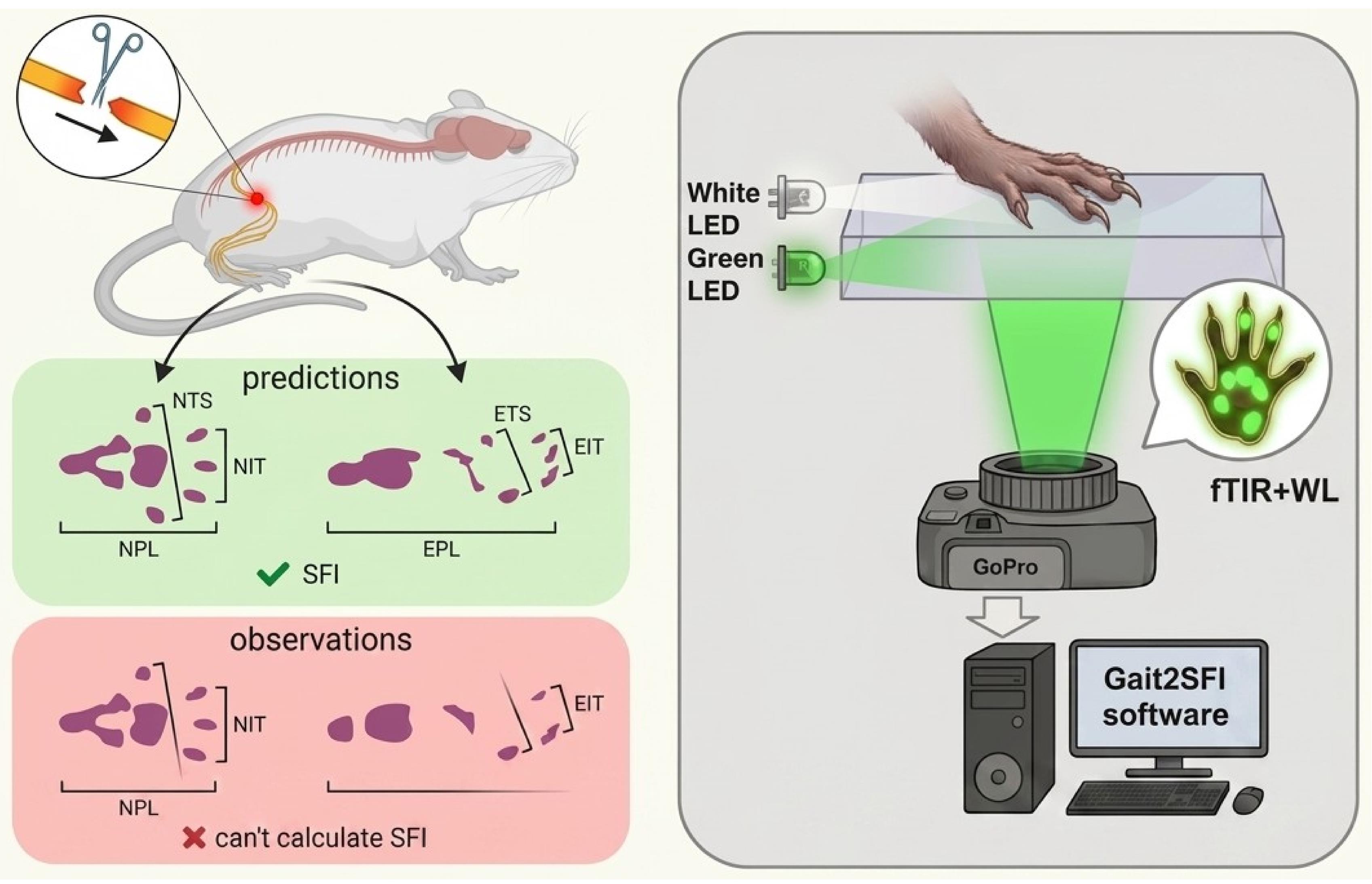
Principle of hybrid frustrated total internal reflection (fTIR)+white-light (WL) imaging for Sciatic Functional Index (SFI) measurement. Conventional fTIR and paper–ink recordings may fail to reveal one or more plantar pads and may show contacts from other paw structures that complicate anatomical interpretation, even in healthy animals, with these limitations becoming more pronounced after sciatic nerve injury (left). Print length, toe spread, and intermediary toe spread are shown for the experimental (EPL, ETS, EIT) and contralateral normal (NPL, NTS, NIT) hindpaw footprints. Hybrid fTIR+WL imaging retains fTIR information on genuine paw–glass contacts (green in the footprint, right), while WL identifies the anatomical structures corresponding to fTIR-positive contacts and reveals pads and plantar regions that do not contact the glass (brown in the footprint, right). The combined information enables calculation of the classical SFI when fTIR alone is insufficient.
Background
Functional gait analysis remains one of the principal methods for evaluating recovery following peripheral nerve injury in experimental rodents. Among the available behavioral tests, the Sciatic Functional Index (SFI), originally introduced by de Medinaceli and subsequently modified by Bain and colleagues, has become the most widely used quantitative index for assessing recovery after sciatic nerve injury because it integrates several aspects of hindlimb motor function into a single numerical parameter [1–3]. Owing to its non-invasive nature, simplicity, and sensitivity to functional deficits, SFI has been extensively employed to evaluate surgical repair techniques, nerve guidance conduits, biomaterials, electrical stimulation, pharmacological interventions, and rehabilitation strategies [4,5]. Recent advances in peripheral nerve regeneration, particularly the development of engineered hydrogels and multifunctional nerve-guidance conduits, further emphasize the importance of reliable functional assessment for evaluating therapeutic efficacy [6–8]. Nevertheless, conventional paper–ink walking-track analysis remains in use even in this technologically advanced field, highlighting the need for accessible, cost-effective approaches that improve the reliability of SFI measurements without requiring expensive specialized equipment.
The classical SFI is calculated from three geometric parameters extracted from hindpaw footprints: print length (PL), toe spread (TS), and intermediary toe spread (ITS). Although these parameters appear straightforward, their reliable determination becomes increasingly difficult following sciatic nerve injury. Loss of toe spreading, altered paw posture, foot dragging, joint contractures, and dorsum walking frequently produce incomplete or distorted footprints, making these parameters difficult or impossible to determine [3–5]. In severe injuries, conventional walking-track analysis often becomes unmeasurable, and arbitrary SFI values have historically been assigned to represent complete functional loss [2].
During the last two decades, digital gait-analysis systems have largely replaced conventional paper–ink walking tracks. Among them, frustrated total internal reflection (fTIR)-based systems, such as CatWalk XT, have become the most widely used because they automatically detect paw contact with the walking surface while simultaneously providing numerous spatiotemporal gait parameters [9,10]. More recently, ventral video-based systems, such as DigiGait, have provided an alternative approach by imaging the projected contour of the plantar surface during locomotion on a transparent treadmill [11]. Although these systems have substantially improved automation and experimental throughput, reliable estimation of the classical SFI remains strongly dependent on the method used to acquire paw footprints. Existing approaches differ not only in the way footprints are recorded but also in the physical and biomechanical information they capture, resulting in systematic differences in the determination of SFI parameters and limiting direct comparison between studies.
The classical paper–ink walking-track method records an integrated footprint accumulated throughout the entire stance phase. While this approach formed the basis of the original SFI, the resulting footprint does not correspond to any specific moment of gait but rather represents the cumulative contact pattern produced throughout stance. Moreover, footprint quality is influenced by smearing, dragging, incomplete transfer of digit pads, and variations in the animal's walking speed. Importantly, the clearest footprints are frequently obtained when animals slow down, hesitate, or briefly stop. Such footprints no longer represent physiological locomotion but instead reflect static or quasi-static paw placement. Consequently, they often contain extensive posterior plantar contact, including heel and tarsal regions, producing artificially increased PL values compared with footprints generated during continuous walking. Because only the final footprint is available for analysis, it is impossible to determine retrospectively whether the animal was walking normally, accelerating, decelerating, or standing still when the footprint was produced.
FTIR-based systems overcome many limitations of paper–ink methods by recording genuine load-bearing plantar contact during locomotion with high temporal resolution. However, they visualize only load-bearing contact areas rather than the entire plantar surface. Consequently, anatomical landmarks required for estimation of the classical SFI are detected only when they physically contact the walking surface. This limitation becomes particularly important because individual plantar pads contact the substrate asynchronously during the stance phase. As a result, all digit and metatarsal pads are frequently not visible simultaneously within a single fTIR frame, even during physiological locomotion. In practice, SFI is commonly estimated either from the frame corresponding to the maximum contact area or from another frame in which the required digit pads happen to be visible. However, these approaches are not equivalent. The maximum contact area represents one well-defined biomechanical instant within the stance cycle but may lack critical digit contacts, whereas frames selected to maximize pad visibility may correspond to different phases of stance. Consequently, individual SFI parameters may be measured at different biomechanical moments, reducing standardization and increasing variability.
An alternative representation available in fTIR-based systems is the total print area, obtained by accumulating all plantar contacts throughout the stance phase. Unlike a single frame, the total print area integrates contact information from the entire stance and therefore more closely resembles the classical ink footprint. However, it no longer represents a single biomechanical instant and may be influenced by paw sliding, paw rotation, or progressive changes in paw position during stance. Furthermore, even the total print area may fail to contain all digit pads required for SFI estimation when individual digits never contact the walking surface, as frequently occurs after peripheral nerve injury. Consequently, there is currently no consensus regarding which footprint representation—maximum contact area, another individual contact frame, or total print area—is most appropriate for estimation of the classical SFI parameters.
A conceptually different approach is implemented in ventral video systems such as DigiGait, which image the plantar contour while animals walk on a transparent treadmill at a constant speed. Although treadmill locomotion improves the reproducibility of gait, DigiGait does not measure genuine plantar contact areas. Instead, SFI is estimated from ventral images of the paw rather than from contact footprints, making the resulting measurements conceptually different from the classical SFI originally developed using paper–ink walking tracks. Consequently, values obtained using contour-based and contact-based methods should not necessarily be considered directly equivalent.
Another unresolved methodological issue concerns the determination of print length (PL). Classical SFI defines PL as a footprint-derived parameter; however, following peripheral nerve injury, the posterior border of the footprint may correspond to different anatomical structures, including metatarsal pads, tarsal pads, the anatomical heel, plantar skin, or even joints contacting the walking surface. Furthermore, while some studies define PL anatomically as the distance from the heel to the third toe [3,4], contact-based gait-analysis systems determine it from the most posterior contact point. Consequently, different studies effectively measure PL from different anatomical landmarks, introducing additional variability and complicating direct comparison of SFI values obtained using different gait-analysis methodologies.
These methodological limitations become considerably more pronounced after sciatic nerve injury. Abnormal gait may include loss of toe spreading, posterior displacement of plantar loading, heel walking, dragging, joint contractures, dorsum walking, or complete absence of contact by individual digits. Under these conditions, one or more of the parameters required for SFI calculation frequently cannot be determined, resulting in exclusion of individual gait cycles or even entire animals from analysis [3,4,12]. Consequently, both the reliability and efficiency of SFI estimation decrease precisely in those experimental models where accurate functional assessment is most critical.
To overcome these limitations, we developed a hybrid gait-analysis system. The system combines fTIR-derived plantar contact information with simultaneous visualization of selected plantar anatomical landmarks required for robust estimation of the classical Sciatic Functional Index using additional oblique white-light illumination. This hybrid acquisition strategy provides complementary contact and anatomical information while preserving compatibility with conventional SFI methodology. Rather than introducing a new functional index, the hybrid approach was designed to improve the robustness and efficiency of estimating the classical SFI by enabling reliable determination of PL, TS, and ITS even when some anatomical landmarks are absent from conventional contact-based footprints. The accompanying Gait2SFI software supports estimation of the classical SFI using different footprint representations, including the maximum contact area, the total print area, or a composite footprint generated by combining the minimum number of consecutive contact frames beginning with the maximum contact area frame that is sufficient to visualize all anatomical landmarks required for SFI estimation. This latter representation preserves a well-defined temporal reference while minimizing smearing, dragging, and changes in paw position associated with integrating contact information over the entire stance phase. In the present study, we describe the hybrid gait-analysis system and validate its performance under both physiological and pathological conditions, including direct comparison with the classical paper–ink walking-track method.
Materials and reagents
Biological materials
1. Male Wistar rats (2–5 months old, 200–450 g), bred in the vivarium of the O.O. Bogomolets Institute of Physiology, NAS of Ukraine (Kyiv, Ukraine). Animals were used in multiple gait-analysis experiments, with some rats contributing to more than one dataset. Data were obtained from a total of 29 rats, including 15 with experimentally induced sciatic nerve injury (SNI). The number of animals included in each validation experiment is specified in the corresponding section.
Reagents
1. Non-toxic watercolor paint (KITE, catalog number: BF_3789335): used to obtain rat paw prints (any other watercolor paint can be used instead)
2. Glass-cleaning solution without ethanol (Clin, catalog number: MD_218910); applied between recordings to remove paw marks, debris, and residues (can be replaced with any glass cleaner without ethanol)
Equipment
1. Custom fTIR walking-track walkway: built in-house (glass platform with green and white LED edge lighting + red tunnel ceiling lighting (Figure 1); assembly described in Procedure, steps A1–3)
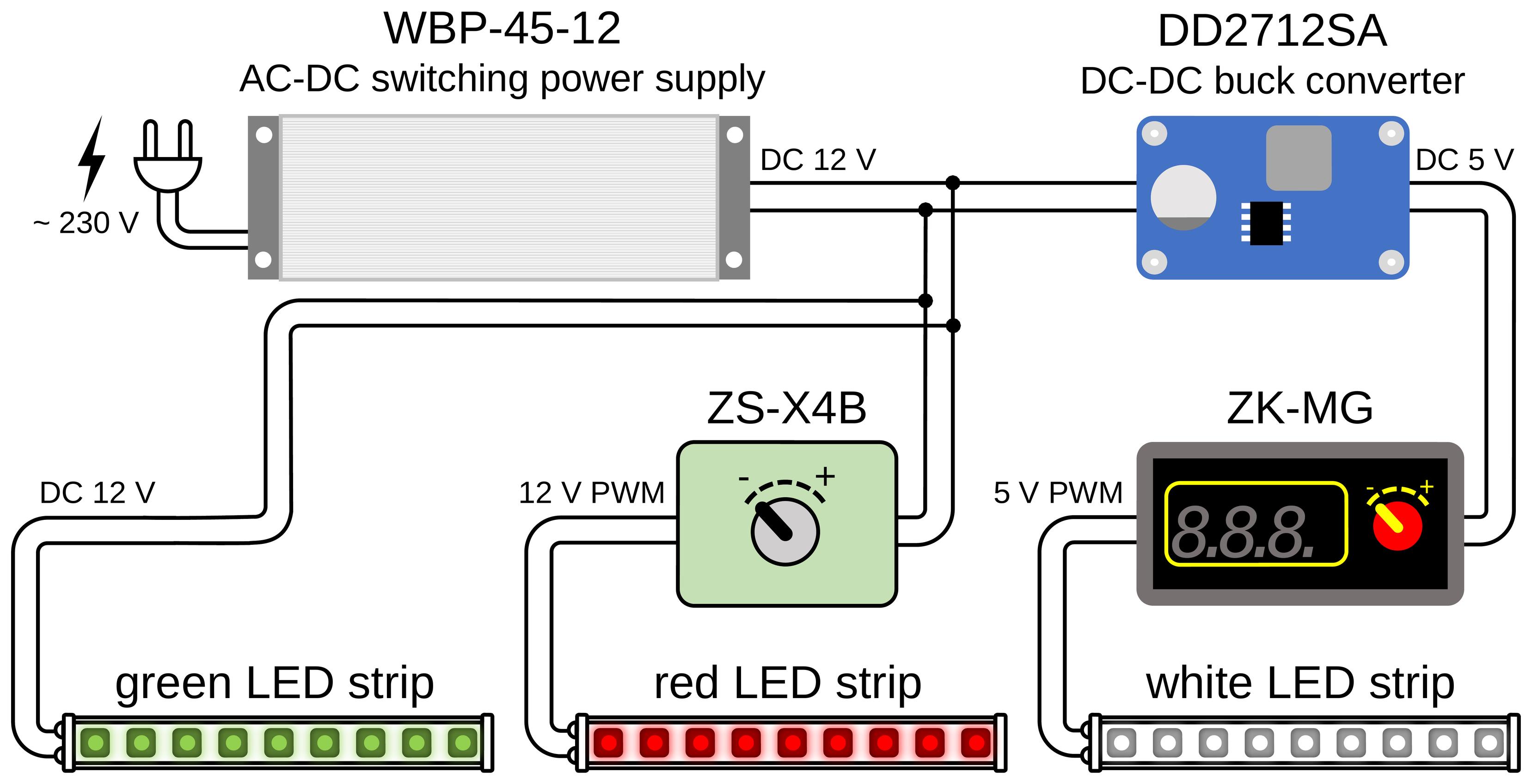
Figure 1. Block diagram of the electrical connections of the LED illumination. A single sealed 12 V, 45 W power supply (Biom WBP-45-12) drives the three LED channels. The green LED strip (fTIR edge illumination of the walking platform) is connected directly to the 12 V output. The red LED strip (tunnel-ceiling illumination) is powered through a ZS-X4B controller that sets its intensity. The white LED strip (oblique white-light illumination) is supplied through a DD2712SA step-down (buck) converter that reduces the voltage to 5 V, followed by a ZK-MG controller that regulates its brightness.
2. High-resolution camera (GoPro, model: HERO10 Black; 5,312 × 2,988 pixels, 50 frames/s)
3. LED power supply; BIOM Professional 12 V DC, 45 W, WBP-45-12, 3.7 A 02095
4. Buck step-down voltage regulator module (Eletechsup, catalog number: DD2712SA)
5. Regulator ZK-MG; PWM controller module (generic)
6. Regulator ZS-X4B; PWM speed controller/LED dimmer (Excellway)
7. LED green (SMD2835, 120 LEDs/m, 9.6 W/m, IP20, 12 V)
8. LED white (ROLUM SX, 6 × 12 mm, 6 W, 5 V, IP67, neutral white 3800–4300 K)
9. LED red (SMD2835, 120 LEDs/m, 12 V, IP20, 1012089)
10. Support stand/darkened enclosure (in-house)
11. Computer for video acquisition and analysis (HP 250 G10)
Software and datasets
1. Gait2SFI.py: Footprint detection and parameter-measurement GUI (this study; https://github.com/olbmv/Gait2SFI; accessed: 17 August 2026)
2. SFI.py: SFI calculation tool with logging to data.csv (this study; https://github.com/olbmv/Gait2SFI; accessed: 17 August 2026)
3. Python 3.10.11 (Python Software Foundation)
4. All data and code have been deposited in GitHub: https://github.com/olbmv/Gait2SFI (accessed: 17 August 2026)
Procedure
登录 / 注册后阅读全文
文章信息
稿件历史记录
提交日期: Aug 27, 2026
接收日期: Oct 25, 2026
在线发布日期: Nov 12, 2026
出版日期: Nov 20, 2026
版权信息
© 2026 The Author(s); This is an open access article under the CC BY-NC license (https://creativecommons.org/licenses/by-nc/4.0/).
如何引用
Bomikhov, O., Sheremet, Y., Chizhmakov, I., Yarynka, V., Petriv, T., Melikov, Z., Medvediev, V., Voitenko, N. and Belan, P. (2026). A Hybrid Gait Analysis System for Robust and Efficient Estimation of the Sciatic Functional Index in Rats. Bio-protocol 16(22): e5889. DOI: 10.21769/BioProtoc.5889.

 Download Citation in RIS Format
分类
神经科学 > 感觉和运动系统
生物科学 > 生物技术
您对这篇实验方案有问题吗?
在此处发布您的问题,我们将邀请本文作者来回答。同时,我们会将您的问题发布到Bio-protocol Exchange,以便寻求社区成员的帮助。
 发布问题


