


Immobilization of Intact Ex Vivo Gonads From Caenorhabditis elegans for High-Resolution Live Microscopy

发布: 2026年12月05日第16卷第23期 DOI: 10.21769/BioProtoc.5884 浏览次数: 30
评审: Anonymous reviewer(s)
 PDF
 Q&A

参见作者原研究论文
The authors used this protocol in:
Dec 2024





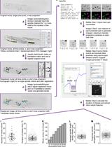
Abstract
C. elegans has emerged as a key model organism for microscopy of intact meiotic nuclei, owing to its small number of chromosomes, transparency, ease of genetic manipulation, and linearly ordered stages of meiotic progression along its gonad. Most studies use either immunofluorescence of extruded and fixed gonad tissue or live imaging of fluorescently tagged proteins within the gonad of the living animal. These techniques enable high-resolution imaging and preserve native cellular dynamics, respectively. To combine these advantages, this protocol describes the methodology for ex vivo imaging of the C. elegans gonads for high-resolution live imaging, including laser-scanning confocal microscopy and single-molecule tracking microscopy. By using a physiologically matched medium, maintaining the gonad’s connection to the carcass, and providing mechanical support with a thin layer of petroleum jelly, we consistently maintain gonad integrity and meiotic function, as described by dynein-driven chromosome motion, in >70% of individual preparations and for >30 min. Advantages of this approach compared to intravital imaging include the ability to add membrane-permeable dyes, reduced aberrations and autofluorescent background, and convenient preparation.
Key features
• Enables fast (<15 min) preparation time for imaging C. elegans meiotic chromosomes.
• Compatible with native imaging of dim fluorescent protein transgenes in live cells.
• Allows addition of small molecules such as fluorescent dyes tagged with HaloTag or SNAP tag ligands.
• Maintains gonad structure and function for 30–60 min of live imaging.
Keywords: C. elegans Microscopy
Live imaging
Single-molecule imaging
Reproductive biology
Meiosis
Graphical overview
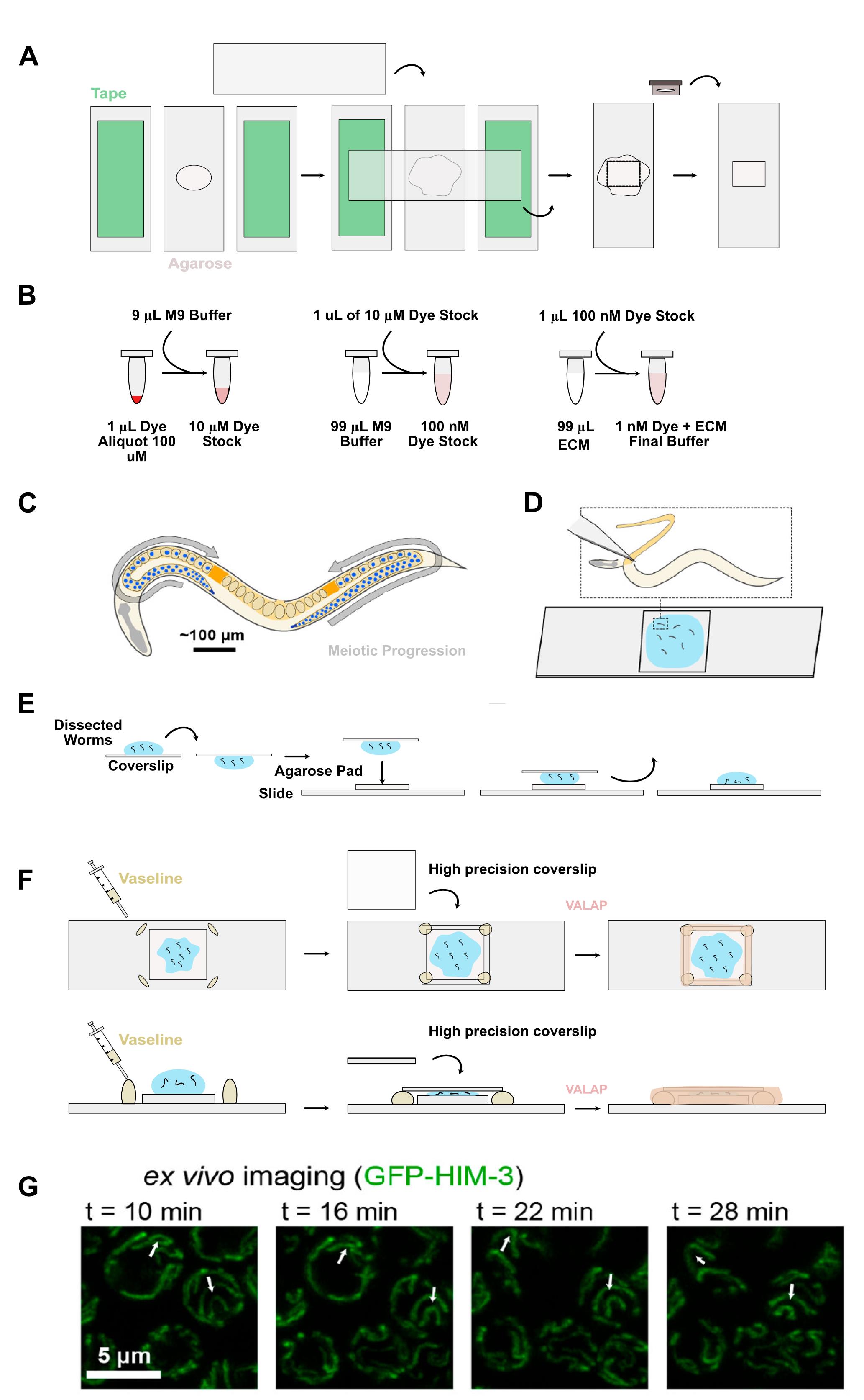
Ex vivo preparation of C. elegans gonad maintains functioning meiotic nuclei. (A) Setup for creation of agarose pads. (B) Illustration of the steps for creating the ECM buffer and serial dilution of fluorescent dye. (C) C. elegans meiosis. (D) Dissections of C. elegans worms for imaging on a glass slide. (E) Illustration of the inversion of the coverslip for transferring worms onto the agarose pad. (F) Using Vaseline to prevent crushing the gonads while still sealing them in place with a coverslip, and the addition of VALAP for the final seal. (G) Meiotic nuclei marked with a fluorescently tagged synaptonemal complex axis protein HIM-3 showing dynein-driven motion. White arrows denote landmarks along chromosomes that are visibly displaced during imaging independent of nuclear rotation. This independent chromosomal movement confirms meiotic function. Panels C, D, and G: Adapted with permission from [1]. Copyright 2021 American Chemical Society.
Background
Meiotic division underlies the formation of germ cells (spores, eggs, sperm) and has been studied in a variety of model organisms with varying advantages. The small nematode roundworm C. elegans is one of the most common and convenient animal models in which to study meiosis by fluorescence microscopy owing to its translucent body, genetic tractability, and linear temporal arrangement of nuclei within its gonad. In contrast to systems including yeast, mice, and humans, in which developing germ cell chromosomes are typically prepared as “spreads” in vitro [2], meiotic nuclei in C. elegans can be imaged within intact gonad tissue via extrusion, fixation, and staining with fluorescent dye-functionalized antibodies [3], allowing researchers to trace the progression of meiosis throughout the gonad. While this technique has advantages, it cannot be used to observe live dynamic processes, and the process of fixation can disrupt delicate molecular structures or subcellular organization. As an alternative, meiotic proteins can be imaged inside the living C. elegans animal using levamisole or other anesthetics, serotonin, and/or physical immobilization [4–6]. In this case, fluorescence imaging can be achieved by conjugating fluorescent proteins to a protein of interest. However, due to the higher autofluorescence background and lower photostability of fluorescent proteins, these labels bleach readily during high-resolution imaging. Imaging within the live worm also requires a specific three-dimensional orientation or “roll” of the worm body for the gonad to be easily visible, and images can be aberrated by the worm cuticle and tissue layers within the animal.
Our technique addresses many of these concerns. With gonad extrusion, we can image clearly without any additional aberrations while simultaneously ensuring meiotic function for 30 min. This technique takes approximately 15 min once practiced, and because the gonad remains intact in a physiologically matched medium, there is less disruption of the sample compared to fixation-based techniques. While it is possible to use exogenous fluorescent dyes in whole-worm preparations such as feeding fluorescent dyes [6,7], their efficiency in terms of time and reagent usage is comparatively limited. The introduction of dyes during the dissection step of our protocol avoids multi-hour incubation steps and allows rapid and efficient staining of enzymatic labels such as the HaloTag or SNAP tag using minimal quantities of total dye (≤0.1 nmol per experiment) compared to fixed gonad or whole-worm preparations [6]. Similarly, small molecule drugs such as nocodazole should be simple to introduce at this stage, as described by another recent study using gonad explants [8]. Because the gonad is no longer encased by the worm cuticle or surrounded by autofluorescent tissues like the gut, this method is less sensitive to optical aberrations and more amenable to imaging of constitutively expressed fluorescent proteins such as GFP than whole-worm preparations. To help achieve reproducible results, the physiological viability of dissected gonads can be validated using the dynein-driven motion of meiotic chromosomes [1], following mitotic germ cell divisions, and/or imaging the structure of the gonad rachis with beta-tubulin [8].
Altogether, this method represents a convenient approach for imaging the C. elegans gonad that is low cost and consistent once practiced. While we have primarily employed this approach to study nuclei in meiotic prophase I (specifically, pachytene), we have achieved preparations with intact tissue from the mitotic tip to the spermatheca, suggesting our technique should allow high-resolution imaging of germ cells and somatic gonad tissues. While we have confined our studies to adult hermaphrodites to date, the explant study reported successful results in L3 and L4 hermaphrodites and late larval males [8], suggesting dissections using younger hermaphrodites and males should also be tractable with our approach.
Materials and reagents
Biological materials
The standard C. elegans strains used in our experiments contain a protein of interest fused to a HaloTag or SNAP tag and a chromosomal/synaptonemal complex marker, typically a GFP-labeled HIM-3 fusion protein. To validate the preparation, HIM-3, GFP-SYP-3, or another chromosomal marker should be used to confirm that meiotic function is maintained (Graphical overview, G). One appropriate strain to use for testing this protocol is CA1218, which expresses the GFP-SYP-3 protein in the germline and is available from the Caenorhabditis Genetics Center (CGC) (https://cgc.umn.edu/strain/CA1218).
Reagents
1. Vaseline petroleum jelly (Unilever), consumer grade, stored at room temperature indefinitely
2. Lanolin (Kyabo Organics), consumer grade, stored at room temperature indefinitely
3. Paraffin wax (Blended Waxes Inc.), consumer grade, stored at room temperature indefinitely
4. Fluorescent dyes functionalized with Halo or SNAP tag ligands, e.g., JF549-HTL (Promega, catalog number: GA111A); typically stored in 1 μL single-use aliquots of 100 μM dye in DMSO at -20 °C for 2 years
5. Levamisole (Thermo Scientific, catalog number: 187870100); stored at room temperature. Can be stored as a 10% stock solution in Milli-Q water at -20 °C for 1 year. Store in 500 μL or less aliquots to avoid more than 10 freeze/thaw cycles per tube
6. Tween-20 (optional) (Sigma-Aldrich, catalog number: 8.22184); stored at room temperature; stock solution of 10% Tween in Milli-Q water for 2 years
7. Leibowitz L-15 w/o phenol red (Gibco, catalog number: 21083-027), stored at 4 °C until manufacturer expiration
8. Fetal bovine serum (Gibco, catalog number: A5256701), stored in aliquots of 50 mL at -20 °C until use; discard or re-aliquot to avoid multiple freeze/thaw cycles after first thaw
9. EGTA (Thermo Scientific, catalog number: 428570500), 100 mM in Milli-Q H2O, stored as a 50 mL solution of 100 mM in Milli-Q H2O kept at 4 °C until use
10. Sucrose (Fisher, catalog number: AA3650830), stored at room temperature as a 1 M solution in Milli-Q water
11. Potassium dihydrogen phosphate (KH2PO4) (Thermo Scientific, catalog number: 011594.36), stored at room temperature
12. Sodium phosphate dibasic anhydrous (Na2HPO4) (Fisher, catalog number: S374-500), stored at room temperature
13. Sodium chloride (NaCl) (Macron, catalog number: 7532-06), stored at room temperature
14. Magnesium sulfate anhydrous (MgSO4) (Fisher, catalog number: M65-500), stored in a 1 M solution at room temperature, filter sterilized
Solutions
1. VALAP sealant (see Recipes)
2. Embryonic culture medium (ECM buffer) (see Recipes)
3. M9 buffer (see Recipes)
Recipes
1. VALAP sealant
| Reagent | Final concentration | Quantity or volume |
|---|---|---|
| Vaseline petroleum jelly | 33.3% | 10 g |
| Lanolin | 33.3% | 10 g |
| Paraffin wax | 33.3% | 10 g |
Typically stored in 1.5 mL aliquots at room temperature.
2. ECM buffer
| Reagent | Final concentration | Quantity or volume (20 mL) |
|---|---|---|
| Leibowitz L-15 w/o phenol red | 84% | 16.8 mL |
| Fetal bovine serum | 9.3% | 1.86 mL |
| Levamisole 10% solution in Milli-Q H2O | 0.01% | 20 μL |
| EGTA 100 mM in Milli-Q H2O | 2 mM | 400 μL |
| 1 M Sucrose in Milli-Q H2O | n/a | Dependent on starting mOsm |
| Milli-Q H2O | n/a | Final to 20 mL |
Check buffer osmolarity using an osmometer and adjust using 1 M sucrose as needed to reach 330 ± 2 mOsm. Gonads are more susceptible to shrink or burst if the osmolarity is outside this range, though we have found in some studies they can tolerate up to 340 mOsm ECM with acceptable gonad health. Store prepared ECM as 200 μL (or other volume as convenient) aliquots in 1.5 mL tubes and at -20 °C until use for up to 2 years. Discard any remaining thawed ECM at the end of each day to avoid freeze/thaw cycles.
3. M9 buffer [9]
| Reagent | Final concentration | Quantity or volume (1 L) |
|---|---|---|
| KH2PO4 | 0.022 M | 3 g |
| Na2HPO4 | 0.042 M | 6 g |
| NaCl | 0.0856 M | 5 g |
| 1 M MgSO4 | 100 μM | 1 mL |
| DI H2O | n/a | 1 L |
M9 buffer can be prepared in a 1 L batch and aliquoted into 50 mL bottles before autoclaving. Once sterile, bottles can be sealed and stored at room temperature indefinitely or until contaminated.
Laboratory supplies
1. Scalpel blades (Feather #11, 2976 #11)
2. Scalpel handle (Feather #3)
3. Worm picks (handmade or, e.g., Tritech Research TWPH1)
4. High-precision (“No. 1.5H” or “HT”, 170 ± 5 μm) coverslips/cover glasses, 22 mm × 22 mm2 (ThorLabs, catalog number: CG15CH2) (18 × 18 mm2 also work well)
5. Microscope slides, 25 mm × 75 mm (1” × 3”) × 1 mm (VWR, catalog number: 16005-106)
6. Razor blades
7. P1000, P200, P20, P10 pipette tips
8. Eyelash brush or other gentle manipulation tool; made by attaching a single fake eyelash to the end of a toothpick with nail polish
9. Fine (<5 mm tip width) paintbrush
10. Empty 30 mm Petri dish or lid
11. Vaseline inside a syringe
Note: We recommend scooping Vaseline into a 5 mL syringe (BD, catalog number: 14-829-45) with a 20-gauge needle (Sol Millenium, catalog number: V111810).
12. Agar plates with nematode growth medium (NGM) and seeded with OP50 E. coli bacteria [9]
Equipment
1. Dissection stereomicroscope (e.g., Zeiss, model: Stemi 305)
2. High-resolution microscope (e.g., Zeiss, model: 880 confocal with Airyscan or Bruker Vutara 352 single-molecule epifluorescence microscope)
3. Humid chambers (sealable containers with a tray that can hold prepared agarose pads above wet paper towels; these are often available at consumer-grade for food storage)
4. Vortexer
5. Heat block
6. Pipettes (P1000, P200, P20, P10)
7. High-precision (mg) scale
8. Incubator or climate-controlled room at the desired temperature for worm maintenance, typically 20 °C
9. Osmometer (e.g., Wescor Vapro Model 5600 or Advanced Instruments Microosmometer 3300)
Procedure
登录 / 注册后阅读全文
文章信息
稿件历史记录
提交日期: Jul 22, 2026
接收日期: Oct 19, 2026
在线发布日期: Nov 12, 2026
出版日期: Dec 5, 2026
版权信息
© 2026 The Author(s); This is an open access article under the CC BY-NC license (https://creativecommons.org/licenses/by-nc/4.0/).
如何引用
Readers should cite both the Bio-protocol article and the original research article where this protocol was used:
- Hughes, A. J. and von Diezmann, L. (2026). Immobilization of Intact Ex Vivo Gonads From Caenorhabditis elegans for High-Resolution Live Microscopy. Bio-protocol 16(23): e5884. DOI: 10.21769/BioProtoc.5884.
- von Diezmann, L., Bristow, C. and Rog, O. (2024). Diffusion within the synaptonemal complex can account for signal transduction along meiotic chromosomes. Mol Biol Cell. 35(12): ee24–05–0225. https://doi.org/10.1091/mbc.e24-05-0225

 Download Citation in RIS Format
分类
生物物理学 > 显微技术 > 单分子定位显微技术
细胞生物学 > 细胞成像 > 活细胞成像
您对这篇实验方案有问题吗?
在此处发布您的问题,我们将邀请本文作者来回答。同时,我们会将您的问题发布到Bio-protocol Exchange,以便寻求社区成员的帮助。
 发布问题


