


Time-Course Analysis of Polyploidy Dynamics in TPA-Induced MEG-01 Cells

发布: 2026年11月20日第16卷第22期 DOI: 10.21769/BioProtoc.5882 浏览次数: 34
评审: Alexander DupuyGiovanni de NolaNikhil Mittal
 PDF
 Q&A






Abstract
Megakaryocyte (MK) polyploidization is a dynamic process regulated by cell cycle regulators, endoreplication switches, survival signals, and cytoskeletal rearrangements. Conventional single-time-point assays [e.g., ploidy measurement at day 3 or 5 after 12-O-tetradecanoylphorbol-13-acetate (TPA) induction] capture only one stage of this process and may miss stage-specific or transient changes. To provide a more complete view, we developed a time-course protocol that measures ploidy at sequential time points (days 0, 1, 5, 7, and 9) post-TPA treatment in MEG-01 cells. This approach adds temporal information to standard ploidy analysis and can help indicate when a regulatory factor acts, during initiation, progression, or the final stage of polyploidization. Comparing multiple time points may also help distinguish consistent trends from variability restricted to specific windows and can suggest candidate regulatory nodes. Collectively, our protocol provides a practical framework for studying the temporal regulation of MK differentiation and other endoreplication-related processes.
Key features
• Monitor ploidy trajectories across multiple points to assess stage-specific gene function.
• Compare trends across multiple time points to help distinguish consistent effects from time-window-specific variability.
• Identify transient or stage-specific differentiation changes that may point to candidate regulatory nodes.
Keywords: Flow cytometry Polyploidy
Megakaryocytes
TPA
Cell differentiation
Graphical overview
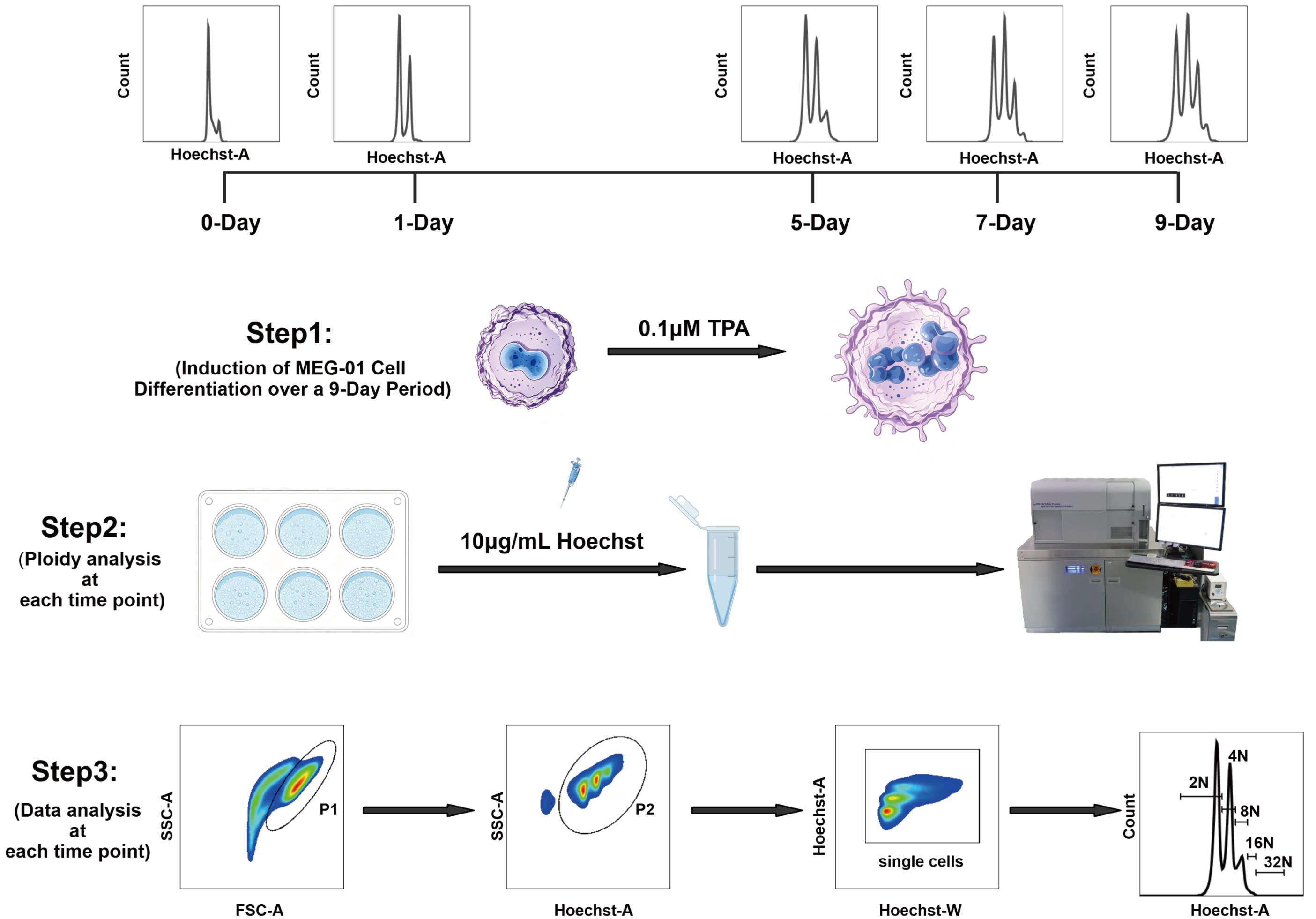
Schematic overview of time-course analysis of polyploidy dynamics in MEG-01 cells. MEG-01 cells were induced with 0.1 μM TPA and harvested at days 0, 1, 5, 7, and 9. Cells were stained with Hoechst 33342 (10 μg/mL) and analyzed by flow cytometry. Ploidy was determined via Hoechst staining. Cells were first gated on forward scatter area (FSC-A) vs. side scatter area (SSC-A) to exclude debris, followed by gating on SSC-A vs. Hoechst-A to exclude debris; singlet gating was then set on the Hoechst-A vs. Hoechst-W plot. Subsequently, the ploidy status was analyzed from the Hoechst-A fluorescence histogram within the singlet gate, resolving peaks corresponding to 2N, 4N, 8N, 16N, and 32N populations.
Background
Megakaryocyte (MK) polyploidization is essential for MK maturation and platelet production. Unlike typical cell division, it is not a one-time event but a dynamic, multistage process controlled by a network of molecular regulators, including cell cycle modulators, endoreplication switch factors, survival signaling pathways, and cytoskeletal rearrangements. Different regulators can act at distinct stages: initiating the mitosis-to-endoreplication switch, promoting endoreplicative cycles, or determining the final ploidy level [1,2].
Conventionally, MK polyploidization is assessed by ploidy measurement at a single endpoint, often day 3 or 5 post-12-O-tetradecanoylphorbol-13-acetate (TPA) treatment. Such endpoint measurements provide limited temporal information and may not capture stage-specific gene functions or regulatory dynamics. To complement this approach, we developed a time-course analysis of ploidy dynamics in MEG-01 cells, a widely used megakaryoblastic cell line [3]. Ploidy is measured at multiple time points—days 0, 1, 5, 7, and 9—following TPA induction. By adding temporal resolution to conventional ploidy analysis, our protocol can provide a more detailed view of MK polyploidization. It can also be adapted to other endoreplicating cell systems, offering a flexible approach for cell biology research.
Materials and reagents
Biological materials
1. MEG-01 (ATCC, catalog number: CRL-2021)
Reagents
1. RPMI-1640 medium (Hyclone, catalog number: SH.30809.01)
2. Fetal bovine serum (FBS) (Gibco, catalog number: 16000044)
3. Penicillin-streptomycin (100×, 10,000 U/mL) (Gibco, catalog number: 15140122)
4. Phosphate buffered saline (PBS) (Cytiva HyClone, catalog number: SH30256.FS)
5. Trypsin-EDTA (0.25%), phenol red (Gibco, catalog number: 25200072)
6. 12-O-tetradecanoylphorbol-13-acetate (TPA) (Cell Signaling Technology, catalog number: 4174)
7. Hoechst 33342 (Thermo Fisher Scientific, catalog number: 62249)
8. Dimethyl sulfoxide (DMSO) (Sigma-Aldrich, catalog number: D2650)
9. Modified Dulbecco’s PBS (DPBS) (Thermo Fisher Scientific, catalog number: 28374)
Solutions
1. RPMI-1640 complete medium (see Recipes)
2. TPA-induced differentiation medium (see Recipes)
3. Hoechst staining medium (see Recipes)
4. TPA + Hoechst co-incubation medium (see Recipes)
Recipes
1. RPMI-1640 complete medium
| Reagent | Proportion (% v/v) | Quantity or volume |
|---|---|---|
| RPMI-1640 medium | 89% | 44.5 mL |
| FBS | 10% | 5 mL |
| Penicillin-Streptomycin (100×) | 1% | 500 μL |
| Total | 100% | 50 mL |
2. TPA-induced differentiation medium
| Reagent | Final concentration | Quantity or volume |
|---|---|---|
| RPMI-1640 complete medium | base | quantum sufficit (q.s.) to 50 mL |
| TPA (200 μM stock in DMSO) | 0.1 μM | 25 μL |
| Total | 50 mL |
TPA is supplied as a 1 mg powder. Store at -20 °C. Before use, dissolve powder in 1 mL of DMSO to yield a 1.6 mM stock solution. For the working concentration of 200 μM, add 125 μL of stock solution to 875 μL of DMSO. Store in solution at -20 °C, desiccated. Protect from light. In lyophilized form, the chemical is stable for 24 months. Once in solution, use within 3 months to prevent loss of potency. Aliquot to avoid multiple freeze/thaw cycles.
Prepare TPA-induced differentiation medium fresh prior to use. Protect TPA from light and avoid repeated freeze/thaw cycles.
3. Hoechst staining medium
| Reagent | Final concentration | Quantity or volume |
|---|---|---|
| RPMI-1640 complete medium | base | q.s. to 1 mL |
| Hoechst 33342 (1 mg/mL stock in DPBS) | 10 μg/mL | 10 μL |
| Total | 1 mL |
Hoechst 33342 is supplied as an aqueous solution at 12.3 mg/mL (20 mM). Hoechst stock solution may be stored at 2–6 °C for up to 6 months or at ≤-20 °C for longer periods, protected from light. Before use, dilute it in DPBS to 1 mg/mL Hoechst 33342 intermediate stock solution; for the preparation method, refer to the manufacturer’s instructions.
Prepare fresh prior to use. Protect from light during incubation.
4. TPA + Hoechst co-incubation medium
| Reagent | Final concentration | Quantity or volume |
|---|---|---|
| RPMI-1640 complete medium | base | q.s. to 1 mL |
| TPA (200 μM stock in DMSO) | 0.1 μM | 5 μL |
| Hoechst 33342 (1 mg/mL stock in DPBS) | 10 μg/mL | 10 μL |
| Total | 1 mL |
Prepare fresh prior to use. Protect from light during incubation.
Laboratory supplies
1. 60 mm TC-treated culture dish (CORNING, catalog number: 430166)
2. Cell culture multi-well plates (6-well plate) (CORNING, catalog number: 3516)
3. 1.5 mL microcentrifuge tubes (CORNING, catalog number: MCT-150-C)
4. 15 mL centrifuge tubes (CORNING, catalog number: 430791)
5. 50 mL centrifuge tubes (CORNING, catalog number: 430829)
6. BD FACSAria fusion cell sorter (85 μm nozzle) (BD Biosciences, catalog number: 647340)
7. Olympus Cell Counter Model R1 counting slide (Olympus, catalog number: R1-SLI)
8. 100 μm nylon cell strainer (FALCON, catalog number: 352360)
Equipment
1. Heracell VIOS 160i CO2 Incubator (Thermo Fisher Scientific, catalog number: 51033565)
2. Thermo Scientific 1300 Series A2 Class II Biological Safety Cabinet (Thermo Fisher Scientific, catalog number: 1356)
3. Benchtop Low-Speed Centrifuge L500-A (Xiangyi, catalog number: 0205209000)
4. Eppendorf Centrifuge 5810 R (Eppendorf, catalog number: 5811000061)
5. Olympus Cell Counter Model R1 (Olympus, catalog number: K23009240-12)
6. Nikon H550S Light Microscope (Nikon, Japan, catalog number: H550S)
7. BD FACSAria Fusion Cell Sorter (BD, catalog number/model: 656700)
8. Thermostatic water bath (Jiaxing Junsi Electronics Co., Ltd., catalog number/model: HH-1)
Software and datasets
1. FlowJo software (BD Biosciences, version 10.8.1)
Procedure
登录 / 注册后阅读全文
文章信息
稿件历史记录
提交日期: Aug 17, 2026
接收日期: Oct 26, 2026
在线发布日期: Nov 12, 2026
出版日期: Nov 20, 2026
版权信息
© 2026 The Author(s); This is an open access article under the CC BY license (https://creativecommons.org/licenses/by/4.0/).
如何引用
Ai, J., Li, T., Li, W. and Yuan, Y. (2026). Time-Course Analysis of Polyploidy Dynamics in TPA-Induced MEG-01 Cells. Bio-protocol 16(22): e5882. DOI: 10.21769/BioProtoc.5882.

 Download Citation in RIS Format
您对这篇实验方案有问题吗?
在此处发布您的问题,我们将邀请本文作者来回答。同时,我们会将您的问题发布到Bio-protocol Exchange,以便寻求社区成员的帮助。
 发布问题


