


HUVEC Isolation—A Robust Protocol to Obtain Human Umbilical Vein Endothelial Cells From Fresh Preparations
从新鲜脐带中分离人脐静脉内皮细胞的可靠方法

(*Contributed equally to this work, §Contributed equally to this work) 发布: 2026年11月20日第16卷第22期 DOI: 10.21769/BioProtoc.5877 浏览次数: 49
评审: Komuraiah MyakalaAnthony FlamierSalah BoudjadiAnonymous reviewer(s)
 PDF
 Q&A






Abstract
Research on angiogenesis necessitates in vitro models that accurately replicate the in vivo vascular system. Human umbilical vein endothelial cells (HUVECs) are widely used in this context due to their stable phenotype, reproducibility, and minimal ethical concerns. Despite the established utility of HUVECs, variability exists among protocols for their isolation from umbilical cords (UCs). These protocols differ in the choice of enzymatic agents, incubation times, mechanical disruption techniques, and post-isolation cell culture strategies. This protocol describes the timely isolation of these primary endothelial cells from UCs and demonstrates its applicability across a range of UC types, thereby enhancing sample availability. The isolation process employs collagenase IV for cell detachment, followed by gentle massage of the UC to facilitate cell collection; cells are then cultured in Endopan 3 medium. Cells are frozen after the first passage or used directly in experiments. The identity and endothelial phenotype of the isolated HUVECs were confirmed through immunofluorescence staining with an endothelial-specific marker (CD31) and a cell junction integrity marker (ZO-1). Flow cytometry further validated the endothelial phenotype by CD31 expression. This protocol offers a cost-effective and reproducible method for isolating HUVECs, particularly advantageous due to the use of a more affordable enzyme and the elimination of pre-coating requirements for cell culture flasks. The protocol is designed to be straightforward, making it accessible to researchers with limited experience in endothelial cell isolation.
Key features
• Versatile application to a broad range of preparations: This protocol accommodates umbilical cords of varying lengths and thicknesses, enhancing sample availability and reducing experimental variability.
• Efficient and selective enzymatic detachment: The use of Collagenase IV ensures effective detachment of endothelial cells with minimized isolation of non-endothelial cell types.
• Enhanced contamination control: The inclusion of amphotericin B and penicillin/streptomycin in the culture medium significantly reduces the risk of contamination, promoting a healthy HUVEC culture.
• Cost-effective and simple cell culture: This protocol eliminates the need for expensive pre-coated culture flasks, making it more accessible to laboratories with limited resources.
Keywords: Human umbilical vein endothelial cells (HUVECs) (人脐静脉内皮细胞) Endothelial cell isolation protocol (内皮细胞分离方法)
Collagenase IV enzymatic detachment (Ⅳ型胶原酶消化分离)
Immunofluorescence staining (免疫荧光染色)
HUVEC differentiation and phenotype confirmation (人脐静脉内皮细胞分化与表型鉴定)
CD31 as an endothelial cell marker (内皮细胞标志物CD31)
Graphical overview
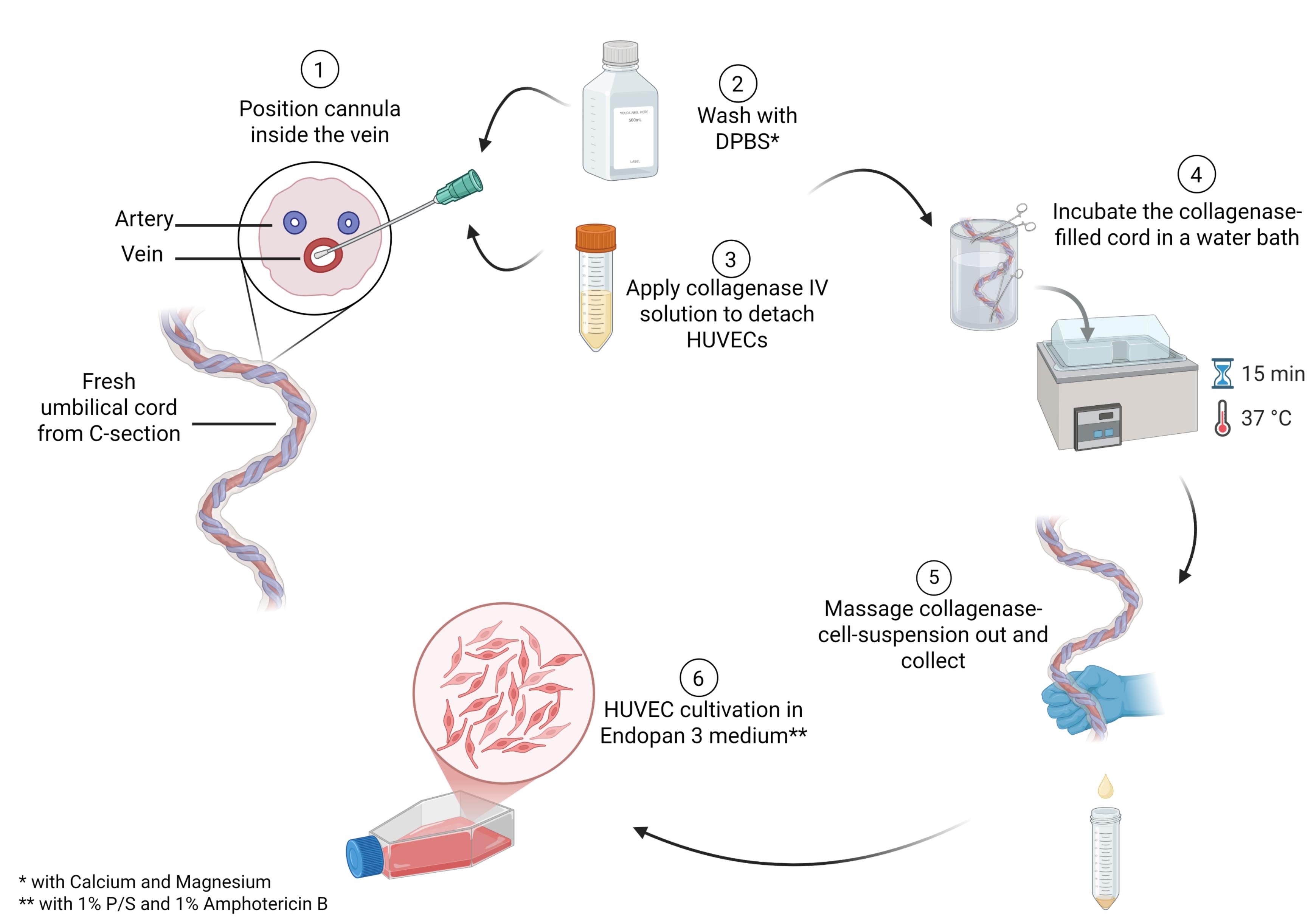
Main steps in the process of human umbilical vein endothelial cell (HUVEC) isolation
Background
In vascular biology, endothelial cells are essential for modeling angiogenesis, blood–brain barrier function, and tumor cell migration [1–4]. While human endothelial cells can be isolated from tissues such as the aorta, liver sinusoids, or the brain, ethical and practical challenges limit their use in laboratory research [5–7]. Therefore, some studies rely on animal-derived endothelial cells, which may not fully replicate human cell behavior. The human umbilical cord (UC), which connects the fetus to the placenta, provides a readily available and ethically acceptable source of primary endothelial cells. The UC is typically discarded after birth, presenting a low ethical barrier for research use [8]. Over the past six decades, the isolation of human umbilical vein endothelial cells (HUVECs) from UCs has become a cornerstone in vascular biology, providing a robust in vitro model for studying endothelial cell biology [8]. HUVECs are valued for accessibility and their ability to mimic physiological properties of human vessels in in vitro assays, making them ideal for a wide range of biomedical applications, including angiogenesis assays, blood–brain barrier modeling, and tumor cell migration studies [2–4].
Despite the shared core principles of cell isolation across published protocols, methodological details vary considerably. Pre-selection criteria for UCs differ across protocols, with differences in factors such as cord length, the presence of blood clots, gestational age, and other relevant donor health indicators [9–11]. This variability can lead to inconsistent results and highlights the need for standardized protocols in HUVEC isolation. For example, some protocols recommend collecting and storing UCs in saline solution at 4 °C [7,9], whereas others do not specify storage conditions [9,11]. The time between UC collection and HUVEC isolation ranges from less than 3 h to up to 48 h [10,12], further contributing to experimental variability. After separation from the donor, UC cells are no longer sustained by the placental environment, which may lead to a progressive decline in cell integrity and yield. Further key differences among existing protocols include the choice of enzymatic detachment agents (e.g., collagenase IV, trypsin, dispase), incubation times, mechanical disruption techniques (e.g., gentle massaging to collect cells or using syringe pressure to expel the enzyme–cell mixture), and post-isolation cell culture conditions [9–14]. Although longer incubation times may increase cell detachment, they may also reduce cell viability or increase contamination with non-endothelial cell types such as fibroblasts.
Additionally, post-isolation culture conditions vary considerably among published protocols. Many protocols require pre-coated flasks [9–12,14] and high concentrations of fetal bovine serum (FBS) [9,11], increasing both the cost and complexity of HUVEC culture. A variety of cell culture media can be used for these endothelial cells, such as Medium 200, EBM-2, or Medium 200 with LVES [9–12,14].
Our protocol addresses the challenges in HUVEC isolation by providing a versatile, efficient, and reproducible method for isolating HUVECs. It can be applied to a broad range of umbilical cords varying in length (minimum of 10 cm) and thickness. The protocol emphasizes timely isolation to preserve cell viability and quality, with isolation ideally performed within hours of delivery to enhance sample availability and reduce experimental variability. Efficient enzymatic detachment is achieved using collagenase IV, which ensures effective and selective detachment of endothelial cells from the umbilical vein, minimizing the isolation of non-endothelial cell types and reducing the risk of contamination. Enhanced contamination control is achieved through the inclusion of amphotericin B and penicillin/streptomycin in the culture medium, significantly reducing the risk of contamination and promoting a healthy and stable HUVEC culture. The protocol also offers a cost-effective and straightforward cell culture approach by eliminating the need for pre-coated culture flasks and reducing the requirement for FBS concentrations, making it more accessible to laboratories with limited resources. The standard cell culture conditions (without pre-coating) also simplify the workflow and reduce experimental variability, further enhancing the protocol's practicality and reproducibility.
Materials and reagents
Biological materials
1. Umbilical cord (from C-section, isolation preferably within 24 h)
Reagents
1. Amphotericin B (Fungizone) (PAN Biotech, catalog number: P06-01100)
2. Collagenase IV, ≥125 CDU/mg, solid, 0.5–5.0 FALGPA units/mg solid (Sigma-Aldrich, catalog number: C5138-100MG, Lot: 0000519267)
3. Dimethyl sulfoxide (DMSO) (Th. Geyer, catalog number: A994.2)
4. Dulbecco’s phosphate-buffered saline (DPBS) w/o calcium and magnesium (PAN Biotech, catalog number: P04-36500)
5. Dulbecco’s phosphate-buffered saline (DPBS*) w/ calcium and magnesium (PAN Biotech, catalog number: P04-35500)
6. Endopan 3 Medium kit (PAN Biotech, catalog number: P04-0010K)
7. Ethanol (99.9%) (Chemsolute, catalog number: 2212.5000)
8. Fetal bovine serum (FBS) (PAN Biotech, catalog number: P30-3302)
9. Penicillin/streptomycin (Biowest, catalog number: L0022-100)
10. Trypsin-EDTA (0.05%) (PAN-Biotech, catalog number: P10-027500)
Additional reagents for validation
1. APC anti-human CD31 mouse IgG (BioLegend, catalog number: 303116)
2. Bovine serum albumin (BSA) (Roth, catalog number: 3737.3)
3. DAPI, 1:1,000 (Novus Biologicals, catalog number: NBP2-31156)’
4. Endothelial cell growth medium 2 (ECGM2) (PromoCell, catalog number: C-22011)
5. Glycerol (Sigma-Aldrich, catalog number: 49767)
6. Hydrochloric acid (HCl) (Merck, catalog number: 109057)
7. Medium 199 (PAN Biotech, catalog number: P04-07050)
8. Mowiol 4-88 (Calbiochem, catalog number: 9002-89-5)
9. Paraformaldehyde 4.5% (PFA) (Chemsolute, catalog number: 2135.5000)
10. Primary antibody CD31 monoclonal antibody (mAb) mouse anti-human, 1:100 (DAKO, catalog number: M0823)
11. Primary antibody ZO-1 (D7D12) mAb rabbit anti-human, 1:100 (Cell Signaling Technology, catalog number: 8193)
12. Secondary antibody donkey anti-mouse IgG, Alexa Fluor 488, 1:400 (Thermo Scientific, catalog number: A-21202)
13. Secondary antibody donkey anti-rabbit IgG, Alexa Fluor 555, 1:400 (Thermo Scientific, catalog number: A-31572)
14. Triton X-100 (Merck, catalog number: 1086031000)
15. Tris hydrochloride (Roth, catalog number: 9090.3)
16. Tween-20 (ITW Reagents, catalog number: A4974)
17. ZO-1 polyclonal antibody (Proteintech, catalog number: CL488-21773)
Solutions
1. Collagenase IV solution (see Recipes)
2. Endopan 3 medium (see Recipes)
3. Endopan 3 medium with supplements (Endopan 3 medium**) (see Recipes)
4. Ethanol (70%) (see Recipes)
5. Freezing medium (see Recipes)
Additional solutions for validation
1. Blocking buffer (see Recipes)
2. ECGM2 medium (see Recipes)
3. Endopan 20 medium (see Recipes)
4. FACS buffer (see Recipes)
5. M199 medium (see Recipes)
6. Mixed medium for endothelial cells (see Recipes)
7. Mowiol mounting medium (see Recipes)
8. PFA (0.04%) (see Recipes)
9. PBS-T (see Recipes)
10. Tris-HCl (see Recipes)
11. Triton X-100 (1%) (see Recipes)
Recipes
1. Collagenase IV solution
| Reagent | Final concentration | Volume for one bottle |
|---|---|---|
| DPBS* | n/a | 15 mL |
| Collagenase IV | 0.1% | 15 mg |
2. Endopan 3 medium
| Reagent | Final concentration | Volume for one bottle |
|---|---|---|
| Endopan 3 | n/a | 500 mL |
| 8 supplements from kit | 1× | 800 μL |
| FBS from kit | 1× | 15 mL |
3. Endopan 3 medium**
| Reagent | Final concentration | Volume for one bottle |
|---|---|---|
| Endopan 3 | n/a | 500 mL |
| 8 supplements from kit | 1× | 800 μL |
| FBS from kit | 1× | 15 mL |
| Amphotericin B | 1% | 5.21 mL |
| Penicillin/streptomycin | 1% | 5.21 mL |
4. Ethanol (70%)
| Reagent | Final concentration | Volume for 50 mL |
|---|---|---|
| Sterile water | n/a | 15 mL |
| Ethanol (99.9%) | 70% | 35 mL |
5. Freezing medium
| Reagent | Final concentration | Volume for 50 mL |
|---|---|---|
| Endopan 3 medium | 50% | 25 mL |
| FBS | 40% | 20 mL |
| DMSO | 10% | 5 mL |
Additional recipes for validation
1. Blocking buffer
| Reagent | Final concentration | Volume for 50 mL |
|---|---|---|
| DPBS | n/a | 50 mL |
| BSA | 5% | 2.5 g |
Stir for 1 h at room temperature. Aliquot and use or store at -20 °C.
2. ECGM2 medium
| Reagent | Final concentration | Volume for one bottle |
|---|---|---|
| ECGM2 | n/a | 500 mL |
| 7 supplements from kit | 1× | 700 μL |
| FBS from kit | 1× | 15 mL |
3. Endopan 20 medium
| Reagent | Final concentration | Volume for one bottle |
|---|---|---|
| Endopan 3 | n/a | 500 mL |
| 8 supplements from kit | 1× | 800 μL |
| FBS from kit | 1× | 15 mL |
| FBS | 20% | 110 mL |
4. FACS buffer
| Reagent | Final concentration | Volume for 50 mL |
|---|---|---|
| DPBS | n/a | 45 mL |
| FBS | 10% | 5 mL |
5. M199 medium
| Reagent | Final concentration | Volume for one bottle |
|---|---|---|
| Medium 199 | n/a | 500 mL |
| FBS | 20% | 126.6 mL |
| Penicillin/streptomycin | 1% | 6.4 mL |
6. Mixed medium for endothelial cells
| Reagent | Final concentration | Volume for one bottle |
|---|---|---|
| ECGM2 medium | 50% | 250 mL |
| M199 medium | 50% | 250 mL |
7. Mowiol mounting medium
| Reagent | Final concentration | Volume or quantity |
|---|---|---|
| Mowiol | 9.6% | 4.8 g |
| Glycerol | 24% | 12 g |
| Sterile water | 1× | 15 mL |
| Tris-HCl (0.2 M, pH 8.5) | 20% | 85 mL |
Mix Mowiol and glycerol with frequent agitation for 1 h at room temperature. Add sterile water and use a stir bar and magnetic stirrer to mix for 1 h at room temperature. Add Tris-HCl and incubate for 2 h at 50 °C with occasional stirring (e.g., stir for 2 min every 20 min). Centrifuge at 5,000× g for 15 min. Aliquot the supernatant and store at -20 °C.
8. PFA (0.04%)
| Reagent | Final concentration | Volume for 50 mL |
|---|---|---|
| DPBS | n/a | 49.6 mL |
| PFA 4.5% | 0.04% | 444 μL |
9. PBS-T
| Reagent | Final concentration | Volume or quantity |
|---|---|---|
| DPBS | n/a | 50 mL |
| Tween-20 | 0.1% | 50 μL |
10. Tris-HCl (0.2 M pH 8.5)
| Reagent | Final concentration | Volume for 1 L |
|---|---|---|
| Sterile water | n/a | 1 L |
| Tris hydrochloride | 0.2 M | 24.2 g |
| HCl | 1× | Few drops |
Dissolve Tris hydrochloride in 800 mL of sterile water. While stirring with a stir bar on a magnetic stirrer, dropwise add HCl to adjust the pH to 8.5. Then, add sterile water to a final volume of 1 L. Store at room temperature.
11. Triton X-100 (1%)
| Reagent | Final concentration | Volume for 50 mL |
|---|---|---|
| DPBS | n/a | 45 mL |
| Triton X-100 | 1 ka% | 5 mL |
Laboratory supplies
1. 3-way stopcock (BD Connecta, catalog number: 394602)
2. 50 mL conical tube, sterile (Greiner Bio-One, catalog number: 227271)
3. Aluminum foil (Labsolute, catalog number: 7696857)
4. Buttoned cannula, sterile (Roth, catalog number: HPX9.1)
5. 130 mm clamps, sterile (Roth/Hammacher, catalog number: TC97.1)
6. 300 mL CorningTM sample containers, sterile (Fisher Scientific, catalog number: 10763394)
7. 22 mm diameter coverslips (Sarstedt, catalog number: HPX1.1)
8. Disposable nitrile gloves (Nitril NextGen, catalog number: 1283)
9. 2 mL NalgeneTM cryovials, sterile (Thermo Scientific, catalog number: 5000-0020)
10. Pasteur pipette (Labsolute, catalog number: 7691061)
11. 150 × 20 mm oversized Petri dish (Sarstedt, catalog number: 82.1184.500)
12. Pipette controller (INTEGRA-Biosciences, catalog number: 155016)
13. Scalpel, sterile (Swann-Morton, catalog number: 0507)
14. 100 mL screw container, sterile (Sarstedt, catalog number: 75.562.005)
15. 10 mL serological pipette (Sarstedt, catalog number: 861.254.001)
16. 30 mL syringe, Luer lock (B. Braun, catalog number: 4717304F)
17. Syringe filter, PA, 0.2 μm (Roth, catalog number: XH57.1)
18. Tape (Durapore, 3 M, catalog number: 1538-1)
19. TC flask T75 (Sarstedt, catalog number: 83.3911.002)
20. Tweezer, sterile, straight, blunt (Roth/Roth Selection, catalog number: 2691.1)
Additional laboratory supplies for validation
1. 1.6 mL Eppendorf tubes (Eppendorf, catalog number: 0030120086)
2. FACS tubes (Sarstedt, catalog number: 55.526.006)
3. Glass slides (Marienfeld, catalog number: 08-104-01)
4. 100 mM Parafilm M (Amcor, catalog number: PM999)
5. 200 μL pipette tips (Sarstedt, catalog number: 70.3030-020)
6. TC flask T75 (Sarstedt, catalog number: 83.3910.002)
7. 24 well TC inserts, 8 μM pores (Sarstedt, catalog number: 83.3932.800)
8. 6-well TC plate (Sarstedt, catalog number: 83.3920)
9. 12-well TC plate (Sarstedt, catalog number: 83.39219)
10. 24-well TC plate (Sarstedt, catalog number: 83.3922)
11. Tweezers, blunt and curved (Roth, catalog number: PX30.1)
Equipment
1. Bead bath (Thermo Fisher, model: precision GP10)
2. Centrifuge (Heraeus Instruments, model: Labofuge 400)
3. CO2 incubator (Thermo Scientific, model: HERAcell 150i, catalog number: 51032719)
4. Inverted microscope with integrated camera (Nikon, model: Eclipse TS100)
5. Laboratory balance, 10 mg to 160 g (Sartorius, model: BP161P)
6. Cell culture hood (Thermo Scientific, model: Herasafe 2025 1.2)
7. Water bath (Thermo Scientific, model: TSGP02)
Additional equipment for validation
1. Water purification system (Sartorius, Arium Mini Plus, catalog number: H2O-MA-UV-T)
2. Benchtop centrifuge (Eppendorf, model: 5415D)
3. Brightfield cell counter (DeNovix, model: CellDrop BF, catalog number: I-20103)
4. Chemical fume hood (Köttermann, model: 2-401-DAAAGY)
5. Flow cytometer (Beckman Coulter, model: CytoFLEX S, BE17054)
6. Inverted fluorescence microscope (Olympus, model: IX83)
7. Live-cell analysis system (Sartorius, model: Incucyte® S3, catalog number: 4647)
8. Magnetic stir bars (Roth, catalog number: X171.1)
9. Magnetic stirrer (IKA, RCT basic, catalog number: Z645060)
10. pH meter (Schott Instruments, model: Lab 860)
11. TEER electrode (Merck Millipore, model: Millicell ERS-2 Replacement Electrode, catalog number: MERSSTX01)
12. TEER measurement device (Millicell, model: ERS-2)
Note: Equivalent products from other manufacturers may be used but should be validated before use.
Software and datasets
1. Microsoft Excel (Microsoft Office 2019, version 16.0) for quantitative analyses
2. Fiji (ImageJ, version 1.54) for visual and quantitative analyses
3. FlowJo v10 (LLC.) for FACS data analyses
4. GraphPad Prism 8 for statistical analyses
Procedure
登录 / 注册后阅读全文
文章信息
稿件历史记录
提交日期: Jul 5, 2026
接收日期: Oct 14, 2026
在线发布日期: Nov 18, 2026
出版日期: Nov 20, 2026
版权信息
© 2026 The Author(s); This is an open access article under the CC BY license (https://creativecommons.org/licenses/by/4.0/).
如何引用
Willer, N. A., Koperski, A., Rieß, A. L., Andag, R., Fischer, A., Hasanov, M. F., Wegwitz, F. and Gallwas, J. (2026). HUVEC Isolation—A Robust Protocol to Obtain Human Umbilical Vein Endothelial Cells From Fresh Preparations. Bio-protocol 16(22): e5877. DOI: 10.21769/BioProtoc.5877.

 Download Citation in RIS Format
分类
细胞生物学 > 细胞分离和培养 > 细胞分离
发育生物学 > 细胞生长和命运决定 > 脐带
细胞生物学 > 基于细胞的分析方法 > 免疫细胞化学
您对这篇实验方案有问题吗?
在此处发布您的问题,我们将邀请本文作者来回答。同时,我们会将您的问题发布到Bio-protocol Exchange,以便寻求社区成员的帮助。
 发布问题


