


Measuring the ADP-Ribosylation-Dependent Release of PARP2 From DNA Using Stopped-Flow Fluorescence Polarization

(*contributed equally to this work) 发布: 2026年12月05日第16卷第23期 DOI: 10.21769/BioProtoc.5874 浏览次数: 39
评审: Elena A. OstrakhovitchSumeet NayakAntara SenguptaVipin K. Yadav
 PDF
 Q&A







Abstract
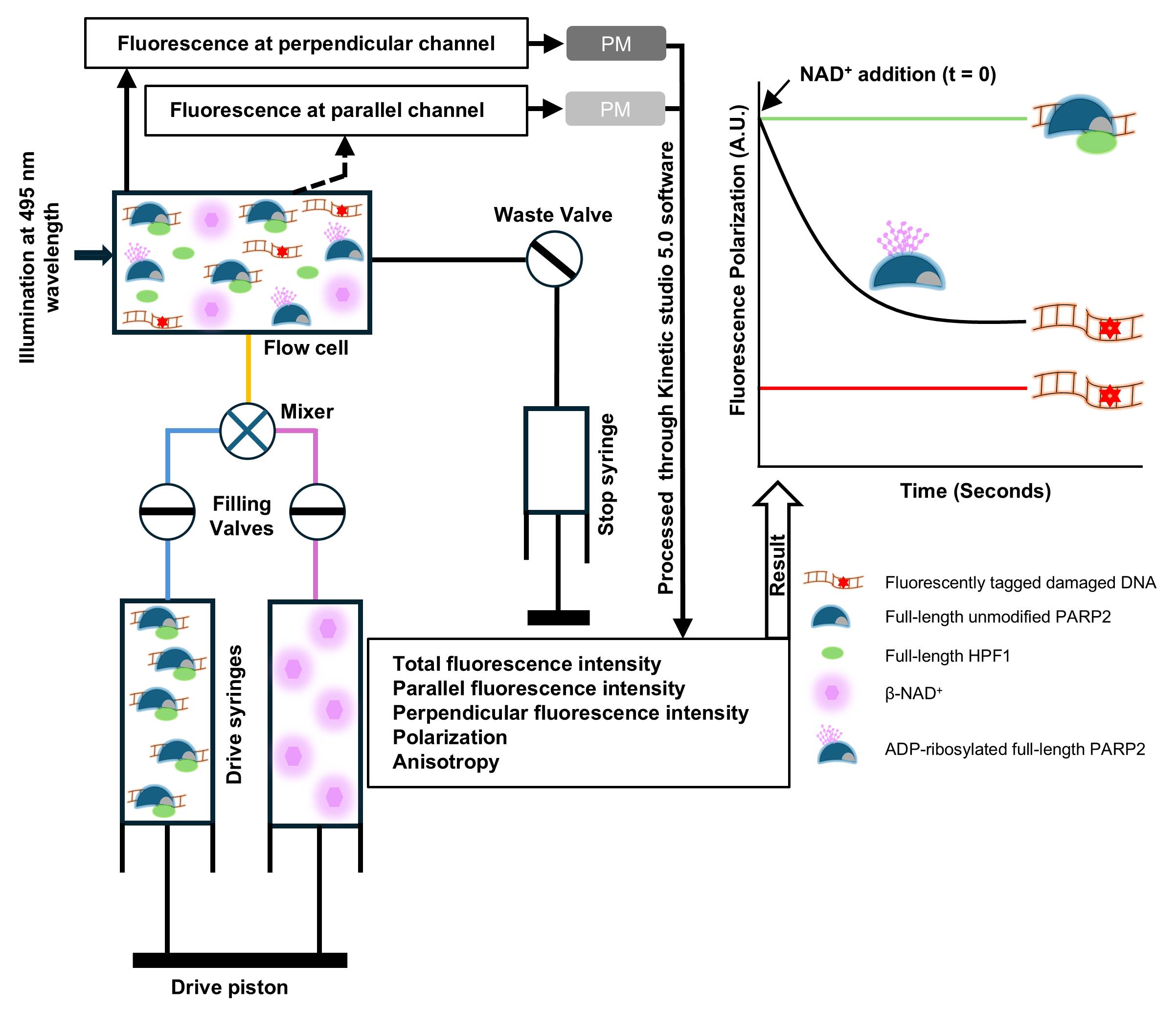
The ADP-ribosyltransferase PARP2 plays a major role in DNA damage repair mechanisms. It recognizes damaged DNA, resulting in poly-ADP-ribosylation (PARylation) of target proteins and itself. The automodification of PARP2, especially at N-terminal serine residues in the presence of histone PARylation factor 1 (HPF1), leads to its release from DNA. Here, we describe a stopped-flow-based fluorescence polarization method for the real-time measurement of PARP2 release from DNA damage by monitoring the change in fluorescence anisotropy of fluorescein-labeled DNA. The method is based on rapid mixing of a preformed DNA-PARP2-HPF1 complex with NAD+ and monitoring the release of PARP2 from DNA. The stopped-flow-based fluorescence anisotropy method provides a fast and sensitive measurement of PARP2 release from DNA that can be widely used to study the effect of PARP2 mutants or inhibitors on PARP2 activity and dissociation from DNA.
Key features
• Complete description of the application of a fluorescence polarization (FP)-based method to measure PARP2 DNA binding and release kinetics.
• Detailed procedures for measuring PARylation-dependent release of PARP2 from DNA using fluorescence polarization.
• Development of a sensitive, rapid, and easy-to-set-up method for measuring PARP2 release from DNA.
• The method is useful for measuring the effects of drugs on PARylation activity through PARP2 release from DNA.
Keywords: DNA damage DNA–protein interaction
PARP2
Poly ADP-ribosylation
Fluorescence polarization
Graphical overview
Background
PARP2 is involved in DNA damage repair mechanisms [1]. It recognizes DNA damage sites and activates the DNA repair cascade by producing poly-ADP-ribosyl moieties (PAR) from NAD+ to itself (automodification) and other target proteins [1–4]. PARP2 activity is modulated by a protein interacting with the PARP2 catalytic domain, Histone Parylation Factor 1 (HPF1), increasing the amount of automodification and targeting it to the serine residues [5,6]. PARP2 automodification results in an increased electrostatic repulsion due to the negative charge of PAR, which leads to PARP2 release from DNA [7,8]. To measure automodification-dependent PARP2 release from DNA, we used fluorescence polarization/fluorescence anisotropy (FP/FA). The fluorescently labeled DNA molecules rotate quickly in solution but, upon protein binding, the large-size DNA–protein complex exhibits lower rotational diffusion, thus increasing anisotropy. Therefore, changes in anisotropy are used to measure protein–DNA interactions and binding dynamics.
First, FP measuring conditions for DNA-PARP2 binding studies were optimized in 96-well plates using a Tecan Infinity M1000pro plate reader. To measure the automodification-dependent PARP2 release from DNA, a saturating (Kd = 4.8 ± 2.9 nM) concentration of PARP2 (200 nM) was mixed with 5 nM oligo-DNA, resulting in an increase in FP signal compared to oligo alone. The automodification reaction was initiated by the addition of 75 μM NAD+, and a decrease in FP signal was observed, reflecting PARP2 release from DNA. In the presence of HPF1, the FP signal decreased more rapidly, supporting the increased automodification of PARP2. However, in plate reader–based FP measurements, due to PAR generation, an initial increase in FP and subsequent PARP2 release was observed [9]. This complicates the monitoring and measurement of PARP2 release rates from DNA, especially when comparing release kinetics for multiple protein constructs. Therefore, to monitor comparative PARP2 release kinetics, we optimized a fluorescence polarization assay using stopped-flow for real-time monitoring of PARP2 release from DNA.
Materials and reagents
Reagents
1. Fluorescein sodium salt (Sigma-Aldrich, catalog number: F6377-100G, CAS: 518-47-8)
2. Fluorescein-labeled nicked double-stranded DNA with a 5′-phosphate oligo (5′ p GGA AGT CTT iFluorT TTG ACT TCC TCG AAG CTT TTG CTT CGA 3′) (Integrated DNA Technologies)
3. 4-(2-hydroxyethyl)-1-piperazineethanesulfonic acid (HEPES) (Fisher Bioreagents, catalog number: BP310-1, CAS: 7365-45-9)
4. Sodium chloride (NaCl) (Fisher Bioreagents, catalog number: BP358-1, CAS: 7647-14-5)
5. Bovine serum albumin (BSA) (Millipore, catalog number: 81-053-3, CAS: 9048-46-8)
6. Tris(2-carboxyethyl)phosphine (TCEP) (Apollo Scientific, catalog number: BIT0122, CAS: 51805-45-9)
7. Ethylenediaminetetraacetic acid (EDTA) (Amresco, catalog number: M101-1, CAS: 6381-92-6)
8. β-nicotinamide adenine dinucleotide (β-NAD+) (Sigma Life Science, catalog number: N0632-1G, CAS: 20111-18-6)
Solutions
1. Fluorescence polarization buffer (see Recipes)
2. Fluorescein solution (see Recipes)
3. Oligo stock preparation buffer (pH 8.0) (see Recipes)
4. Protein purifications (see Recipes)
5. 10 mM fluorescein (sodium salt) solution (see Recipes)
6. β-NAD+ in Milli-Q water (see Recipes)
Recipes
1. Fluorescence polarization buffer (pH 8.0)
| Reagent | Final concentration | Quantity or volume |
|---|---|---|
| HEPES (1 M) | 10 mM | 500 μL |
| NaCl (5 M) | 150 mM | 1.5 mL |
| TCEP (0.5 M) | 0.1 mM | 10 μL |
| EDTA (100 mM) | 0.1 mM | 50 μL |
| BSA (100 mg/mL) | 0.1 mg/mL | 50 μL |
| Distilled water | - | up to 50 mL |
Prepare fresh prior to use. Adjust to pH 8.0 using an electronic pH meter and filter the buffer using a 0.45-μm syringe filter.
2. Fluorescein solution (1 mL)
| Reagent | Final concentration | Quantity or volume |
|---|---|---|
| Fluorescein (10 mM) | 10 μM | 1 μL |
| Fluorescence polarization buffer | - | 999 μL |
3. Oligo stock preparation buffer (pH 8.0)
| Reagent | Final concentration | Quantity or volume |
|---|---|---|
| HEPES (1 M) | 10 mM | 100 μL |
| NaCl (5 M) | 100 mM | 200 μL |
| EDTA (100 mM) | 1 mM | 100 μL |
| Distilled water | - | up to 10 mL |
Store at 4 °C. Adjust to pH 8.0 using an electronic pH meter.
4. Protein purifications
| Protein | Length (amino acid) | Uniprot id | Purification reference |
|---|---|---|---|
| Full-length PARP2 | 583 | Q9UGN5 | Obaji et al. [10] |
| Full-length HPF1 | 346 | Q9NWY4 | Dhakar et al. [11] |
5. 10 mM fluorescein (sodium salt) solution
Prepare in absolute ethanol by mixing the powder in the solvent.
6. β-NAD+ in Milli-Q water
Prepare by mixing the powder in the solvent.
Laboratory supplies
1. 96-well polypropylene black microplates (Grenier Bio-One, catalog number: 650209)
2. 0.45 μm Minisart PES syringe filter (Sartorius, catalog number: 16533)
3. 5 mL plastic syringe (Fisherbrand, catalog number: 14955452)
4. 520 nm bandpass filter, FWHM 10 nm, 25 mm diameter (Andover Corporation, catalog number: 520FS10-25)
Equipment
1. Hi-Tech Scientific Kinet Asyst Stopped-Flow Spectrometer (TgK Scientific, model: SHU-61DX2)
2. Fisher brand water bath (Unity Lab Services, model: FSGPD05)
Software and datasets
1. GraphPad Prism 10.0 (Siemens ecosystem, 10.0)
2. Kinetic studio 5.0 (TgK Scientific, 5.0)
Procedure
登录 / 注册后阅读全文
文章信息
稿件历史记录
提交日期: Aug 14, 2026
接收日期: Oct 19, 2026
在线发布日期: Nov 11, 2026
出版日期: Dec 5, 2026
版权信息
© 2026 The Author(s); This is an open access article under the CC BY license (https://creativecommons.org/licenses/by/4.0/).
如何引用
Dhakar, S. S., Galera-Prat, A., Kiema, T. R. and Lehtiö, L. (2026). Measuring the ADP-Ribosylation-Dependent Release of PARP2 From DNA Using Stopped-Flow Fluorescence Polarization. Bio-protocol 16(23): e5874. DOI: 10.21769/BioProtoc.5874.

 Download Citation in RIS Format
您对这篇实验方案有问题吗?
在此处发布您的问题,我们将邀请本文作者来回答。同时,我们会将您的问题发布到Bio-protocol Exchange,以便寻求社区成员的帮助。
 发布问题


