


Hybridization Chain Reaction and Microscopy to Visualize Feeding Site–Specific Gene Expression in Arabidopsis Roots Infected by Plant-Parasitic Cyst Nematodes

发布: 2026年11月20日第16卷第22期 DOI: 10.21769/BioProtoc.5872 浏览次数: 33
评审: Nicolás M. CecchiniAnonymous reviewer(s)
 PDF
 Q&A





Abstract
Plant-parasitic cyst nematodes affect crop growth and cause substantial yield losses. Upon invading plant roots, cyst nematodes convert a few hundred cells from the vascular tissues into a feeding structure, called the syncytium. The nematodes use the syncytium to withdraw nutrients from the host plant, which serves as their sole source of nutrition. The molecular mechanisms underlying feeding site initiation and development remain poorly understood. One of the key factors involved in this process is transcriptional changes in host plants induced by nematode infection. To date, various transcriptomic techniques, including sc-RNAseq and RNA tomography, have been used to study host transcriptional changes during nematode infection. Although these studies provide valuable information, confirmation of the spatial gene expression of candidate genes during syncytium development remains limited. Therefore, we optimized hybridization chain reaction (HCR) and microscopy for high-resolution spatial analysis of gene expression and simultaneous analysis of feeding site morphology. Here, we provide a proof of principle of the protocol, using confocal microscopy to visualize the expression of target genes Protein Phosphatase 2A subunit A3 (AtPP2AA3) and α-Expansin 4 (AtEXPA4), followed by 2-photon excitation microscopy revealing feeding site morphology. This combination enables high-resolution visualization of gene expression in a spatial context, revealing that both AtPP2AA3 and AtEXPA4 are enriched in the feeding structure. We anticipate that this combination of methods can also be applied to other (plant) studies requiring high spatial resolution and can be used to validate transcriptomic data.
Key features
• Infection of Arabidopsis thaliana seedlings with the plant-parasitic cyst nematode Heterodera schachtii.
• Fixation of (infected) seedlings and hybridization chain reaction to study the expression of putative syncytium/feeding site-specific genes.
• Analysis of the samples by confocal microscopy to visualize the expression of candidate genes.
• Analysis of the samples by 2-photon excitation microscopy to unravel the morphology of the feeding site.
Keywords: Arabidopsis thaliana Plant-parasitic nematodes
Cyst nematodes
Heterodera schachtii
Feeding site
Syncytium
Hybridization Chain Reaction
Gene expression analysis
Microscopy
Graphical overview
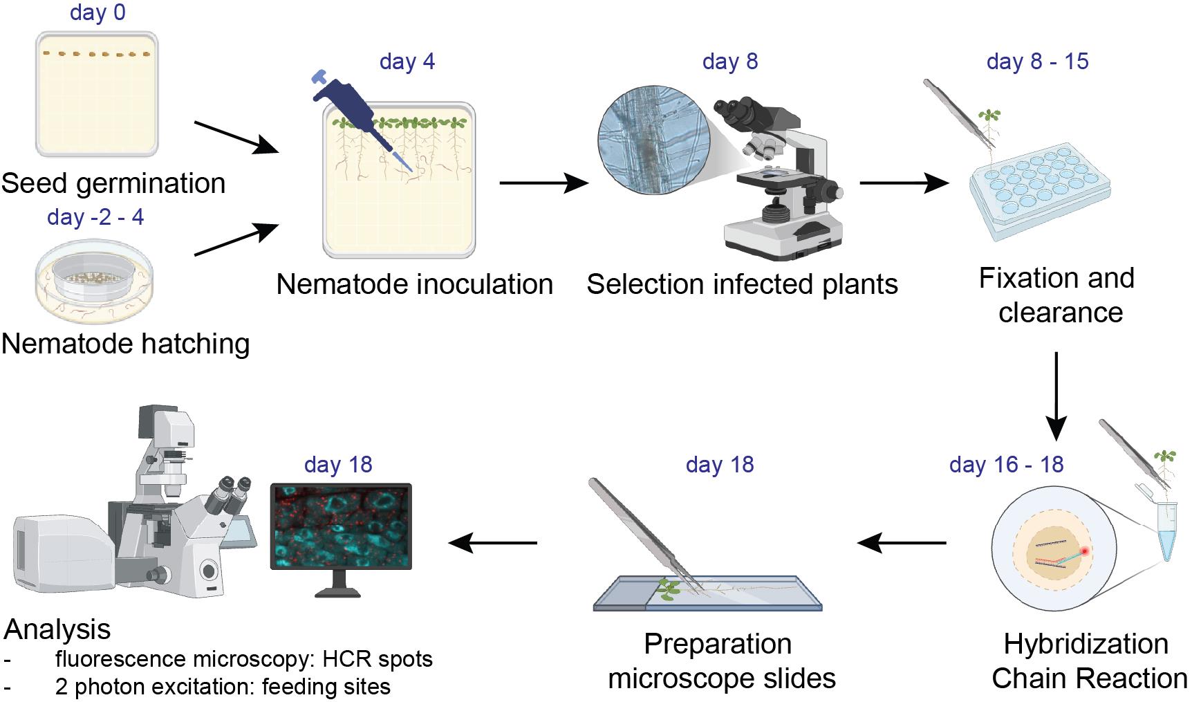
Hybridization chain reaction and microscopy to visualize feeding site–specific gene expression in Arabidopsis roots infected by plant-parasitic cyst nematodes. Workflow diagram of the different steps of the protocol. Image created using BioRender.
Background
Plant-parasitic cyst nematodes, such as Heterodera schachtii, induce the formation of feeding sites, called syncytia, in the root vascular cylinder tissues upon infection. Cyst nematodes withdraw nutrients from the plant via these syncytia, causing substantial crop losses, both in quality and quantity [1,2]. The initiation and expansion of the syncytium is often accompanied by massive transcriptional changes in the host root cells [1]. In recent years, various studies employed transcriptomic techniques, including RNA microarray, microdissection combined with RNAseq, sc-RNAseq, and spatial transcriptomics, to dissect the molecular mechanisms of syncytium development. Single-molecule gene expression could shed light on the spatial expression of genes upon nematode infection. Whole-mount single-molecule RNA fluorescence in situ hybridization (smFISH) has been standardized to visualize and quantitatively measure messenger RNA molecules in Arabidopsis roots [3]. However, upon nematode infection, we found it impossible to image smFISH punctae in feeding cells induced by cyst nematodes near the vascular tissues of the maturation zone in plant roots. Autofluorescence and limited transparency reduced the signal-to-noise ratio in the inner tissues of the root. Additionally, the fluorescence intensity from smFISH punctae was low. To overcome these limitations, we employed hybridization chain reaction (HCR), another powerful RNA-FISH technique, which is an alternative (or improvement) to smFISH [4–7]. The principle is essentially the same: by adding fluorescently labeled probes directed against a specific transcript of interest, this transcript can be visualized in the cell. But in HCR, instead of directly labeling the probes with a fluorescent tag, the probes contain an adaptor to which fluorescently tagged hairpins are added in a second incubation step. This initiates a chain reaction leading to ~10× brighter spots compared to regular smFISH spots (Figure S1). Tissue clearing for fluorescence imaging [8] and HCR [9,10] have been applied to plant roots before but, to date, these methods have not been published for studying feeding site–specific gene expression upon nematode infection.
Here, we present a detailed protocol describing Arabidopsis root infection with cyst nematodes, tissue fixation, HCR, and microscopy to study and spatially resolve gene expression in the syncytium. The localization of syncytia close to vascular tissues and autofluorescence due to tissue damage at the infection site prevent visualization of feeding site morphology with regular confocal microscopy. We report that a combination of sample clearing and 2-photon excitation (2PE) allows a deeper z-acquisition and 3D reconstruction at higher resolution, enabling analysis of feeding site morphology induced by plant-parasitic nematodes. Regular confocal microscopy and 2PE can be combined to first visualize HCR spots and then the feeding site morphology.
Materials and reagents
Biological materials
1. Heterodera schachtii second-stage juveniles (J2) hatched from sand/clay-mix cyst samples [Woensdrecht population from Instituut voor Rationele Suikerproductie (IRS), the Netherlands]
2. Arabidopsis thaliana Columbia-0 (Col-0) accession
Reagents
1. Sodium azide solution (Sigma-Aldrich, CAS: 26628-22-8); 1.0% (w/v) in demi-water
2. Gentamicin sulfate (Duchefa Biochemie, 1405-41-00)
3. Nystatin (Duchefa Biochemie, CAS: 1400-61-9)
4. Zinc chloride (Supelco, CAS: 7646-85-7); 30 mM in tap water
5. Sucrose (food-grade, local retail store); 70% (w/v) in demi-water, dissolve using a magnetic stirrer and autoclave at 121 °C for 20 min
6. Tween-20 (SAFC., 9005-64-5); 10% (v/v) in demi-water and autoclave
7. Triton X-100 (Santa Cruz Biotechnology, CAS: 9002-93-1); 0.4% (v/v) in demi-water
8. Mercury(II) chloride (Sigma-Aldrich, CAS: 7487-94-7); 0.8% (w/v) in demi-water
9. Boric acid (Duchefa Biochemie, CAS: 10043-35-3)
10. Cobalt chloride (Duchefa Biochemie, CAS: 7791-13-1)
11. Copper sulfate (Duchefa Biochemie, CAS: 7758-99-8)
12. Manganese (II) chloride (Sigma-Aldrich, CAS: 13346-34-9)
13. FeNaEDTA (Duchefa Biochemie, CAS: 15708-41-5)
14. Sodium chloride (Duchefa Biochemie, CAS: 7647-14-5)
15. Sodium molybdate (Duchefa Biochemie, CAS: 10102-40-6)
16. Zinc sulfate (Supelco, CAS: 7446-20-0)
17. Nicotinic acid (Duchefa Biochemie, CAS: 59-67-6)
18. Pyridoxine HCl (Duchefa Biochemie, CAS: 58-56-0)
19. Calcium nitrate (Duchefa Biochemie, CAS: 13477-34-4)
20. Magnesium sulfate (Duchefa Biochemie, CAS: 10034-99-8)
21. Potassium nitrate (Acros Organics, CAS: 7757-79-1)
22. Potassium phosphate (Duchefa Biochemie, CAS: 7778-77-0)
23. Myo-inositol (Duchefa Biochemie, CAS: 87-89-8)
24. Thiamine HCl (Duchefa Biochemie, CAS: 67-03-8)
25. Potassium hydroxide, 5 M (Duchefa Biochemie, CAS: 1310-58-3)
26. Sodium hypochlorite (Boom, CAS: 2102 9499); 5%, not commercial bleach
27. Gelrite, 0.7% (Duchefa Biochemie, CAS: 71010-52-1)
28. Daichin agar (Duchefa Biochemie, CAS: 9002-18-0)
29. Nuclease-free H2O (Ambion, catalog number: AM9932)
30. Paraformaldehyde (PFA) (Biolyst Scientific, catalog number: 15710)
31. Phosphate-buffered saline (PBS) (Invitrogen, catalog number: AM9625); diluted in nuclease-free H2O
32. Xylitol powder (Sigma-Aldrich, catalog number: X3375-100g)
33. Sodium deoxycholate (Sigma-Aldrich, catalog number: 30970)
34. Urea (Sigma-Aldrich, catalog number: 5378)
35. HCR probes (directed against your transcript of interest) (Molecular Instruments)
36. HCR hairpins [h1 and h2 (X3-647)] (Molecular Instruments)
37. Sodium chloride (Boom, catalog number: 76021267.5000)
38. Sodium citrate (Boom, catalog number: 76051272.1000)
39. Glucose (Sigma-Aldrich, catalog number: G8270-1kg); 10% w/v in nuclease-free H2O
40. TRIS (Invitrogen, catalog number: 15504-020); 1 M in nuclease-free H2O
41. Catalase (Sigma-Aldrich, catalog number: 18634-1kg)
42. Glucose oxidase, lyophilized from Aspergillus niger (Sigma-Aldrich, catalog number: G2133-10KU)
43. Sodium acetate (VWR, CAS: 6131-90-4); 50 mM in nuclease-free H2O
44. Vaselin (Vaseline Original, 100 mL, catalog number: 8566195)
45. Lanolin (Thermo Scientific, catalog number: A16902.30)
46. Paraffin wax (Sigma-Aldrich, catalog number: 18634-1kg)
47. Ultrapure (type I) water (ELGA PURELAB Felx)
48. 70% ethanol (EtOH) (Boom, CAS: 64-17-5); diluted in nuclease-free H2O
49. HCR HiFi probe hybridization buffer (Molecular Instruments, catalog number: BPH02323)
50. HCR HiFi probe wash buffer (Molecular Instruments, catalog number: BPH01923)
51. HCR gold amplifier buffer (Molecular Instruments, catalog number: BAM02323)
52. HCR gold amplification wash buffer (Molecular Instruments, catalog number: BAM01923)
Solutions
1. Antibiotic solution (see Recipes)
2. KNOP solution I (see Recipes)
3. KNOP solution II (see Recipes)
4. Modified KNOP medium (see Recipes)
5. Seed surface sterilization solution (see Recipes)
6. 4% paraformaldehyde (PFA) (see Recipes)
7. ClearSee (see Recipes)
8. 20× saline sodium citrate (SSC) (see Recipes)
9. Imaging buffer (see Recipes)
10. Glucose oxidase in sodium acetate (see Recipes)
11. Valap (see Recipes)
Recipes
1. Antibiotic solution
| Reagent | Final concentration | Quantity or volume |
|---|---|---|
| Gentamicin sulfate (100 mg/mL) | 1.5 mg/mL | 450 μL |
| Nystatin (0.75 mg/mL) | 0.05 mg/mL | 2 mL |
| Zinc chloride (30 mM) | 3 mM | 27.55 mL |
| Total | n/a | 30 mL |
First, prepare stock concentrations: For gentamicin (100 mg/mL), dissolve 1 mg of gentamicin sulfate in 10 mL of ultrapure water and filter-sterilize using a 0.2 μm filter and a 15 mL syringe. For nystatin (0.75 mg/mL), dissolve 7.5 mg of nystatin in 10 mL of tap water. Aliquot antibiotics into 2 mL Eppendorf tubes and store at -20 °C in a container (in the dark). For zinc chloride (30 mM), dissolve 4.089 g of zinc chloride in 1 L of tap water in a 1 L SCHOTT DURAN bottle and autoclave for 20 min at 121 °C. Store at room temperature.
Note: Although nystatin dissolves in DMSO, make a stock of nystatin in tap water, as DMSO harms the nematode juveniles.
2. KNOP solution I [11]
| Reagent | Quantity or volume |
|---|---|
| Boric acid | 57 mg |
| Cobalt chloride | 0.6 mg |
| Copper sulphate | 1.4 mg |
| Manganese chloride | 57 mg |
| FeNaEDTA | 0.29 mg |
| Sodium chloride | 40 mg |
| Sodium molybdate | 1 mg |
| Zinc sulfate | 7.2 mg |
| Nicotinic acid | 100 mg |
| Pyridoxine HCl | 100 mg |
| Ultrapure H2O | 100 mL |
Prepare a 1,000× stock of KNOP solution I in ultrapure water in a 1 L SCHOTT DURAN bottle. Autoclave for 20 min at 121°C and store at room temperature. Open the bottle in a sterile flow cabinet to prevent contamination.
3. KNOP solution II [11]
| Reagent | Quantity or volume |
|---|---|
| Calcium nitrate | 24 mg |
| Magnesium sulfate | 3.94 mg |
| Potassium nitrate | 24.2 mg |
| Potassium phosphate | 5.4 mg |
| Myo-inositol | 10 mg |
| Thiamine HCl | 1 mg |
| Ultrapure H2O | 1,000 mL |
Prepare a 100× stock of KNOP solution II in ultrapure water in a 1 L SCHOTT DURAN bottle. Autoclave for 20 min at 121 °C and store at room temperature. Open the bottle in a sterile flow cabinet to prevent contamination.
4. Modified KNOP medium [11]
| Reagent | Final concentration | Quantity or volume |
|---|---|---|
| KNOP solution I (1,000×) | 1% | 1 mL |
| KNOP solution II (100×) | 1% | 10 mL |
| Sucrose | 10 g/L | 10 g |
| Daichin agar | 8 g/L | 8 g |
| Total | n/a | 1 L |
To prepare 1 L of modified KNOP medium, add KNOP solution I (1,000×) and KNOP solution II (100×) to a 1 L SCHOTT DURAN bottle in a clean bench. Then, add the sucrose and ~900 mL of ultrapure water. Adjust the pH to 6.4 with potassium hydroxide. Finally, add Daichin agar and top up to a final volume of 1 L. Autoclave for 20 min at 121 °C and pour ~50 mL of sterile media into square Petri dishes. Store plates at room temperature until use.
5. Seed surface sterilization solution
| Reagent | Final concentration | Quantity or volume |
|---|---|---|
| Sodium hypochlorite (5%) | 2.5% | 25 mL |
| Triton X-100 | 0.05% | 25 μL or 2 drops |
| Ultrapure H2O | n/a | Up to 50 mL |
Make a seed-sterilizing solution in a 50 mL Falcon tube, wrap it with aluminum foil, and store at 4 °C. Before every use, invert the Falcon tube several times; the formation of bubbles indicates thorough mixing and should give a chlorine-like odor. Prepared bleach solution can be stored for up to three months, as long as it is opened on a clean bench or until the chlorine-like odor dissipates.
6. 4% PFA
| Reagent | Final concentration | Quantity or volume |
|---|---|---|
| PFA | 4% | 10 mL |
| 10× PBS | 1× | 4 mL |
| Nuclease-free H2O | n/a | 26 mL |
| Total | 40 mL |
Prepare a 4% PFA solution in a 50 mL Falcon tube. Prepared PFA can be used up to one week. Store at room temperature.
7. ClearSee [8]
| Reagent | Final concentration | Quantity or volume |
|---|---|---|
| Xylitol | 10% w/v | 2.5 g |
| Sodium deoxycholate | 15% w/v | 3.75 g |
| Urea | 25% w/v | 6.25 g |
| Nuclease-free H2O | n/a | 15 mL |
| Total | 25 mL |
Prepare ClearSee solution in a 50 mL Falcon tube. The solution, stored at 4 °C, can be used for 2 months. We have not tested longer storage.
Caution: Add 15 mL of nuclease-free water to dissolve the chemicals. If needed, the volume can be topped up with nuclease-free water afterward to a final volume of 25 mL.
8. 20× SSC
| Reagent | Final concentration | Quantity or volume |
|---|---|---|
| Sodium chloride | 3 M | 175.30 g |
| Sodium citrate | 0.3 M | 88.20 g |
| Nuclease-free H2O | 1 L | |
| Total | 1 L |
First, dissolve sodium chloride and sodium citrate in a small volume of nuclease-free water. Adjust the pH to 7.00. The volume can then be topped up with nuclease-free water to a final volume of 1 L.
9. Imaging buffer
| Reagent | Final concentration | Quantity or volume |
|---|---|---|
| Nuclease-free H2O | n/a | 850 μL |
| 20× SSC (see Recipe 8) | n/a | 100 μL |
| 10% glucose in nuclease-free H2O | 0.4% | 40 μL |
| TRIS 1 M | 10 mM | 10 μL |
| 10% Tween | 0.05% | 5 μL |
| Total | 1 mL |
Freshly prepare imaging buffer in a 1.5 mL Eppendorf tube on the day of imaging.
10. Glucose oxidase in sodium acetate
| Reagent | Final concentration | Quantity or volume |
|---|---|---|
| Sodium acetate | 50 mM | 30 mL |
| Glucose oxidase | 3.7 mg/mL | 34 mg |
| Total | 30 mL |
Prepare glucose oxidase solution in a 50 mL Falcon tube. After preparation, aliquot into Eppendorf tubes and store at -20 °C.
Critical: The final solution should be pH 5.0. Adjust if needed.
11. Valap
| Reagent | Final concentration | Quantity or volume |
|---|---|---|
| Vaselin | 50 g | |
| Lanolin | 50 g | |
| Paraffin | 50 g |
Prepare Valap in a glass bottle that can be closed with a lid. Store at room temperature.
Laboratory supplies
1. 50 mL sterile tubes (Sarstedt, catalog number: 62.559.001)
2. 2 mL microcentrifuge tubes (Sarstedt, catalog number: 72.695.201)
3. 10 mL serological pipettes (Sarstedt, catalog number: 86.1254.001)
4. 100–1,000 μL filter tips (Corning, catalog number: 4809)
5. 200 μL filter tips (Sarstedt, catalog number: 70.3031.355)
6. 20 μL filter tips (Sarstedt, catalog number: 70.3030.365)
7. 10 μL filter tips (Sarstedt, catalog number: 70.3010.275)
8. Squared Petri dishes, 120 × 120 mm, plastic (Greiner Bio-One, catalog number: 688102)
9. 1 L Duran laboratory bottle (Sigma-Aldrich, catalog number: Z305200)
10. Magnetic stirrer
11. Toothpicks (Carl Roth, catalog number: EC48.1)
12. Centrifuge glass DURAN with conical bottom, 50 mL (Carl Roth, catalog number: K222.1)
13. Nitrile gloves (Romed HOLLAND, catalog number: NTP805)
14. Duran super duty beaker (Sigma-Aldrich, catalog number: Z680893)
15. Pencil (Staples, catalog number: 3158285)
16. Transparent cling film, unperforated PVC, 30 cm × 300 m roll (universal plastic kitchen foil) (local supermarket)
17. Glass Pasteur pipette (WU Mainz, Supplier Dispolab, catalog number: 200760)
18. Borosilicate glass narrow-neck Erlenmeyer flask 250 mL (Fisher Scientific, catalog number: 15429103)
19. Spatula (Sigma-Aldrich, catalog number: Z648299)
20. Plastic shoe box (Tupperware)
21. 24-well plate (Greiner, catalog number: 662 160)
22. Aluminum foil (Carl Roth, catalog number: AA76.1)
23. DNA LoBind tube 0.5 mL (Eppendorf, catalog number: 022431005)
24. Microscope slides (Avantar, VWR, catalog number: 631-1551)
25. Coverslips (Menzel-Gläser, 24 × 60 mm, #1.5)
Equipment
1. Growth cabinet (Snijders Labs, Micro Clima-series Economic lux chamber, ECL02) set to 21 °C with 16/8 h light/dark cycles using cool white light (MASTER TL-D Super 80 36 W1840; Philips)
2. Pipetboy (Gilson MACROMAN, catalog number: F110120)
3. Pipette (Gilson)
4. Thermo Multifuge 3S-R refrigerated centrifuge swinging-bucket rotor
5. Tabletop centrifuge (Eppendorf, model: 5415C)
6. Flow cabinet (CleanAir, CA/RSV3)
7. Fume hood (INTOS Lab Solutions, catalog number: EN14175)
8. Vortex-Genie 2 mixer (Sigma-Aldrich, catalog number: Z258423)
9. Freezer (-20 °C) (Liebherr Comfort, model: LBH-FKDv-4213-T)
10. Refrigerator (4 °C) (Veriport)
11. Sink with sand filter
12. Sieves for rinsing (500–1,000 μm, 350–375 μm, 175 μm, 100 μm)
13. Sieves for hatching (100 μm)
14. Containers for toxic waste
15. Binocular microscope (Leica, model: MZ12)
16. Racks for conical glass tubes, 50 mL tubes, and Eppendorf tubes (Sigma-Aldrich, catalog number: HS120683)
17. Desiccator (Sigma-Aldrich, model: SYNB135124)
18. Glass Petri dishes (Boeco, Germany)
19. Autoclave (CertoClav Multicontrol, catalog number: L23128502312A)
20. Tokyo RIKAKIKAI EYELA A-3s Vacuum Aspirator
21. Carrousel (Agar Scientific, catalog number: AR384)
22. Forceps (Sigma-Aldrich, catalog number: Z168793)
23. Heat block (Eppendorf, model: Thermomixer compact)
24. Thermocycler (Bio-Rad, model: Thermo Cycler T100)
25. Nikon Eclipse Ti2 inverted microscope with Yokogawa CSU-X1 spinning disk confocal [pinhole 50 μm (fixed)] and Hamamatsu Orca-Flash4.0 V3 digital CMOS C13440-20CU camera
26. Plan Apochromat Lambda objective 10× air, NA = 0.45 (Nikon, model: MRD70170)
28. Plan Apochromat Lambda S objective 40× C Silicon oil, NA = 1.25 (Nikon, model: MRD73400)
29. Leica TCS SP5 on a Leica DMI6000 CS inverted multiphoton/confocal microscope
30. HCX IR Apochromat Lambda objective 25× water, NA = 0.95, WD = 2,500 μm (Leica)
Software and datasets
1. Imaging software Leica Application Suite Advanced Fluorescence (LAS AF) 2.7.2.9586
Procedure
登录/注册后免费查看全文
文章信息
稿件历史记录
提交日期: Jul 16, 2026
接收日期: Sep 29, 2026
在线发布日期: Oct 28, 2026
出版日期: Nov 20, 2026
版权信息
© 2026 The Author(s); This is an open access article under the CC BY license (https://creativecommons.org/licenses/by/4.0/).
如何引用
Lathe, R. S., Pijnacker, A., de Ruijter, N. C., Lozano-Torres, J. L., Korswagen, H. C. and Bruggeman, C. W. (2026). Hybridization Chain Reaction and Microscopy to Visualize Feeding Site–Specific Gene Expression in Arabidopsis Roots Infected by Plant-Parasitic Cyst Nematodes. Bio-protocol 16(22): e5872. DOI: 10.21769/BioProtoc.5872.

 Download Citation in RIS Format
分类
植物科学
您对这篇实验方案有问题吗?
在此处发布您的问题,我们将邀请本文作者来回答。同时,我们会将您的问题发布到Bio-protocol Exchange,以便寻求社区成员的帮助。
 发布问题


