


Monitoring Protein Secretion in Living Cells With a HiBiT-Integrated RUSH System

(*contributed equally to this work, § Technical contact) 发布: 2026年11月05日第16卷第21期 DOI: 10.21769/BioProtoc.5871 浏览次数: 23
评审: Medhat S ElhalawanyAnonymous reviewer(s)
 PDF
 Q&A

参见作者原研究论文
The authors used this protocol in:
Sep 2025




Abstract
The cellular secretory pathway is a fundamental physiological process; however, precise, efficient, and quantitative methods for monitoring this process remain rarely explored. Although several classic approaches have been developed to investigate secretory trafficking, the RUSH (Retention Using Selective Hooks) system currently represents the most advanced technique. Here, we introduce a method termed the HiBiT-integrated RUSH system and provide a detailed protocol for its application. This approach enables real-time monitoring of the secretion of cell membrane receptors such as ACE2, PDL1, NTCP, and CD209. By converting the cellular secretion process into a quantitative curve, our method offers an objective means for result interpretation. The HiBiT-integrated RUSH system is particularly effective for studying the transport dynamics of various cell membrane receptors, thereby facilitating the identification of both common and specific regulators of the cellular secretory pathway.
Key features
• This protocol converts cellular secretion into quantitative curves, enabling objective, real-time assessment of secretory protein transport.
• The protocol is validated for monitoring ACE2 and PDL1 secretion and is applicable to diverse cell membrane receptors.
• The protocol retains the spatiotemporal control of the classic RUSH system while adding HiBiT-based sensitive, live-cell detection.
• The protocol facilitates the discovery of both common and specific regulators of the cellular secretory pathway across different cargo proteins.
Keywords: HiBiT tag Cellular secretory pathway
RUSH (Retention Using Selective Hooks)
Real-time monitoring
Quantitative secretion assay
Live-cell detection
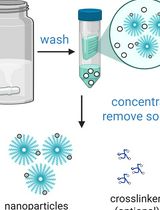
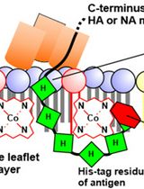
Graphical overview
Background
The cellular secretory pathway is a fundamental physiological process that refers to the transport of newly synthesized proteins to their target compartments. In eukaryotes, conventional protein secretion is primarily mediated by secretory vesicles, which transport cargo through the endoplasmic reticulum (ER) and the Golgi apparatus to the plasma membrane (PM) or extracellular space. This process is essential because it directs proteins, such as cytokines, chemokines, hormones, and receptors, to their sites of action, where they perform crucial physiological and pathological functions [1].
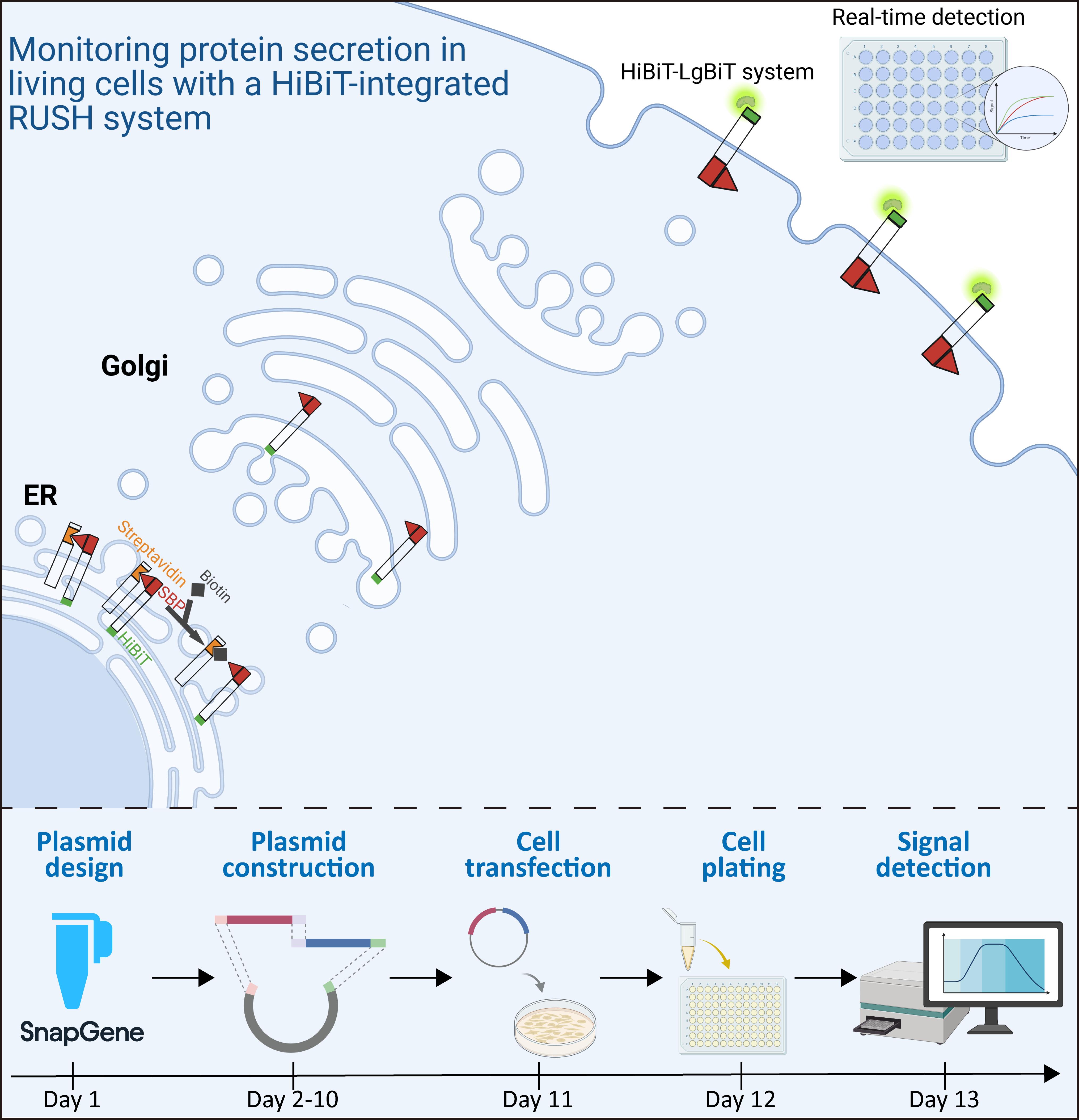
Several classic methods have been developed to investigate secretory trafficking, with the RUSH (Retention Using Selective Hooks) system being the most advanced technique [2–5]. The RUSH system exploits the high-affinity interaction between streptavidin (Str) and a streptavidin-binding peptide (SBP) to reversibly retain an enhanced GFP (EGFP)-tagged reporter within the ER. Subsequent addition of biotin, which competitively displaces SBP from Str, triggers reporter release. This controlled release enables quantification of secretion kinetics and status by monitoring EGFP localization dynamics via fluorescence microscopy [3].
Researchers have conducted impressive studies using the RUSH system, driving significant progress in the field of cellular trafficking [6–8]. However, the classic RUSH assay requires the acquisition and quantification of numerous fluorescence images, which poses challenges for achieving precision and efficiency, particularly constraining its use in high-throughput screening applications. Boncompain et al. [9] utilized the classic RUSH system to screen and validate three molecules from over 4,000 compounds that inhibit HIV infection by targeting CC chemokine receptor 5 (CCR5) secretion. To our knowledge, this is the only reported study to apply the RUSH technique to high-throughput screening. HiBiT is an 11-amino-acid peptide tag developed by Promega in recent years. When HiBiT-tagged proteins on the cell surface or in the extracellular space bind to the exogenously added LgBiT, a NanoBiT luciferase is generated, enabling the detection and quantification of HiBiT-tagged proteins in living cells [10,11]. Building on this advantage, we propose substituting EGFP in the RUSH reporter with HiBiT, thereby converting the fluorescent signal into a quantifiable luciferase signal, which would facilitate real-time, quantitative monitoring of reporter trafficking.
Here, we combined the HiBiT technology with the classic RUSH system to investigate the secretion process of angiotensin-converting enzyme 2 (ACE2) as a model, which is the receptor for SARS-CoV-2. Using a plate reader, we achieved real-time monitoring of the secretion dynamics, enabling a precise and efficient identification of the trafficking regulators. Notably, this strategy was also applied to monitor the secretion process of programmed death-ligand 1 (PDL1), sodium taurocholate cotransporting polypeptide (NTCP), and CD209 (also known as DC-SIGN), all of which are cell membrane receptors with important physiological functions, highlighting the significant potential of this approach in advancing the field of cellular trafficking research.
Since its introduction in 2012, RUSH has been widely used by researchers and has become a cornerstone method in the field of cellular secretion studies [3]. However, its application still faces several limitations. One of the main challenges is achieving a perfect hook, as fluorescence leakage can compromise result interpretation [12]. In addition, quantitative bioimaging remains a difficult task, especially since a single two-dimensional image cannot accurately reflect the three-dimensional nature of cells [13]. Furthermore, cellular transport is a highly dynamic process, necessitating rapid and precise data acquisition.
Denus et al. also utilized the combination of RUSH and HiBiT to quantitatively measure unconventional protein secretion, demonstrating the feasibility and compatibility of the HiBiT-integrated RUSH system [14]. However, their approach relied on endpoint measurements rather than real-time monitoring, which is essential for capturing the highly dynamic nature of cellular secretion. In this study, we employed the HiBiT-integrated RUSH system, which converts the cellular secretion process into a quantitative curve without the need for stable cell line construction or achieving a perfect hook, thereby providing an objective means for result interpretation. It is particularly effective for studying the transport dynamics of various cell membrane receptors such as ACE2, PDL1, NTCP, and CD209, facilitating the identification of both common and specific regulators of the cellular secretory process. Ongoing studies are focused on screening compound libraries using this strategy to identify potential interventions targeting the secretion of disease-associated proteins.
Materials and reagents
Biological materials
1. 293T cells (American Type Culture Collection, catalog number: CRL-3216)
2. Str–Ii_SBP-EGFP-Golgin84 plasmid (Addgene, catalog number: 65303)
3. Str-KDEL_SBP-EGFP-Ecadherin plasmid (Addgene, catalog number: 65286)
4. Str–Ii_HiBiT-ACE2-SBP-FLAG (generated in-house)
5. Str–Ii_HiBiT-PDL1-SBP-FLAG (generated in-house)
6. Str-KDEL_CD209-SBP-FLAG-HiBiT (generated in-house)
7. Str-KDEL_HiBiT-FLAG-SBP-NTCP (generated in-house)
Reagents
1. Restriction enzyme AscI (Gene-Protein Link, catalog number: G05F023)
2. Restriction enzyme XbaI (Gene-Protein Link, catalog number: G05F265)
3. Seamless Cloning kit (Beyotime, catalog number: D7010)
4. Dulbecco’s modified Eagle’s medium (DMEM) (Eallbio, catalog number: 03.1002C)
5. Fetal bovine serum (FBS) (ExCell Bio, catalog number: FSP500)
6. Penicillin–streptomycin solution (Eallbio, catalog number: 03.12001A)
7. jetPRIME transfection reagent (Polyplus, catalog number: 101000027)
8. Anti-FLAG M2 affinity gel (Merck Millipore, catalog number: A2220)
9. Nano-Glo HiBiT extracellular detection reagent (Promega, catalog number: N2421)
10. Biotin (Solarbio, catalog number: D8150)
11. Brefeldin A (BFA) (Beyotime, catalog number: S1536)
12. Trypsin-EDTA solution (Beyotime, catalog number: C0201)
13. Phosphate-buffered saline (PBS) (Beyotime, catalog number: C0221B)
Solutions
1. 100× biotin stock solution (see Recipes)
2. HiBiT detection solution (see Recipes)
Recipes
1. 100× biotin stock solution
| Reagent | Final concentration | Quantity or volume |
|---|---|---|
| Biotin (powder) | 0.96 mg/mL (100×) | 24 mg |
| DMEM | 100% | 25 mL |
Weigh 24 mg of biotin and dissolve in 25 mL of DMEM. When biotin is completely dissolved, sterilize by filtration with a 0.22 μm membrane. Store at 4 °C for 3 months.
2. HiBiT detection solution
| Reagent | Final concentration | Quantity or volume |
|---|---|---|
| LgBiT (Nano-Glo HiBiT extracellular detection reagent) | 0.5% | 0.5 μL/well |
| Furimazine (Nano-Glo HiBiT extracellular detection reagent) | 1.0% | 1 μL/well |
| DMEM | 98.5% | 98.5 μL/well |
For each well to be measured, prepare the HiBiT detection solution fresh in DMEM at the following final concentrations: 0.5% (v/v) LgBiT and 1.0% (v/v) furimazine substrate. For a single 96-well plate (100 μL per well), prepare a sufficient volume by scaling the volumes accordingly. Prepare the solution immediately before use and do not store it for later assays.
Laboratory supplies
1. Cell culture dishes, 10 cm (Servicebio, catalog number: CCD-100H)
2. 15 mL conical tubes (Servicebio, catalog number: EP-1501-J)
3. 96-well white cell culture plates (Beyotime, catalog number: FCP968)
4. 50 mL conical tubes (Servicebio, catalog number: EP-5001-J)
5. 10 μL pipette tips (Servicebio, catalog number: TP-10-C)
6. 200 μL pipette tips (Servicebio, catalog number: TP-200-C)
7. 1,000 μL pipette tips (Servicebio, catalog number: TP-1000-C)
8. 10 mL serological pipettes (Servicebio, catalog number: SP-10-SF)
Equipment
1. Plate reader capable of maintaining 37 °C (Tecan, model: Spark)
Software and datasets
1. GraphPad (GraphPad Software, Inc., Version 8.4.2)
2. SnapGene (Dotmatics, Version 4.2.4)
3. Tecan SparkControl
Note: This protocol does not contain omics data or code that need to be publicly shared. Relevant datasets will be made available upon reasonable request.
Procedure
登录/注册后免费查看全文
文章信息
稿件历史记录
提交日期: Jul 26, 2026
接收日期: Oct 14, 2026
在线发布日期: Oct 27, 2026
出版日期: Nov 5, 2026
版权信息
© 2026 The Author(s); This is an open access article under the CC BY-NC license (https://creativecommons.org/licenses/by-nc/4.0/).
如何引用
Readers should cite both the Bio-protocol article and the original research article where this protocol was used:
- Yang, M., Wang, M., Liu, M., Zhang, C., Peng, R., An, Y., Zhong, H., Wei, C., Yan, X. and Wan, L. (2026). Monitoring Protein Secretion in Living Cells With a HiBiT-Integrated RUSH System. Bio-protocol 16(21): e5871. DOI: 10.21769/BioProtoc.5871.
- Zhang, C., Yang, M., An, Y., Liu, M., Li, H., Zhao, R., Yang, X., Huang, L., Zhang, Y., Peng, R., et al. (2025). Real-time monitoring of secretory protein traffic for investigating trafficking regulators of ACE2 in living cells. J Biol Chem. 301(9): 110593. https://doi.org/10.1016/j.jbc.2025.110593

 Download Citation in RIS Format
分类
生物工程 > 生物医学工程 > 药物递送
细胞生物学 > 基于细胞的分析方法 > 蛋白质分泌
您对这篇实验方案有问题吗?
在此处发布您的问题,我们将邀请本文作者来回答。同时,我们会将您的问题发布到Bio-protocol Exchange,以便寻求社区成员的帮助。
 发布问题


