


Myoclone Assay: Clonogenic Expansion of Activating Muscle Stem Cells

发布: 2026年11月20日第16卷第22期 DOI: 10.21769/BioProtoc.5869 浏览次数: 20
评审: Alexander DupuyHsih-Yin Tan
 PDF
 Q&A






Abstract
Muscle regeneration depends on the function of adult muscle stem cells (MuSCs). During homeostasis, MuSCs reside in a quiescent state of prolonged reversible cell cycle exit. Upon muscle injury, MuSCs exit the quiescent state and activate to enter the cell cycle. Subsequently, they undergo rounds of clonogenic expansion before differentiating to form new muscle fibers. Any impairment to the MuSCs’ ability to switch between these cell states can impair muscle regeneration. To study MuSC function, it is therefore important to quantitatively assess MuSCs’ capacity to switch between cell states. Numerous robust methods have been developed that assess the activation and proliferation capacity of MuSCs. However, these methods often require thousands of cells, and MuSCs are rare. Here, we present a protocol to assess MuSC activation using only 60 cells per sample by monitoring the parallel clonogenic expansion of single MuSCs. Single primary MuSCs are seeded into 96-well half-area microplates at a 1-cell-per-well density, which takes as little as 2 min per sample. The cells are then incubated for 8 days in the presence of growth factors, stained with Hoechst, and imaged by inverse fluorescence microscopy. We demonstrate that relative clonogenic expansion data provide results that are similar to published data obtained using other methods, such as cumulative EdU incorporation assays. This myoclone protocol enables the quantitative assessment of MuSC activation using only 60 cells, in a time-efficient and cost-effective manner, and thereby can provide new insights into MuSC function.
Key features
• Requires a minimum of 60 cells to assess clonogenic expansion of a MuSC population.
• Can easily be included in any workflow that involves FACS.
• Uses an easy-to-score phenotype of clonogenic expansion that does not require specific software.
• Enables downstream assays with clonally expanded cell cultures.
Keywords: Clonogenic expansion Muscle stem cells
Activation
Proliferation
Quiescence
Graphical overview
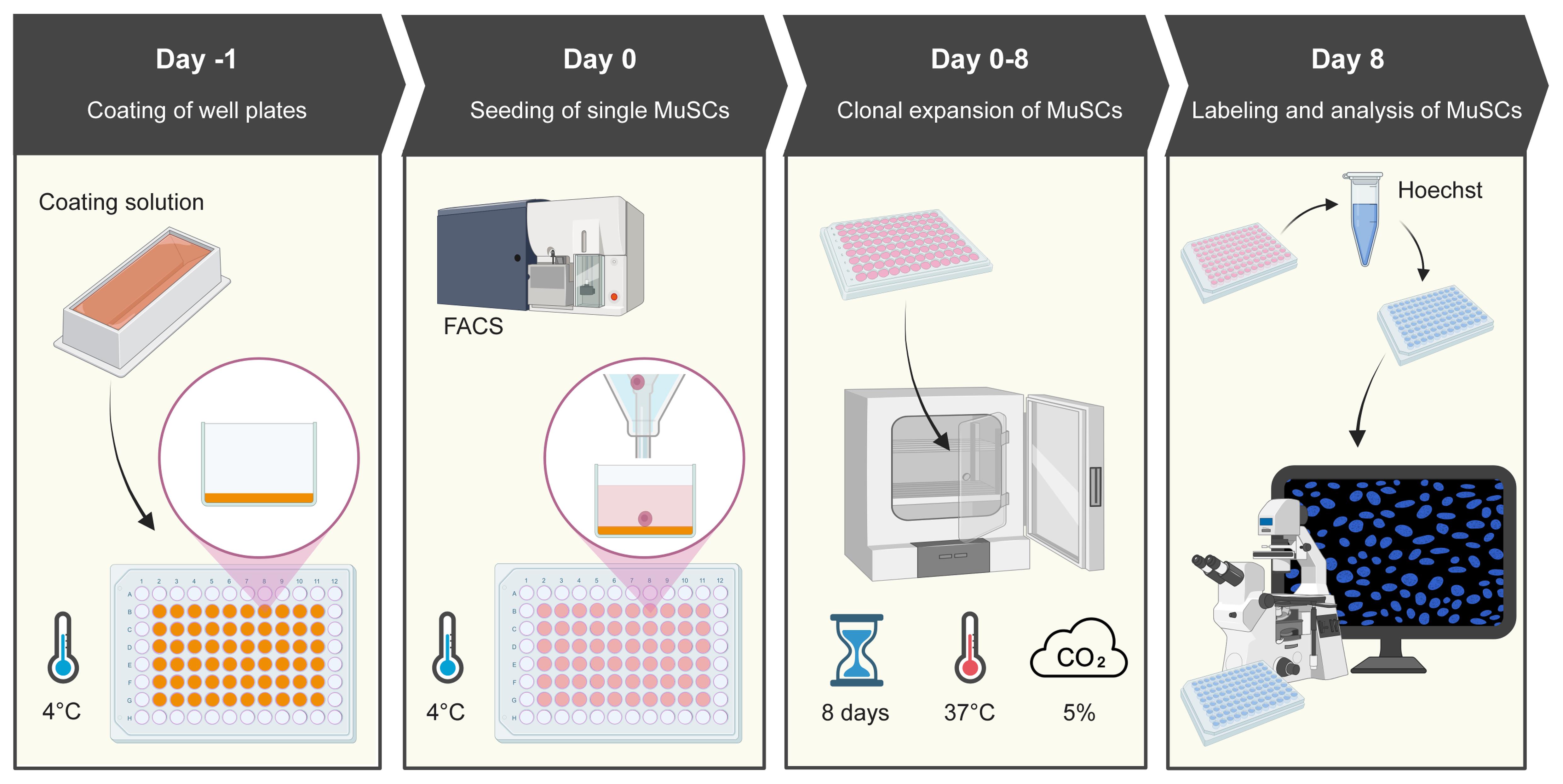
Procedure schematics. The schematic shows the four main procedures of the protocol.
Background
Adult stem cells are paramount for tissue regeneration, given their ability to rapidly expand into large numbers of differentiation-competent cells that can rebuild tissue [1,2]. One well-studied adult stem cell model is the muscle stem cell (MuSC). MuSCs preserve skeletal muscle function and drive its regeneration. MuSCs are rare cells that reside in a quiescent state of prolonged cell cycle exit. Upon injury, MuSCs activate to exit the quiescent state, proliferate, and differentiate to make new myofibers [2–4]. Some MuSCs self-renew and re-enter the quiescent state to repopulate the niche in anticipation of future muscle damage [5].
Understanding MuSC function requires assays to quantitatively assess the processes of activation and proliferation. Nucleotide analogs are the gold standard for assessing MuSC activation and proliferation. However, such experiments require thousands of cells, and MuSCs are rare [6,7]. In other adult stem cell models, the ability of stem cells to activate and replicate into undifferentiated daughter cells is commonly tested using clonogenic expansion assays [8]. These assays use low numbers of single cells as input and can reveal altered stem cell activation, tissue function, and impacted outcomes of tissue regeneration following transplants [9,10]. Here, we present the myoclone assay, a protocol for the quantitative assessment of MuSC activation and clonogenic growth based on our recent publication [11].
The myoclone assay requires 60 cells per sample, a much lower number than in other assays that require thousands of cells per sample [7,12–14]. MuSCs are commonly isolated from skeletal muscles using fluorescence-activated cell sorting (FACS) [7,15–19]. Given the low cell number required, the myoclone assay can easily be added to any experimental plan involving FACS. Moreover, the low cell number greatly cuts the required sort time, which results in cost savings and the ability to analyze more samples in parallel. Finally, since the MuSCs remain alive in the microwells at the end of the assay, the myoclone assay can be combined with downstream protocols such as cell culture and immunostaining.
Here, we demonstrate the sensitivity of the myoclone assay using MuSCs isolated from diaphragm and hindlimb muscles of young mice. MuSCs isolated from these two muscle groups are known to have different activation and proliferation dynamics [6].
Materials and reagents
Biological materials
1. Primary muscle stem cells (MuSCs) (e.g., isolated from Mus musculus in-house)
Reagents
1. Laminin mouse protein, natural (Gibco, catalog number: 23017015)
2. Bovine collagen type 1 (Sigma-Aldrich, catalog number: C8919)
3. Anhydrous acetic acid (Merck, catalog number: 1.00063.1000)
4. PBS tablets (Gibco, catalog number: 18912014)
5. Ham’s F-10 nutrient mix (Gibco, catalog number: 11574436)
6. Horse serum heat-inactivated, New Zealand origin (Thermo Fisher, catalog number: 26050088)
7. Penicillin-streptomycin (Gibco, catalog number: 15140122)
8. Human FGF-basic (FGF-2/bFGF) (154 aa) recombinant protein, PeproTech (Gibco, catalog number: 100-18B)
9. Hoechst 33342 (Thermo Scientific, catalog number: 62249)
10. Biotin anti-mouse CD106 antibody, clone 429 (BioLegend, catalog number: 105704)
11. PE/Cyanine7 Streptavidin (BioLegend, catalog number: 405206)
12. FITC anti-mouse CD45 antibody, clone 30-F11 (BioLegend, catalog number: 103108)
13. FITC anti-mouse CD31 antibody, clone MEC13.3 (BioLegend, catalog number: 102506)
14. Pacific Blue anti-mouse Ly-6A/E (Sca-1) antibody, clone D7 (BioLegend, catalog number: 108120)
Solutions
1. Basal myoclone medium (see Recipes)
2. 1× PBS (see Recipes)
3. Laminin stock solution (see Recipes)
4. Collagen stock solution (see Recipes)
Recipes
1. Basal myoclone medium
| Reagent | Final concentration | Quantity or volume |
|---|---|---|
| Ham’s F-10 nutrient mix | NA | 500 mL |
| Horse serum heat-inactivated | 20% | 100 mL |
| Penicillin-streptomycin | 1% Penicillin (100 units/mL) 1% Streptomycin (0.1 mg/mL) | 5 mL |
Thaw 100 mL of horse serum (HS) and 5 mL of penicillin-streptomycin (Pen/Strep, 10,000 units/mL / 10 mg/mL) at 37 °C in a water bath. Once thawed, bring the HS and Pen/Strep into a laminar flow hood (LAF) together with an unopened bottle of Ham’s F-10 (500 mL). Add 100 mL of HS and 5 mL of Pen/Strep to the 500 mL bottle of Ham’s F-10 after taking out and discarding 105 mL of the F-10. Mix the medium by gently inverting the bottle to avoid the formation of bubbles. Store the bottle of basal myoclone medium at 4 °C.
2. 1× PBS
| Reagent | Final concentration | Quantity or volume |
|---|---|---|
| PBS tablets | 1× (10 g/L) | 5 g |
| Milli-Q water | NA | 500 mL |
Open the vial of PBS tablets in the LAF. Dissolve one 5 g sterile PBS tablet in 500 mL of Milli-Q water in a 500 mL borosilicate glass bottle. Mix using a stir bar and a magnetic rotator. Alternatively, leave the PBS tablet to dissolve in the Milli-Q water for 1 h and swirl to mix afterward. Filter the PBS using a 0.45-μm, 500-mL polyethersulfone filter unit attached to an autoclaved 500 mL borosilicate bottle. Store the sterile PBS at 4 °C.
3. Laminin stock solution
| Reagent | Final concentration | Quantity or volume |
|---|---|---|
| Laminin mouse protein | 0.02 mg/mL | 1 mg |
| Sterile 1× PBS | NA | 50 mL |
Retrieve Laminin mouse protein from the -80 °C freezer and thaw. Once thawed, keep on ice until use to prevent gel formation. Bring filter-sterilized 1× PBS and the laminin to the LAF. Transfer the total 1 mL volume of Laminin mouse protein (1 mg/mL) from its vial into a 50 mL borosilicate glass bottle with 49 mL of sterile 1× PBS to obtain a laminin stock solution at a final concentration of 0.02 mg/mL. Swirl to mix. Aliquot as desired (e.g., 5 mL aliquots) and store at -80 °C until use. Note that the vendor can provide Laminin mouse protein at varying concentrations, and the volumes may need adjustment to obtain a final concentration of 0.02 mg/mL.
4. Collagen stock solution
| Reagent | Final concentration | Quantity or volume |
|---|---|---|
| Anhydrous acetic acid | 100 mM | 5.15 mL |
| Milli-Q water | NA | 895 mL |
Caution: This step involves working with highly concentrated acid. Make sure to wear personal protective equipment (lab coat, gloves, closed-toe shoes, protective eyewear) and work in a chemical fume hood.
Collect filtered Milli-Q water from a Milli-Q water dispenser. Bring anhydrous acetic acid and Milli-Q water to the fume hood. To prepare acidified water, transfer 895 mL of sterile Milli-Q water to a borosilicate glass bottle and add 5.15 mL of anhydrous acetic acid to the Milli-Q water.
| Reagent | Final concentration | Quantity or volume |
|---|---|---|
| Bovine collagen type 1 | 0.05 mg/mL | 10 mL (1 mg/mL) |
| Acidified water | 95 mM in collagen stock solution | 190 mL |
Transfer 190 mL of acidified water to a 200 mL borosilicate glass bottle. Add 10 mL of bovine collagen type 1 (10 mg) to the 200 mL borosilicate glass bottle to obtain the final collagen stock solution (0.05 mg/mL). Swirl to mix. Filter the collagen stock solution using a 0.45-μm, 500-mL polyethersulfone filter unit attached to an autoclaved 200 mL borosilicate bottle. After filtration, swirl the solution to mix. Store the collagen stock solution at 4 °C until use.
Laboratory supplies
1. Cell culture plates, 96-well, CELLSTAR, half area, black, with lid (VWR, catalog number: 392-0058)
2. VWR® LTS® compatible filter tips, 1,000 μL micropipette tips (VWR, catalog number: 732-3642)
3. VWR® LTS® compatible filter tips, 250 μL micropipette tips (VWR, catalog number: 613-6503)
4. 25 mL serological pipette tips (Hounisen, catalog number: 86.1685.001)
5. 15 mL conical tubes, 120 × 17 mm, PP DNA-, DNAse-, RNase-free, non-pyrogenic, non-cytotoxic (Hounisen, catalog number: 62.554.502)
6. 50 mL conical tubes, 114 × 28 mm, PP DNA-, DNAse-, RNase-free, non-pyrogenic, non-cytotoxic (Hounisen, catalog number: 62.547.254)
7. DURAN® BlueCap bottle, 250 mL borosilicate glass bottle (Hounisen, catalog number: 0610.0250)
8. DURAN® BlueCap bottle, 50 mL borosilicate glass bottle (Hounisen, catalog number: 0610.0050)
9. 50 mL reservoir for multichannel micropipette, sterile (Hounisen, catalog number: 0699.8002)
10. 0.45 μm polyethersulfone (PES) Filter Unit (Fisher Scientific, catalog number: 15923307)
Equipment
1. BD FACSAriaTMIII Sorter (BD Biosciences, catalog number: 648282-24)
2. Widefield Inverted Light Microscope, AxioObserver (Zeiss) with objectives 10×/NA 0.30 Air, Ph1, EC Plan-Neofluar and 5×/NA0.16 Air, Ph1, EC Plan-Neofluar
3. Eppendorf centrifuge 5810 (Eppendorf, catalog number: EP022628188)
4. FisherbrandTM Mini Tube Rotator (Fisher Scientific, catalog number: 15534080)
5. Thermo ScientificTM Series 8000 Direct-Heat CO2 Incubator, 184 L (Fisher Scientific, catalog number: 11862711)
6. LAF: NinoSAFE Class II Biological Safety Cabinet (Nino Labinterior, catalog number: 101861)
7. Corning® StripettorTM Ultra Pipette Controller (Corning, catalog number: 4099)
8. RAININ Pipet-Lite LTS Pipette L-20XLS+ (Mettler Toledo, catalog number: 17014392)
9. RAININ Pipet-Lite LTS Pipette L-200XLS+ (Mettler Toledo, catalog number: 17014391)
10. RAININ Pipet-Lite LTS Pipette L-1000XLS+ (Mettler Toledo, catalog number: 17014382)
11. RAININ Pipet-Lite Multi Pipette L8-200XLS+ (Mettler Toledo, catalog number: 17013805)
12. Graduated cylinder
13. Ice machine
14. Fume hood
15. Water bath (Thermo Fisher Scientific, catalog number: FSSWB15)
16. Ice bucket
17. Freezer (-20 °C)
18. Refrigerator (2–8 °C)
19. Autoclave
20. Arium Pro DI Ultrapure Water System (Sartorius, catalog number: H2OPRO-DI-T)
Software and datasets
1. GraphPad Prism (GraphPad Software Inc., v.10.4.1)
2. Fiji image processing package (ImageJ2, v.1.54p)
Procedure
登录/注册后免费查看全文
文章信息
稿件历史记录
提交日期: May 30, 2026
接收日期: Oct 14, 2026
在线发布日期: Oct 27, 2026
出版日期: Nov 20, 2026
版权信息
© 2026 The Author(s); This is an open access article under the CC BY license (https://creativecommons.org/licenses/by/4.0/).
如何引用
Stella, M. P. J., Damgaard, O., Frimand, Z. and De Morree, A. (2026). Myoclone Assay: Clonogenic Expansion of Activating Muscle Stem Cells. Bio-protocol 16(22): e5869. DOI: 10.21769/BioProtoc.5869.

 Download Citation in RIS Format
您对这篇实验方案有问题吗?
在此处发布您的问题,我们将邀请本文作者来回答。同时,我们会将您的问题发布到Bio-protocol Exchange,以便寻求社区成员的帮助。
 发布问题


