


TurboID-Mediated Proteomic Study in the Primary Cilia of Embryonic Mouse Brain

发布: 2026年11月20日第16卷第22期 DOI: 10.21769/BioProtoc.5868 浏览次数: 44
评审: Sam PatelAnonymous reviewer(s)
 PDF
 Q&A





Abstract
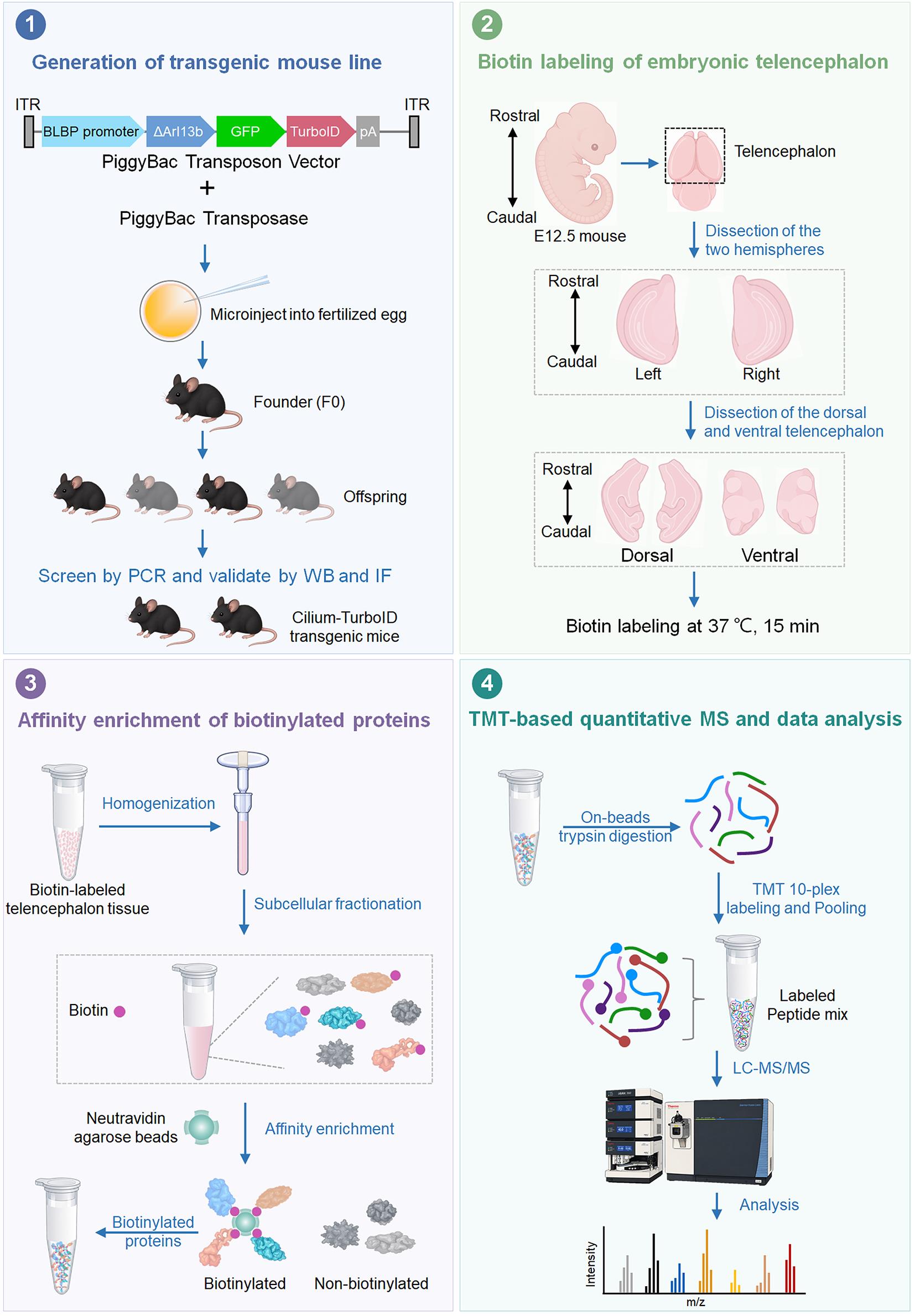
Primary cilia of radial glia are important signaling compartments during embryonic brain development, but their molecular composition in vivo remains difficult to characterize. Conventional ciliary isolation can introduce substantial non-ciliary contamination, whereas APEX-based proximity labeling requires hydrogen peroxide (H2O2) to initiate labeling, and BioID generally requires prolonged labeling times. Here, we describe a step-by-step protocol for in vivo proximity-labeling proteomics of radial glial primary cilia in the embryonic mouse telencephalon using a Cilium-TurboID transgenic mouse model. TurboID is targeted to radial glial cilia through a ΔARL13B-GFP fusion under the BLBP promoter, enabling rapid biotin labeling in freshly dissected embryonic brain tissue. Following labeling, the tissue is subjected to subcellular fractionation, neutravidin-based enrichment of biotinylated proteins, on-bead trypsin digestion, TMT labeling, and quantitative mass spectrometry. This workflow enables spatially targeted profiling of the low-abundance ciliary proteome directly in native embryonic brain tissue, while avoiding the need to physically isolate primary cilia and eliminating the requirement for H2O2 during proximity labeling. The rapid labeling kinetics of TurboID also provide improved temporal resolution compared with conventional BioID, making the workflow well-suited for investigating ciliary protein composition in developing neural tissues.
Key features
• Enables in vivo proteomic profiling of radial glial primary cilia in the embryonic mouse telencephalon.
• Uses a BLBP-driven Cilium-TurboID transgenic mouse for cell type- and cilium-targeted proximity labeling.
• Combines subcellular fractionation with neutravidin enrichment to improve recovery of ciliary proteins from brain tissue.
• Supports region-specific comparison of ciliary proteomes between dorsal and ventral telencephalon.
Keywords: Proximity labeling TurboID
Primary cilium
Radial glia
Embryonic mouse brain
In vivo proteomics
Graphical overview
Background
Primary cilia are specialized, microtubule-based cellular compartments that organize receptors and signaling proteins involved in developmental pathways such as Hedgehog (Hh) [1,2]. Their importance is particularly evident in the developing nervous system, where defects in ciliary assembly or signaling can disrupt neural progenitor behavior and cause brain abnormalities associated with ciliopathies [3–5]. In the embryonic telencephalon, radial glia (RG) are the major neural progenitors and maintain their primary cilia at the ventricular surface, where the cilia are positioned at the interface between progenitor cells and the cerebrospinal fluid (CSF) [6–8]. Identifying the proteins that reside in or transiently associate with these cilia is therefore important for understanding how ciliary signaling contributes to neural development and neurodevelopmental disease.
The molecular characterization of primary cilia in vivo remains technically challenging because the cilium is extremely small and comprises approximately 1/30,000 of the total cellular space [9,10]. Conventional ciliary proteomics has relied on physical isolation or biochemical enrichment of cilia. Although these approaches have successfully identified many ciliary proteins, isolation of primary cilia from complex mammalian tissues can result in substantial contamination from non-ciliary compartments and may preferentially recover abundant proteins [11]. Moreover, disrupting cells and isolating cilia before analysis can compromise weak or transient associations that occur within the native ciliary environments.
Proximity labeling provides an alternative approach in which proteins near a genetically targeted enzyme are covalently labeled before tissue disruption. APEX-based approaches provide rapid labeling but require hydrogen peroxide, whereas conventional BioID uses biotin without oxidative treatment but generally requires prolonged labeling [12]. TurboID, an engineered promiscuous biotin ligase, substantially accelerates biotinylation and enables proximity labeling on a timescale of minutes, making it well-suited to spatial proteomics in living cells and organisms [12–14]. Cilia-targeted proximity labeling has previously been used to characterize ciliary proteomes and to examine changes associated with Hh signaling in cultured cells [13,15,16]. However, applying proximity labeling to primary cilia in the developing brain presents additional challenges, including cell type- and cilium-specific targeting, the low abundance of ciliary proteins, and high background from abundant non-ciliary proteins in the surrounding tissue.
The present protocol addresses these challenges by combining cell type–specific genetic targeting of TurboID with tissue-level biochemical enrichment and quantitative mass spectrometry. In our approach, TurboID is targeted to the primary cilia of RG using a ΔARL13B-GFP-TurboID fusion under control of the BLBP promoter. The resulting transgenic mouse enables cilia-associated proteins to be labeled directly in the developing telencephalon. Rapid biotin labeling is followed by subcellular fractionation to reduce abundant non-target material, neutravidin-based enrichment of biotinylated proteins, and TMT (tandem mass tag)-based quantitative mass spectrometry. In the corresponding Cell Reports study, this workflow enabled profiling of RG ciliary proteins in the E12.5 dorsal and ventral telencephalon and identified 258 candidate ciliary proteins, including previously recognized ciliary proteins and components displaying regional differences between the two telencephalic domains [17].
The major advantage of this approach over physical cilium isolation is that proteins are labeled in the native tissue before biochemical processing, which preserves spatial information without requiring intact primary cilia to be isolated as a separate fraction. Compared with other proximity labeling methods, e.g., APEX and conventional BioID, this protocol using TurboID enables rapid, H2O2-free proximity labeling with shorter labeling times and improved temporal resolution [18,19]. Importantly, this in vivo proximity labeling captures the ciliary proteome within the physiological environment of the developing brain, where interactions among different cell types and developmental cues can influence ciliary protein composition and signaling. This provides an important advantage over cell culture–based proximity-labeling approaches, which may not fully recapitulate the multicellular and tissue-specific environment of the developing telencephalon. In addition, the use of neutravidin for affinity enrichment provides an effective strategy for recovering biotinylated proteins from tissue lysates while reducing nonspecific protein binding relative to streptavidin under the conditions used in this workflow. The combination of cell type–specific genetic targeting, optimized tissue fractionation, and neutravidin-based affinity enrichment therefore makes this workflow particularly suitable for profiling the low-abundance ciliary proteome of neural progenitor populations. However, low-level basal biotinylation was detectable in Cilium-TurboID tissue in the absence of exogenous biotin; this background was mitigated by optimizing the biochemical enrichment workflow and using neutravidin-based affinity capture [17]. In addition, because the protocol relies on a genetically engineered mouse model, establishing the transgenic line requires more time and resources than cell-based proximity-labeling approaches.
This workflow can be applied beyond the RG primary cilia examined here. By replacing the cell type–specific regulatory element or adapting the ciliary targeting strategy, it may be used to study ciliary proteomes in other neural populations and developmental stages, including specialized cilia in the ventricular system and choroid plexus. More broadly, the combination of genetically encoded proximity labeling, tissue fractionation, and quantitative proteomics provides a framework for investigating other spatially restricted protein environments in complex developing tissues.
Materials and reagents
Biological materials
1. Bacterial strains: TOP10 (Thermo Fisher Scientific, catalog number: C404010)
2. Cilium-TurboID transgenic C57BL/6 mouse (not commercially available [17])
Reagents
1. BLBP-GFP plasmid (Addgene, catalog number: 63174); Cilium-TurboID plasmid (not commercially available [17])
2. Ampicillin, sodium salt (Research Products International, catalog number: A40040-5.0)
3. Maxi Fast-Ion Plasmid kit (IBI Scientific, catalog number: Ib47122)
4. Proteinase K solution (20 mg/mL) (Fisher Scientific, catalog number: 25530049)
5. Triton X-100 (Sigma, catalog number: T8787-50 mL)
6. 2× Taq Master Mix (Dye Plus) (Vazyme, catalog number: P112-01)
7. DPBS (Fisher Scientific, catalog number: 14-190-136)
8. Biotin, molecular biology grade (Sigma, catalog number: B4501-1G)
9. DMSO, molecular biology grade (Sigma, catalog number: D8418-50ML)
10. Neurobasal medium (Thermo Fisher Scientific, catalog number: 21103-049)
11. 20% PFA (Fisher Scientific, catalog number: 50-980-493)
12. O.C.T. compound (Fisher Scientific, catalog number: 23-730-571)
13. Donkey serum (Sigma, catalog number: S30-100ML)
14. Chicken anti-GFP (Aves Labs, catalog number: GFP-1020)
15. Rabbit anti-ARL13B (Proteintech, catalog number: 17711–1-AP)
16. Donkey anti-chicken Alexa Fluor 488 (Jackson ImmunoResearch Labs, catalog number: 703-545-155)
17. Donkey anti-rabbit Rhodamine (Jackson ImmunoResearch Labs, catalog number: 711-025-152)
18. Donkey anti-rabbit Alexa Fluor 488 (Jackson ImmunoResearch Labs, catalog number: 711-545-152)
19. Alexa Fluor 647 Streptavidin (Jackson ImmunoResearch Labs, catalog number: 016-600-084)
20. Fluoromount-G mounting medium (SouthernBiotech, catalog number: 0100-01)
21. Hoechst 33258 (Sigma, catalog number: 94403-1ML)
22. 1 M sodium hydroxide (NaOH), molecular biology grade (Sigma, catalog number: 1091371003)
23. 36.5%–38.0% hydrochloric acid (HCl) (Fisher Scientific, catalog number: A144-500)
24. HEPES, molecular biology grade (Fisher Scientific, catalog number: BP310-500)
25. KCI, molecular biology grade (Fisher Scientific, catalog number: P217-500)
26. MgCl2, molecular biology grade (Fisher Scientific, catalog number: AC223211000)
27. 0.5 M EDTA (Fisher Scientific, catalog number: AAJ15694AE)
28. 0.5 M EGTA (Fisher Scientific, catalog number: 50-255-956)
29. Tris base, molecular biology grade (Fisher Scientific, catalog number: BP152-500)
30. NaCl, molecular biology grade (Fisher Scientific, catalog number: S271-500)
31. SDS, molecular biology grade (Fisher Scientific, catalog number: BP166-100)
32. Protease inhibitor cocktail (Roche, catalog number: 11836170001)
33. 10% NP-40 (Thermo Fisher Scientific, catalog number: 28324)
34. BCA Protein Assay kit (Thermo Fisher Scientific, catalog number: 23225)
35. Neutravidin agarose resin (Thermo Fisher Scientific, catalog number: 29201)
36. 2× Laemmli sample buffer (Bio-Rad Laboratories, catalog number: 1610737)
37. Mouse anti-GAPDH (Santa Cruz Biotechnology, catalog number: sc-32233)
38. HRP-conjugated donkey anti-rabbit IgG (Jackson ImmunoResearch Labs, catalog number: 711-035-152)
39. HRP-conjugated donkey anti-mouse IgG (Jackson ImmunoResearch Labs, catalog number: 715-035-151)
40. HRP-conjugated neutravidin (Thermo Fisher Scientific, catalog number: 31030)
41. SuperSignal West Pico PLUS Chemiluminescent Substrate (Thermo Fisher Scientific, catalog number: 34577)
42. 1 M Triethylammonium bicarbonate (TEAB) (Thermo Fisher Scientific, catalog number: 90114)
43. 10plex TMT label reagent set (Thermo Fisher Scientific, catalog number: 90110)
44. Pierce TCEP HCl (Thermo Fisher Scientific, catalog number: 20490)
45. 2-Chloroacetamide ≥98% (Sigma-Aldrich, catalog number: 22790)
46. Trypsin (Promega, catalog number: V5111)
47. Calcium chloride, molecular biology grade (Sigma-Aldrich, catalog number: C5670)
48. Resin, Acquity UPLC® BEH 1.7 μm C18 (Waters, catalog number: 186004690)
49. Water, HPLC grade (Optima LC-MS grade) (Thermo Fisher Scientific, catalog number: W6-4)
50. Acetonitrile, HPLC grade (Honeywell, catalog number: AH015-4)
51. Methanol, HPLC grade (J.T. Baker, catalog number: 9093-33)
52. Chloroform, HPLC grade (Thermo Fisher Scientific, catalog number: C298-4)
53. Trifluoroacetic acid (TFA), HPLC grade (Sigma-Aldrich, catalog number: T6508-10AMP)
54. Polymicro fused silica capillary tubing 100 μm (Molex, catalog number: 1068150023)
55. 11 PP vial crimp/snap 250 μL (Thermo Fisher Scientific, catalog number: C4011-13)
56. Snap-it seal (Thermo Fisher Scientific, catalog number: C4011-53)
57. Pierce HeLa protein digest standard (Thermo Fisher Scientific, catalog number: 88329)
58. Urea (Sigma-Aldrich, catalog number: U5128-110G)
59. Pierce High pH RP Peptide Fractionation kit (Thermo Fisher Scientific, catalog number: 84868)
60. Ethanol absolute (VWR, catalog number: 89125-186)
61. 0.4% Trypan Blue solution (Thermo Fisher Scientific, catalog number: 15250061)
62. Sucrose (Thermo Fisher Scientific, catalog number: ICN821713)
Solutions
1. Mouse tail digestion buffer (see Recipes)
2. 50 mM biotin stock solution (see Recipes)
3. Biotin labeling medium (see Recipes)
4. Blocking buffer (see Recipes)
5. 25× protease inhibitor cocktail buffer (see Recipes)
6. Subcellular fractionation buffer (see Recipes)
7. 10× TBS buffer (see Recipes)
Recipes
1. Mouse tail digestion buffer
| Reagent | Final concentration | Quantity or volume |
|---|---|---|
| KCl | 50 mM | 0.373 g |
| Tris | 10 mM | 0.121 g |
| Triton X-100 | 0.1% | 100 μL |
| Proteinase K | 0.4 mg/mL | 2 mL |
| H2O (MilliQ water) | n/a | * |
| Total | n/a | 100 mL |
*Mix until the reagents are fully dissolved and adjust the pH of the solution to 9.0 by adding HCl (36.5%–38.0%) while measuring with a pH meter. Then, bring the volume up to 100 mL with MilliQ water.
Store at 4 °C.
2. 50 mM biotin stock solution
| Reagent | Final concentration | Quantity or volume |
|---|---|---|
| Biotin | 50 mM | 0.5 g |
| DMSO | n/a | 41 mL |
| Total | n/a | 41 mL |
Store at -20 °C.
3. Biotin labeling medium
| Reagent | Final concentration | Volume |
|---|---|---|
| 50 mM biotin stock (Recipe 2) | 50 μM | 50 μL |
| Neurobasal medium | n/a | 49.95 mL |
| Total | n/a | 50 mL |
Aliquot and store at 4 °C for one month.
4. Blocking buffer
| Reagent | Final concentration | Volume |
|---|---|---|
| Donkey serum | 2% | 2 mL |
| Triton X-100 | 0.2% | 200 μL |
| DPBS | n/a | 97.8 mL |
| Total | n/a | 100 mL |
Store at 4 °C.
5. 25× protease inhibitor cocktail buffer
| Reagent | Final concentration | Quantity or volume |
|---|---|---|
| Protease inhibitor cocktail | 25× | 1 tablet |
| H2O (MilliQ water) | n/a | 2 mL |
| Total | n/a | 2 mL |
*Add the 25× protease inhibitor cocktail buffer to the subcellular fractionation buffer immediately before use to achieve a final concentration of 1×.
Store at -20 °C.
6. Subcellular fractionation buffer
| Reagent | Final concentration | Quantity or volume |
|---|---|---|
| HEPES | 20 mM | 4.77 g |
| KCI | 10 mM | 0.75 g |
| MgCl2 | 2 mM | 0.19 g |
| 0.5 M EDTA | 1 mM | 2 mL |
| 0.5 M EGTA | 1 mM | 2 mL |
| H2O (MilliQ water) | n/a | * |
| Total | n/a | 1 L |
*Mix until the reagents are fully dissolved and adjust the pH of the solution to 7.4 by adding 1 M NaOH while measuring with a pH meter. Then, bring the volume up to 1 L with MilliQ water. Just before use, add and dilute the 25× protease inhibitor cocktail buffer to the subcellular fractionation buffer to achieve a final concentration of 1×.
Store at 4 °C.
7. 10× TBS buffer
| Reagent | Final concentration | Quantity or volume |
|---|---|---|
| Tris | 200 mM | 24.23 g |
| NaCl | 1.5 M | 87.66 g |
| 0.5 M EDTA | 10 mM | 20 mL |
| H2O (MilliQ water) | n/a | * |
| Total | n/a | 1 L |
*Mix until the reagents are fully dissolved and adjust the pH of the solution to 7.4 by adding HCl (36.5% to 38.0%) while measuring with a pH meter. Then, bring the volume up to 1 L with MilliQ water.
Store at 4 °C.
Laboratory supplies
1. 10 cm dish (Fisher Scientific, catalog number: FB012924)
2. Moria ultra-fine forceps (Fine Science Tools, catalog number: 11370-40)
3. Moria ultra-fine curved forceps (Fine Science Tools, catalog number: 11370-42)
4. Extra-fine Bonn 8.5 cm scissors (Fine Science Tools, catalog number: 14085-08)
5. 15 mL conical tube (Corning, catalog number: 430791)
6. Polystyrene round-bottom tube (Falcon, catalog number: 352058)
7. Superfrost Plus microscope slides (Fisher Scientific, catalog number: 12-550-15)
8. Superslip coverslips (Fisher Scientific, catalog number: 12-541-056)
9. Tweezers (Fine Science Tools, catalog number: 11252-00)
10. Parafilm (Fisher Scientific, catalog number: PM999)
11. KIMBLE Dounce tissue grinder set (DWK Life Sciences, catalog number: D8938)
12. 1.5 mL protein LoBind tube (Eppendorf, catalog number: 22431081)
13. 5 mL protein LoBind tube (Eppendorf, catalog number: 30108302)
14. 1 mL syringe (BD Biosciences, catalog number: 309628)
15. 27-gauge needle (BD Biosciences, catalog number: 305136)
16. Plastic cryomolds (TedPella, catalog number: 27110)
17. StainTray with black lid (Simport, catalog number: M920-2)
18. PAP pen liquid blocker (Newcomer Supply, catalog number: NC9204359)
19. 0.2 mL PCR tube strips (Fisher Scientific, catalog number: 14230210)
20. Kimwipes (Fisher Scientific, catalog number: 06-666)
Equipment
1. Vortex mixer (Fisher Scientific, catalog number: 02-215-414)
2. Centrifuge (Eppendorf, model: 5702R)
3. Centrifuge (Eppendorf, model: 5804R)
4. VWR Digital Advanced 2 Block Heater (VWR, catalog number: 75838-282)
5. Leica stereomicroscope (Leica, model: S6E)
6. Confocal microscope (Zeiss, model: LSM 880)
7. Sonicator equipped with a microtip, measuring 3/32 inches in diameter (Branson, model: SLPt)
8. Leica cryostat (Leica, model: CM1860)
9. Orbitrap Fusion Lumos mass spectrometer (Thermo Fisher Scientific, catalog number: IQLAAEGAAPFADBMBHQ)
10. EASY-nLC 1200 (Thermo Fisher Scientific, catalog number: LC140)
11. Laser-based micropipette puller system (Sutter Instrument, catalog number: P-2000)
12. Thermomixer F1.5 (Eppendorf, catalog number: 5384000020)
13. Microfuge18 centrifuge (Beckman Coulter, catalog number: 367160)
14. Vacuum Centrifuge (Labconco, catalog number: 7810016)
15. Thermal Cycler (Bio-Rad, model: T100)
16. Real-Time PCR System (Applied Biosystems, model: QuantStudio 3)
17. Mini Gel Electrophoresis System (Thermo Fisher Scientific, model: Owl EasyCast B2)
18. Imaging Systems for blots and gels (Bio-Rad, model: ChemiDoc MP)
19. Trans-Blot Turbo Transfer Starter System (Bio-Rad, catalog number: 17001917)
Software and datasets
1. ProLuCID (Bruker Scientific LLC, IP2)
2. DTASelect2.0 (Bruker Scientific LLC, IP2)
3. Census2.0 (Bruker Scientific LLC, IP2)
4. RAWConverter (John R. Yates III Lab, Scripps Research, La Jolla)
5. XCalibur (Thermo Fisher Scientific, catalog number: OPTON-30965-7)
Procedure
登录/注册后免费查看全文
文章信息
稿件历史记录
提交日期: Aug 16, 2026
接收日期: Oct 8, 2026
在线发布日期: Oct 23, 2026
出版日期: Nov 20, 2026
版权信息
© 2026 The Author(s); This is an open access article under the CC BY-NC license (https://creativecommons.org/licenses/by-nc/4.0/).
如何引用
Liu, X. and Ge, X. (2026). TurboID-Mediated Proteomic Study in the Primary Cilia of Embryonic Mouse Brain. Bio-protocol 16(22): e5868. DOI: 10.21769/BioProtoc.5868.

 Download Citation in RIS Format
您对这篇实验方案有问题吗?
在此处发布您的问题,我们将邀请本文作者来回答。同时,我们会将您的问题发布到Bio-protocol Exchange,以便寻求社区成员的帮助。
 发布问题


