


Semi-Automated Workflow for Segmenting and Quantifying Ribosomes in Electron Tomograms of High-Pressure Frozen and Resin-Embedded Samples

(*contributed equally to this work) 发布: 2026年11月20日第16卷第22期 DOI: 10.21769/BioProtoc.5866 浏览次数: 44
评审: Matthew RussellElena A. OstrakhovitchAnonymous reviewer(s)
 PDF
 Q&A





Abstract
Ribosomes are the central molecular machines of protein synthesis, and accurate counts of ribosome numbers are essential for linking translational capacity to cellular physiology. Common approaches infer ribosome abundance indirectly, for example, by measuring rRNA or ribosomal proteins, which can obscure spatial heterogeneity and sample-specific differences. This protocol presents an accessible image analysis pipeline for segmenting and counting individual ribosomes in three-dimensional transmission electron microscopy (3D-TEM) data. Established experimental procedures, namely high-pressure freezing (HPF), freeze substitution, resin embedding, serial semi-thick sectioning, and dual-axis tomography, are summarized here with direct references to primary protocols. The core of this work details electron tomography reconstruction and dual-axis stitching in IMOD 5.1, region-of-interest cropping in Fiji, and a semi-automated segmentation pipeline in arivis Pro 4.5. This pipeline inverts and denoises tomograms, detects ribosomal particles with a tuned Blob Finder routine, applies empirically derived voxel-count thresholds alongside a laboratory-specific shrinkage correction, and converts segmented counts to ribosomes per cubic micrometer and, for cultured cells, per cell. Validation shows that tomographic counts agree with independent biochemical rRNA measurements to within the same order of magnitude, and the protocol states the false-positive and false-negative limitations inherent to nonspecific heavy-metal staining.
Key features
• Detailed image analysis pipeline for ribosome segmentation in electron tomograms using IMOD, Fiji, and arivis Pro, with recommended parameter values.
• Condensed but executable sample-preparation section covering high-pressure freezing, freeze substitution, resin embedding, sectioning, and dual-axis tomography for both C. elegans and cultured mammalian (hTERT-RPE-1) cells.
• Shrinkage-corrected quantification that converts voxel counts to ribosomes per cubic micrometer and per cell using an empirically derived shrinkage factor of 1.575.
• Validation of ribosome counts at 25, 50, and 100 voxel thresholds in control and rpoa-1(RNAi) samples using an independent biochemical method.
Keywords: Ribosome quantification Electron tomography
High-pressure freezing
Freeze-substitution
arivis Pro
IMOD
image segmentation
Shrinkage correction
hTERT-RPE-1
C. elegans
Graphical overview
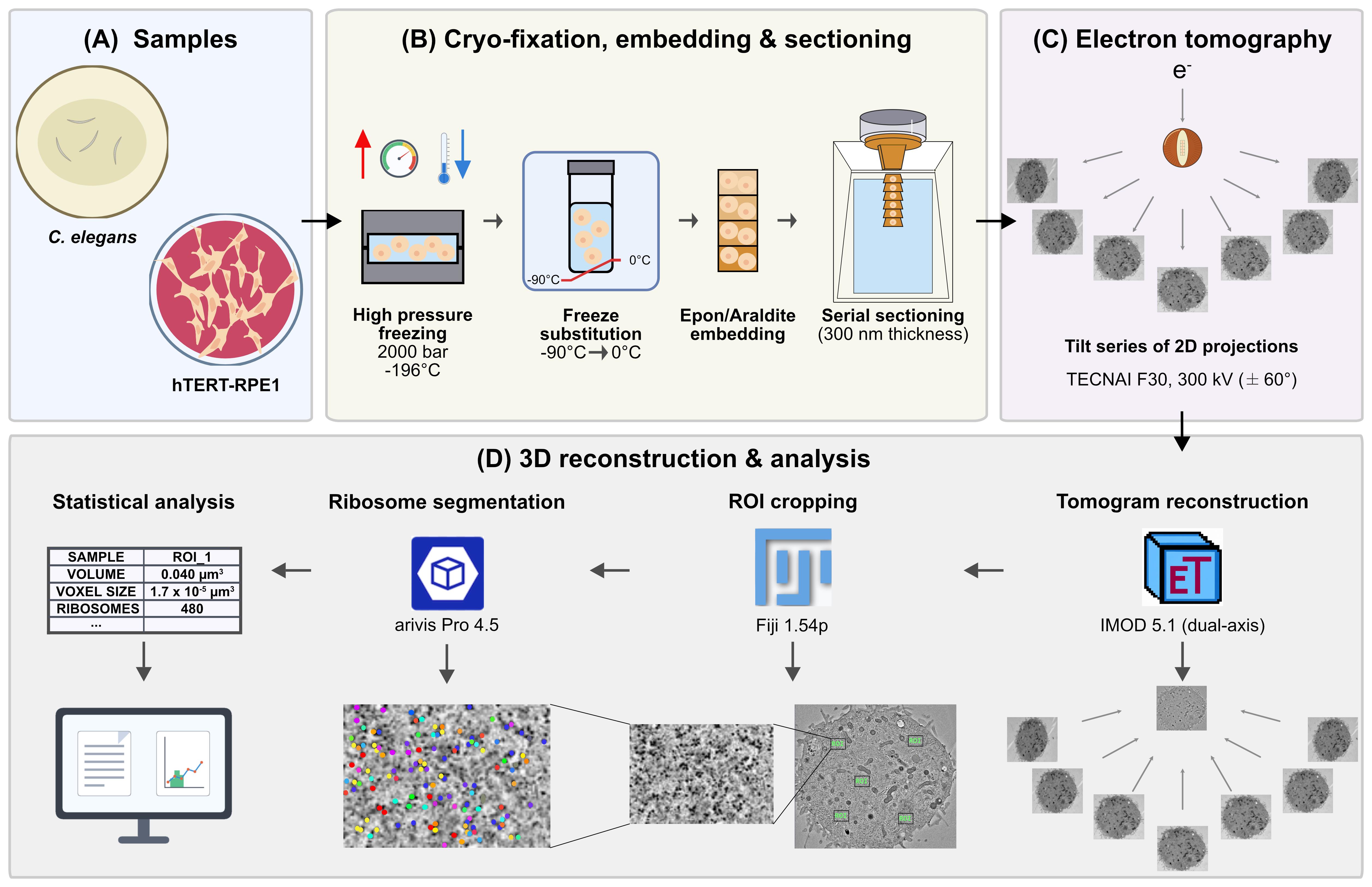
Graphical overview of the global workflow to count ribosomes in cellular sub-volumes. (A) Samples: hTERT-RPE-1 cells and Caenorhabditis elegans. (B) Cryo-fixation, embedding, and sectioning: high-pressure freezing (HPF, approximately 2,000 bar, -196 °C), freeze-substitution (FS, -90 to 0 °C), Epon/Araldite embedding, and serial sectioning (300 nm). (C) Electron tomography: dual-axis tilt-series of 2D projections on a TECNAI F30 at 300 kV (±60°). (D) 3D reconstruction and analysis: tomogram reconstruction in IMOD 5.1 (dual-axis), ROI cropping in Fiji 1.54p, ribosome segmentation in arivis Pro 4.5, and statistical analysis relating voxel size, ROI volume, and ribosome number.
Background
Ribosome abundance profoundly influences growth, development, and proteome output, yet robust in situ quantification across single cells and tissues remains challenging. Traditional biochemical rRNA measurements such as capillary electrophoresis, ribosomal protein quantification, and bulk estimates provide population-level readouts but suffer from extraction losses, protocol-dependent bias, and poor spatial resolution in heterogeneous tissues [1]. Although cryo-electron tomography (cryo-ET) has been used to segment ribosomes in focused ion beam (FIB)-milled lamellae of vitrified cells [2–6], it requires expensive equipment and expert knowledge and is limited to a relatively small field of view. Additionally, cryo-ET entails complications, such as ice contamination, low signal-to-noise ratios, and low throughput. To address these limitations, we developed an electron tomography-based pipeline to count ribosomes directly within preserved cellular volumes [1]. The present protocol focuses on the computational workflow for segmenting ribosomes in resin-embedded samples. The underlying sample preparation uses high-pressure freezing (HPF) and freeze-substitution, building on our established procedures for C. elegans [7] and adherent mammalian cells [8], and the specific implementation for ribosome quantification is documented in the primary study [1]. This workflow makes robust ribosome quantification accessible to a much broader audience without requiring specialized cryo-EM equipment. A limitation of this approach is that heavy-metal staining labels nucleic acids and proteins nonspecifically, so assembled ribosomes cannot be distinguished from similarly sized globular densities on the basis of contrast alone. False positives (e.g., unassembled subunits, proteasomes, major vault proteins, glycogen granules) and false negatives (ribosomes lost in regions of poor contrast) are therefore expected. We mitigate this in three ways: by bracketing the result across several voxel-count thresholds rather than reporting a single value (section D9), by validating against an independent biochemical measurement of rRNA by capillary electrophoresis, and by confirming that a genetic perturbation of ribosome biogenesis, rpoa-1(RNAi), shifts the density in the expected direction.
Materials and reagents
To provide a standalone, end-to-end workflow, all biological materials, reagents, laboratory supplies, and recipes required for the sample preparation steps (section A) and the computational segmentation are listed below. For extended background, rationale, and troubleshooting on the cryo-preparation itself, see Fabig et al. (2019) [7], Section 3.2, for C. elegans, and Kiewisz et al. (2023) for mammalian cells [8].
Biological materials
1. hTERT-RPE-1 cells, human retinal pigment epithelial cells (ATCC, catalog number: CRL-4000) [1]
2. Caenorhabditis elegans, wild type (N2, Bristol) (Caenorhabditis Genetics Center, CGC) [1,7]
3. Escherichia coli OP50 (Caenorhabditis Genetics Center, CGC) [1,7]
4. Escherichia coli HT115(DE3) (Caenorhabditis Genetics Center, CGC) [1]
5. pL4440 RNAi vector (Addgene, plasmid number: 1654). The rpoa-1 construct targets RPOA-1, the largest subunit of C. elegans RNA polymerase I in order to inhibit ribosome biogenesis. It was generated by cloning nucleotides 4073–5115 of transcript Y48E1A.1a.1 into pL4440. Forward primer: 5′-TGCACTTGTTCGTCGATCCA-3′; reverse primer: 5′-GAGAAGACGGTCCTGCAACA-3′ [1]
Reagents
1. Acetone (CH3COCH3), glass-distilled, EM grade (EMS, Electron Microscopy Sciences, catalog number: 10015)
2. Araldite 502, Embed 812, Embedding kit with DMP (Science Services, catalog number: E13940-DMP)
3. BSA (albumin fraction V) (Carl Roth, catalog number: 8076.2)
4. Chloroform (CHCl3) (EMS, catalog number: 12550)
5. Colloidal gold solution, diameter 20 nm (BBI Solutions, catalog number: EM.GC20/4)
6. DAPI (Sigma-Aldrich, catalog number: D9542)
7. Disodium hydrogen phosphate (Na2HPO4) (Carl Roth, catalog number: P030.2)
8. DMEM, high glucose, GlutaMAX Supplement (Thermo Fisher Scientific, catalog number: 10566016)
9. Fetal bovine serum (FBS), heat-inactivated, origin: USA (Thermo Fisher Scientific, catalog number: 10082147)
10. Formvar (Science Services, catalog number: E15800)
11. 1-Hexadecene (Merck, catalog number: 8.22064.0500)
12. Isopropyl-β-D-thiogalactopyranoside (IPTG) (Sigma-Aldrich, catalog number: I1284)
13. Lead citrate (C12H10O14Pb3) (Science Services, catalog number: E17810)
14. Magnesium sulphate heptahydrate (MgSO4·7 H2O) (Carl Roth, catalog number: P027.1)
15. MemBrite Fix 543/560 Cell Surface Staining kit (Biotium, catalog number: 30094-T)
16. Methanol (CH3OH) (VWR, catalog number: 20834.291)
17. Osmium tetroxide (OsO4) (Science Services, catalog number: E19100)
18. Penicillin-streptomycin-glutamine (100×) (Thermo Fisher Scientific, catalog number: 10378016)
19. Poly-L-lysine (0.1% w/v in H2O) solution (Sigma-Aldrich, catalog number: P8920-100ML)
20. Polyvinylpyrrolidone (PVP), Avg. Mol. Wt. 10,000 (Sigma-Aldrich, catalog number: P2307-100G)
21. Potassium dihydrogen phosphate (KH2PO4) (Carl Roth, catalog number: 3904.1)
22. Sodium chloride (NaCl) (Merck, catalog number: 106404)
23. Trypsin-ethylenediaminetetraacetic acid (EDTA) (0.25%) (Thermo Fisher Scientific, catalog number: 25200056)
24. Uranyl acetate [UO2(CH3COO2)] 98%, ACS reagent (Polysciences, catalog number: 21447-25)
Caution: Osmium tetroxide, uranyl acetate, lead citrate, chloroform, and methanol are toxic and/or volatile. Uranyl acetate is additionally weakly radioactive. Handle all of these in a fume hood with appropriate personal protective equipment and dispose of waste according to institutional regulations.
Solutions
1. Automatic freeze-substitution (AFS) cocktail (see Recipes)
2. DMEM-WT (see Recipes)
3. DMEM-HPF cryoprotectant solution (see Recipes)
4. Epon/Araldite resin (see Recipes)
5. Formvar solution (see Recipes)
6. Lead citrate post-staining solution (see Recipes)
7. M9 buffer (see Recipes)
8. Uranyl acetate post-staining solution (see Recipes)
9. Worm cryoprotectant solution (see Recipes)
Note: The essential recipes for these solutions, post-stains, and embedding resin are provided in the Recipes section below to allow for a standalone workflow. For comprehensive background, rationale, and extensive troubleshooting on these specific sample preparation steps, please refer to Fabig et al. (2019) [7] and Kiewisz et al. (2023) [8].
Recipes
1. AFS cocktail
| Reagent | Final concentration | Quantity or volume |
|---|---|---|
| Osmium tetroxide | 1.0% (w/v) | 0.50 g |
| Uranyl acetate | 0.1% (w/v) | 0.05 g |
| Acetone | n/a | to 50 mL |
| Total | n/a | 50 mL |
Note: Aliquot 1 mL each in 2 mL cryotubes, freeze them immediately in liquid nitrogen, and store them in liquid nitrogen until further use.
2. DMEM-WT (standard medium for mammalian cell culture)
| Reagent | Final concentration | Quantity or volume |
|---|---|---|
| FBS | 10% (v/v of DMEM) | 50 mL |
| Penicillin-streptomycin-glutamine | 1% (v/v of DMEM) | 5 mL |
| DMEM | n/a | 500 mL |
| Total | n/a | 555 mL |
3. DMEM-HPF cryoprotectant solution
| Reagent | Final concentration | Quantity or volume |
|---|---|---|
| BSA | 10% (w/v) | 1 g |
| DMEM-WT | n/a | to 10 mL |
| Total | n/a | 10 mL |
4. Epon/Araldite resin
| Reagent | Final concentration | Quantity or volume |
|---|---|---|
| Embed 812 | n/a | 31.02 g |
| Araldite 502 | n/a | 22.20 g |
| DDSA (dodecenyl succinic anhydride, supplied with the kit) | n/a | 61.00 g |
| DMP-30 (2,4,6-tris(dimethylaminomethyl)phenol, supplied with the kit) | n/a | 2.67 g |
| Total | n/a | 116.89 g |
Mix all reagents in a single-use plastic beaker with a magnetic stirrer overnight. The next day, fill up 10 mL syringes with mixed resin, close them with paraffin, and store them at -20 °C until further use.
Note: All four components are supplied in the Araldite 502/Embed 812 Embedding kit with DMP.
5. Formvar solution
| Reagent | Final concentration | Quantity or volume |
|---|---|---|
| Formvar powder | 1% (w/v) | 1 g |
| Chloroform | n/a | 100 mL |
| Total | n/a | 100 mL |
Note: To coat grids, dip a clean glass slide into the Formvar solution, air-dry, and float the resulting film onto the surface of a clean water bath. Place slot grids onto the floating film, collect film with a glass slide, and dry before use.
6. Lead citrate post-staining solution
| Reagent | Final concentration | Quantity or volume |
|---|---|---|
| Lead citrate | 0.4% (w/v) | 0.2 g |
| diH2O | n/a | 50 mL |
| Total | n/a | 50 mL |
7. M9 buffer
| Reagent | Final concentration | Quantity or volume |
|---|---|---|
| KH2PO4 | 22.0 mM | 3 g |
| Na2HPO4 | 42.3 mM | 6 g |
| NaCl | 85.6 mM | 5 g |
| MgSO4·7 H2O | 1.0 mM | 0.25 g |
| diH2O | n/a | to 1,000 mL |
| Total | n/a | 1,000 mL |
8. Uranyl acetate post-staining solution
| Reagent | Final concentration | Quantity or volume |
|---|---|---|
| Uranyl acetate | 2% (w/v) | 0.2 g |
| Methanol | 70% (v/v) | 7 mL |
| diH2O | n/a | to 10 mL |
| Total | n/a | 10 mL |
9. Worm cryoprotectant solution
| Reagent | Final concentration | Quantity or volume |
|---|---|---|
| PVP | 10% (w/v) | 1 g |
| M9 buffer | n/a | 10 mL |
| Total | n/a | 10 mL |
Laboratory supplies
1. Sapphire discs for high-pressure freezing (diameter 3 mm, thickness 0.16 mm) (Wohlwend GmbH, catalog number: Article 500)
2. Aluminum specimen carriers for high-pressure freezing (diameter 3 mm, cavity 40/600 μm, thickness 0.84 mm) (Wohlwend GmbH, catalog number: Article 737)
3. Type-A aluminum specimen carriers for high-pressure freezing (diameter 3 mm, cavity 100/200 μm, thickness 0.50 mm) (Wohlwend GmbH, catalog number: Article 241)
4. Type-B aluminum specimen covers (flat at one side) for high-pressure freezing (diameter 3 mm, cavity 300 μm, thickness 0.50 mm) (Wohlwend GmbH, catalog number: Article 242)
5. Nematode growth medium (NGM) agar plates (prepared in-house according to [9])
6. Gilder single oval 2 mm slot copper TEM support grids (Electron Microscopy Sciences, catalog number: G2010-Cu)
7. Grid storage box, 100-capacity (Electron Microscopy Sciences, catalog number: 71155)
8. Cell culture flask, 250 mL, 75 cm2 (T75), polystyrene, sterile, with red filter screw cap (Greiner Bio-One, catalog number: 658175)
9. Cryotubes, 2 mL (Greiner Bio-One, catalog number: 126261)
10. Disposable syringes, 10 mL (B. Braun, catalog number: 4617100V-02)
11. Parafilm M (Pechiney, catalog number: HS234526A)
12. Filter paper (Sigma-Aldrich, catalog number: WHA1540090)
13. Syringe filters, 0.2 μm (Sarstedt AG, catalog number: 83.1826.001)
Equipment
1. High-pressure freezer (Wohlwend, model: HPF Compact 03) [1,8]
2. Automatic freeze-substitution system (Leica Microsystems, model: EM AFS2) [1,7,8]
3. Ultramicrotome (Leica Microsystems, model: EM UC6) [1,8]
4. Transmission electron microscope for tomography (Thermo Fisher Scientific, model: TECNAI F30, 300 kV) [1,7,8]
5. Transmission electron microscope for screening (Thermo Fisher Scientific, model: Morgagni, 80 kV) [1]
6. sCMOS camera (Gatan, model: OneView, 4k × 4k) [1,7,8]
7. CCD camera (EMSIS, model: Veleta, 2k × 2k) [1]
8. Dual-axis specimen holder (Fischione Instruments, model: Type 2040) [1]
9. Confocal microscope (Leica Microsystems, model: TCS SP5) with a 40×/1.25 HCX PL APO lambda blue oil objective [1]
10. Bioanalyzer (Agilent, model: 2100) with RNA 6000 Nano Kit (Agilent) [1]
11. Centrifuge (5702 R) (Eppendorf, catalog number: 5703000010)
12. Incubator cell culture (TECO 20) (Selutec, catalog number: 600.005)
Software and datasets
1. arivis Pro [10] (segmentation), 4.5, 2024, commercial, paid (license required), https://www.zeiss.com/microscopy/de/produkte/software/arivis-pro.html
2. IMOD [11, 12] (reconstruction, mrc2tif), 5.1, 2024, GPL, free, https://bio3d.colorado.edu/imod/download.html
3. SerialEM [13, 14] (acquisition), 3.x, 2020–2024, free for academic use, https://bio3d.colorado.edu/SerialEM/download.html
4. Fiji [15] (ROI cropping), 1.54p, 2025, GPL, free, https://imagej.net/software/fiji/downloads
5. RStudio (statistics), R 4.4.1; RStudio 2024.12.0, 2024, open source, free, https://posit.co/products/open-source/rstudio
6. Tomogram and segmentation datasets, n/a, 2024–2025, open access, free (OMERO; accession IDs in Supplementary) [1], https://omero.med.tu-dresden.de/webclient/?show=project-1303
Note: The primary study used arivis Vision4D 3.4 and 3.5 [1]. This protocol is written for the current arivis Pro 4.5, with menu terminology updated accordingly, while parameter values remain unchanged. Free alternatives such as Fiji with TrackMate or the 3D ImageJ Suite, or ilastik, can be adapted but require independent re-validation.
Procedure
登录/注册后免费查看全文
文章信息
稿件历史记录
提交日期: Jul 25, 2026
接收日期: Oct 7, 2026
在线发布日期: Oct 22, 2026
出版日期: Nov 20, 2026
版权信息
© 2026 The Author(s); This is an open access article under the CC BY-NC license (https://creativecommons.org/licenses/by-nc/4.0/).
如何引用
El Hankouri, M., Poddar, A., Müller-Reichert, T. and Fabig, G. (2026). Semi-Automated Workflow for Segmenting and Quantifying Ribosomes in Electron Tomograms of High-Pressure Frozen and Resin-Embedded Samples. Bio-protocol 16(22): e5866. DOI: 10.21769/BioProtoc.5866.

 Download Citation in RIS Format
您对这篇实验方案有问题吗?
在此处发布您的问题,我们将邀请本文作者来回答。同时,我们会将您的问题发布到Bio-protocol Exchange,以便寻求社区成员的帮助。
 发布问题


