


Long-Term Neutrophil Depletion and Flow Cytometric Analysis of Brain Neutrophils in the 5xFAD Mouse Model

发布: 2026年11月05日第16卷第21期 DOI: 10.21769/BioProtoc.5864 浏览次数: 61
评审: Sébastien GillotinSreesankar EaswaranAnonymous reviewer(s)
 PDF
 Q&A





Abstract
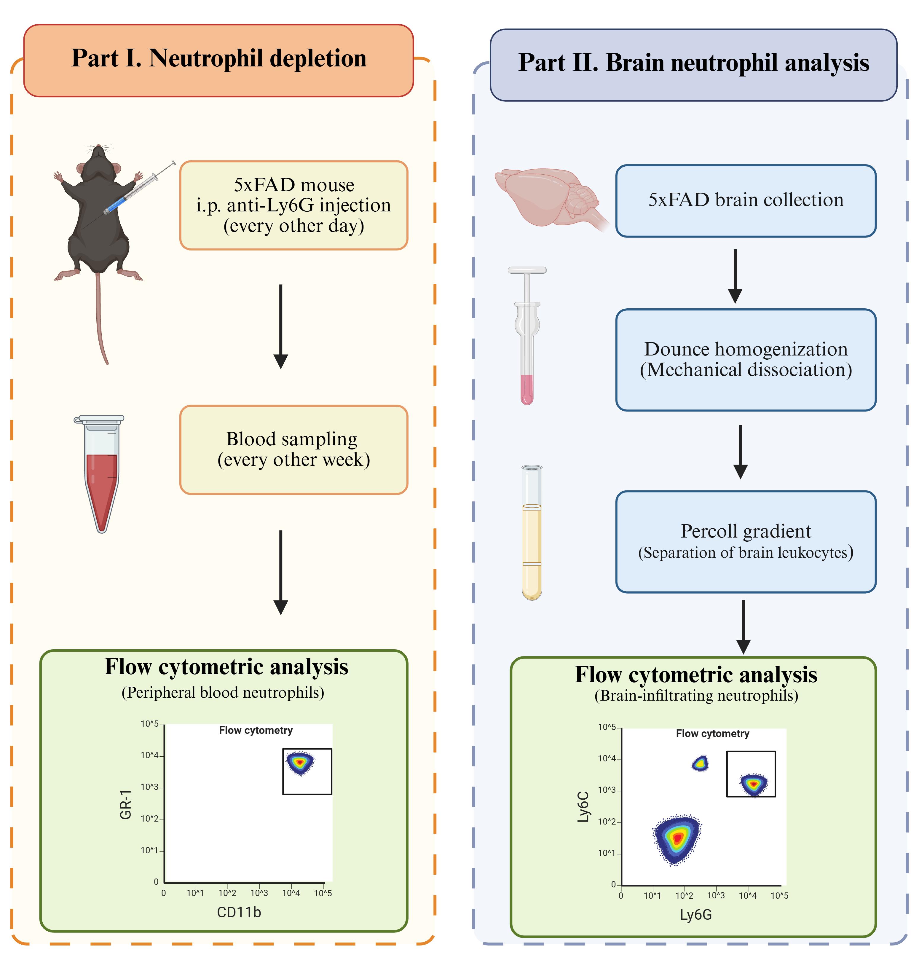
Neutrophils are the most abundant circulating leukocytes in human blood and have been implicated in the progression of Alzheimer’s disease (AD). Elevated neutrophil numbers have been observed in the blood and brain parenchyma of AD patients and mouse models, yet their precise pathological role remains poorly understood. A major barrier to progress is the lack of validated protocols for long-term neutrophil depletion and reliable recovery of brain-infiltrating neutrophils for characterization. Here, we describe a step-by-step protocol for sustained neutrophil depletion in the 5xFAD mouse model using intraperitoneal injection of an anti-Ly6G antibody for six weeks. We used a dose-escalation strategy to maintain depletion efficiency, as neutrophil regeneration increases over time. We also applied a previously established flow cytometry method to monitor neutrophil depletion in peripheral blood, using an antibody panel designed to avoid interference from the depleting anti-Ly6G clone. Furthermore, we present an optimized brain tissue processing workflow that combines mechanical dissociation with Percoll density gradient centrifugation to collect brain-infiltrating neutrophils for downstream flow cytometric analysis. Here, this workflow was applied and validated in the 5xFAD mouse model but is not specific to AD brain tissue and may be applicable to other mouse models.
Key features
• Depletion of circulating neutrophils in mice.
• Assessment of neutrophil depletion efficiency in mouse blood.
• Processing of mouse brain parenchyma and recovery of brain-infiltrating neutrophils.
• Flow cytometric analysis of neutrophils isolated from mouse brain tissue.
Keywords: 5xFAD Neutrophil depletion
Neutrophil isolation
Flow cytometry
Anti-Ly6G
Graphical overview
Background
Alzheimer’s disease (AD) is one of the most serious neurodegenerative disorders worldwide. The pathology of AD is complex and involves multiple pathologies, including amyloid-β accumulation, tau neurofibrillary tangles, abnormal neuroinflammation, and oxidative stress. Recently, increasing evidence has suggested that innate immune cells contribute to the progression of AD. Elevated neutrophil numbers have been observed in patients with AD and in the brains of AD mouse models, including 5xFAD and 3xTg mice, compared with healthy controls [1,2]. Neutrophils may contribute to disease progression by impairing cerebral blood flow and vascular function [3]. However, the precise role of neutrophils in AD pathologies remains unclear. Experimental approaches that enable sustained manipulation of neutrophils and subsequent characterization of brain-associated neutrophils are therefore valuable for investigating their contribution to AD and other chronic neurodegenerative diseases.
Neutrophil depletion using anti-Ly6G antibodies is widely used to investigate neutrophil function in mouse models. Most published depletion protocols, however, use treatment periods of less than two to three weeks [4,5]. The commonly used rat anti-Ly6G clone 1A8 can induce anti-rat antibodies following repeated administration, potentially accelerating clearance of the injected antibody and reducing depletion efficiency during prolonged treatment [6]. The use of murinized anti-Ly6G antibodies provides an approach for overcoming this limitation. Olofsen et al. demonstrated sustained neutrophil depletion using a murinized anti-Ly6G antibody for 28 days [7]. However, chronic neurodegenerative diseases may require neutrophil manipulation over longer experimental periods, and the performance of prolonged depletion strategies in an AD model has not been established. In addition, prolonged anti-Ly6G treatment creates challenges for monitoring depletion because occupancy of Ly6G by the depleting antibody can interfere with subsequent detection of the same epitope. Alternative identification strategies, including forward scatter (FSC)/side scatter (SSC) characteristics in combination with GR-1 and CD11b expression, can therefore be used to evaluate circulating neutrophils during treatment as described before [7].
In addition to sustained depletion, investigation of neutrophil function in neurodegenerative disease requires reliable recovery and characterization of neutrophils from brain tissue. Current studies investigating neutrophils in AD have primarily relied on microscopy to identify neutrophils within the brain parenchyma [8]. Flow cytometry provides an alternative approach that enables phenotypic characterization of individual immune-cell populations, but established protocols for brain immune-cell isolation generally focus on microglia or broader myeloid populations [9–11]. Mechanical tissue dissociation and Percoll density-gradient separation are established methods for recovering leukocytes from brain tissue. Nevertheless, analysis of brain-infiltrating neutrophils remains technically challenging because neutrophils represent a small fraction of brain leukocytes and are short-lived cells that can be sensitive to tissue processing. Thus, a workflow that applies established brain leukocyte isolation approaches specifically to the recovery and flow cytometric characterization of neutrophils would facilitate their investigation in AD mouse models.
Here, we describe two independent procedures for studying neutrophils in the 5xFAD mouse model of AD. Part I and Part II can be performed independently according to the experimental objective. However, they may also be combined when both long-term neutrophil depletion and subsequent analysis of brain-associated neutrophils are required. Part I describes neutrophil depletion. We adopted the use of murine anti-Ly6G and the alternative flow cytometric identification strategy described for long-term depletion [7], while extending the experimental setting from a 28-day timeline to a 6-week treatment in a chronic neurodegenerative disease model. Part II describes the processing of brain tissue for flow cytometric analysis of brain-infiltrating neutrophils. We apply established mechanical dissociation and Percoll-based leukocyte separation procedures [11] to 5xFAD mouse brain, using gentle mechanical homogenization followed by 40%/80% Percoll density-gradient separation and flow cytometric identification of neutrophils. This workflow consistently enabled the identification of a distinct CD45+CD11b+Ly6G+Ly6Cmed population in both wild-type and 5xFAD mouse brains. Thus, rather than introducing the individual depletion, dissociation, or density-gradient techniques themselves, the present protocol extends and integrates these approaches for prolonged neutrophil manipulation and subsequent brain neutrophil analysis in an AD mouse model.
Materials and reagents
Biological materials
1. 5xFAD mice, B6.Cg-Tg(APPSwFlLon,PSEN1*M146L*L286V)6799Vas/Mmjax (The Jackson Laboratory, MMRRC Strain #034848-JAX), 4 months old, male and female
Reagents
1. Bovine serum albumin (BSA) (Sigma-Aldrich, catalog number: A7906)
2. Isoflurane (Piramal, catalog number: ISO-6)
3. Ophthalmic forceps (Med Vet International, catalog number: 50-307-2670)
4. 10× Phosphate-buffered saline (PBS) (Teknova, catalog number: P0195)
5. 10× Hank’s balanced salt solution (HBSS) (Gibco, catalog number: 14185052)
6. Gibco 1× HBSS, no calcium, no magnesium (Gibco, catalog number: 14170112)
7. InVivoPure pH 7.0 dilution buffer (Bioxcell, catalog number: IP0070)
8. RecombiMAb mouse IgG2a isotype control, unknown specificity (Bioxcell, catalog number: CP160)
9. RecombiMAb anti-mouse Ly6G (Bioxcell, catalog number: CP129)
10. BD Pharm LyseTM lysing buffer (BD Pharmingen, catalog number: 555899)
11. Percoll centrifugation media (Cytiva, catalog number: 17089102)
12. Anti-mouse CD16/CD32 (mouse BD Fc block) (BD Pharmingen, catalog number: 553141)
13. UltraComp eBeadsTM Plus compensation beads (Invitrogen, catalog number: 01-3333-42)
14. SYTOX blue nucleic acid stain (Invitrogen, catalog number: S11348)
15. Invitrogen™ EDTA (0.5 M), pH 8.0, RNase-free (Invitrogen, catalog number: AM9260G)
16. Antibodies (Tables 1 and 2)
Table 1. Antibody panel used for evaluating neutrophil depletion
| Antibody | Clone | Fluorophore | Company | Catalog number | Dilution |
|---|---|---|---|---|---|
| CD45 | QA17A26 | FITC | BioLegend | 157608 | 1:100 |
| CD170 (Siglec-F) | S17007L | PE/Cyanine7 | BioLegend | 155527 | 1:100 |
| CD115 (CSF-1R) | AFS98 | PE | BioLegend | 135505 | 1:100 |
| Ly6G/Ly6C (Gr-1) | RB6-8C5 | APC | BioLegend | 108411 | 1:100 |
| CD11b | M1/70 | APC/Cyanine7 | BioLegend | 101225 | 1:100 |
Table 2. Antibody panel used for brain-infiltrating neutrophils
| Antibody | Clone | Fluorophore | Company | Catalog number | Dilution |
| CD45 | QA17A26 | FITC | BioLegend | 157608 | 1:100 |
| Ly6G | 1A8 | APC | BioLegend | 127613 | 1:100 |
| Ly6C | HK1.4 | PE/Dazzle 594 | BioLegend | 128043 | 1:100 |
| CD11b | M1/70 | APC/Cyanine7 | BioLegend | 101225 | 1:100 |
Solutions
1. 1× PBS (see Recipes)
2. Flow buffer (see Recipes)
3. Isotonic Percoll (see Recipes)
4. 80% Percoll (see Recipes)
5. 40% Percoll (see Recipes)
Recipes
1. 1× PBS
| Reagent | Quantity or volume |
|---|---|
| Milli-Q water | 900 mL |
| 10× PBS | 100 mL |
| Total | 1 L |
Note: The 1× PBS used for mouse perfusion is stored at 4 °C for up to 6 months.
2. Flow buffer
| Reagent | Final concentration | Quantity or volume |
|---|---|---|
| BSA | 0.5% | 2.5 g |
| 1× PBS | n/a | 500 mL |
| Total | 500 mL |
Note: The flow buffer is stored at 4 °C for up to 3 months.
3. Isotonic Percoll
| Reagent | Quantity or volume |
|---|---|
| Percoll | 49.5 mL |
| 10× HBSS | 5.5 mL |
| Total | 55 mL |
Note: Isotonic Percoll should be freshly prepared.
4. 80% Percoll
| Reagent | Quantity or volume |
|---|---|
| Isotonic Percoll | 30 mL |
| 1× HBSS | 7.5 mL |
| Total | 37.5 mL |
Note: 80% Percoll should be freshly prepared.
5. 40% Percoll
| Reagent | Quantity or volume |
|---|---|
| Isotonic Percoll | 24 mL |
| 1× HBSS | 36 mL |
| Total | 60 mL |
Note: 40% Percoll should be freshly prepared.
Laboratory supplies
1. 1.5 mL tubes (Eppendorf, catalog number: 05-402-25)
2. 6-well plates (Fisherbrand, catalog number: FB012927)
3. 70 μm mesh (Falcon, catalog number: 352350)
4. 15 mL centrifuge tube (Fisherbrand, catalog number: 07-200-886)
5. Surflo winged infusion set (Terumo, catalog number: SV-25BLK)
6. 60 mL syringe (Axiom, catalog number: MK-1235525)
Equipment
1. Legato syringe pumps (KD Scientific, model: Legato270)
2. Attune NxT Flow Cytometer (Thermo Fisher, model: Attune NxT)
Software and datasets
1. Prism v9.3 (GraphPad, 11/15/2021)
2. FlowJoTM v10.8 Software (BD Life Sciences, 2023)
Procedure
登录/注册后免费查看全文
文章信息
稿件历史记录
提交日期: Jul 28, 2026
接收日期: Oct 2, 2026
在线发布日期: Oct 21, 2026
出版日期: Nov 5, 2026
版权信息
© 2026 The Author(s); This is an open access article under the CC BY-NC license (https://creativecommons.org/licenses/by-nc/4.0/).
如何引用
Huang, H., Bao, X. and Deng, Q. (2026). Long-Term Neutrophil Depletion and Flow Cytometric Analysis of Brain Neutrophils in the 5xFAD Mouse Model. Bio-protocol 16(21): e5864. DOI: 10.21769/BioProtoc.5864.

 Download Citation in RIS Format
您对这篇实验方案有问题吗?
在此处发布您的问题,我们将邀请本文作者来回答。同时,我们会将您的问题发布到Bio-protocol Exchange,以便寻求社区成员的帮助。
 发布问题


