


Visualizing Cell Type–Specific Glycan Profiles in Tissue Sections With Glycan Painting

发布: 2026年10月20日第16卷第20期 DOI: 10.21769/BioProtoc.5859 浏览次数: 18
评审: Durai SellegounderSukrut Chandrashekhar KamerkarKalpana Banumathy Kalyanasundaram
 PDF
PDF  Q&A
Q&A






Abstract
Glycans play critical roles in numerous biological processes. Therefore, visualizing the spatial distribution of glycans within cells and tissues is essential for understanding tissue architecture and cellular identity. Glycan painting is a novel tissue-staining method that employs three fluorescently labeled lectins with different glycan-binding specificities to visualize cell type–specific glycan profiles as unique color patterns. In this study, the protocol was validated using formalin-fixed paraffin-embedded sections of mouse embryos. Although Glycan painting is a qualitative method, each cell population that is difficult to distinguish with a single lectin can be differentiated as unique color patterns generated from the combined images of three fluorescent lectins. Moreover, because it does not require costly reagents or specialized equipment, Glycan painting provides a cost-effective and broadly accessible approach for full-color tissue imaging and cell-type discrimination.
Key features
• Glycan painting enables full-color visualization of cell type–specific glycan profiles through simultaneous staining with three fluorescently labeled lectins with different glycan-binding abilities.
• Applicable to formalin-fixed paraffin-embedded tissue sections.
• Provides comprehensive visualization of tissue architecture while simultaneously facilitating cell-type discrimination.
• Compatible with standard fluorescence microscopy and uses readily available lectins and reagents, enabling rapid adoption without specialized instrumentation.
Keywords: Glycans Lectins
Fluorescent staining
Tissue section staining
Multicolor visualization
Glycan painting
Lectins
Fluorescent staining
Tissue section staining
Multicolor visualization
Glycan painting
Graphical overview
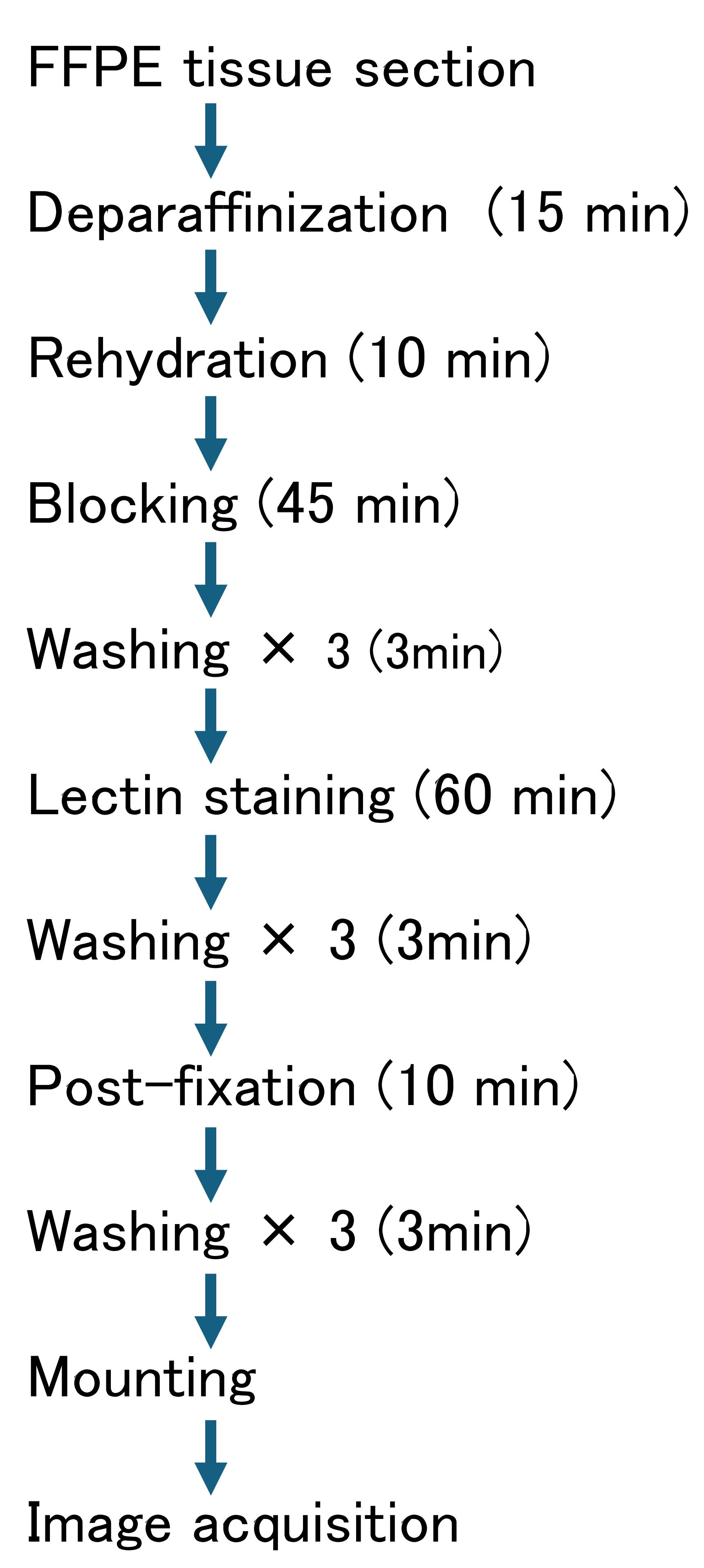
Graphical overview of the Glycan painting protocol. The workflow includes deparaffinization and lectin staining. Approximate incubation times are indicated for each step.
Background
Hematoxylin and eosin (H&E) staining is one of the most fundamental and widely used staining methods and has served as a cornerstone of pathology, histology, and developmental biology for over 100 years [1]. By providing color contrast in otherwise colorless and transparent tissue sections, H&E staining enables visualization of overall tissue architecture and cellular morphology and has long been employed as a fundamental method for histological observation. In H&E staining, the basic dye hematoxylin stains cell nuclei containing RNA and DNA as blue-violet, whereas the acidic dye eosin stains the cytoplasm and extracellular matrix as pink-red. Together, these contrasting dyes reveal a wide range of morphological features, including cell size, nuclear shape, cellular density, and tissue architecture, allowing cell types to be inferred through comprehensive visual assessment.
However, because the identification of cell types in H&E staining relies primarily on morphological features, the ability to distinguish morphologically similar cell populations remains difficult. In addition, H&E staining does not provide direct information regarding cellular function or the state of molecular expression. To address these limitations, immunofluorescent staining using antibodies against cell type–specific proteins has become widely adopted [2]. Immunofluorescent staining enables the visualization of cell type–specific molecular markers, providing information at the molecular expression level that complements conventional morphological observations. While immunofluorescent staining is useful, this staining method also has several limitations, including a restricted multiplex staining capacity, the need to optimize staining conditions, and dependence on predefined molecular markers.
The development of a technology capable of identifying various cell types in biological tissues as distinct color patterns would have broad significance across research fields such as cell and developmental biology, neuroscience, and pathology. In particular, the intuitive visualization of spatial cellular organization and intracellular relationships may provide new perspectives on tissue structure beyond conventional morphology-based analysis. Here, the focus is placed on cell-specific profiles of glycans, which are widely distributed on the cell surface, within cells, and in the extracellular matrix, where they play crucial roles in development and tissue organization [3]. Furthermore, a vast number of glycan-binding molecules, collectively known as lectins, exist throughout the biological world [4]. However, individual glycans do not exhibit strict cell-type specificity, and lectins possess relatively broad and overlapping glycan-binding specificities, limiting their utility as cell identification markers in a conventional single-probe staining approach. In contrast, combinations of glycan profiles exhibit cell-type specificity [5].
We therefore developed a novel staining method termed glycan painting in which differences in glycan profiles are translated into distinct color patterns, enabling cell populations to be distinguished based on their glycan composition [6]. In brief, merged images generated from triplex fluorescence staining of tissue sections using different lectins labeled with distinct fluorescent dyes allow different cell populations to be visualized as characteristic colors. In this report, ConA, WGA, and LEL were selected because they recognize distinct glycan structures and provide distinct staining patterns, enabling diverse RGB color combinations for visual discrimination of different tissue and cell populations. This approach is based on the additive mixing of three primary colors of light (red, green, and blue) and is analogous to the color-generation mechanism of an LCD monitor. In this study, glycan painting was applied to comprehensively visualize mouse embryonic tissues by leveraging the broad distribution of glycans and the overlapping glycan-binding specificity of lectins, features traditionally regarded as limitations, to generate full-color tissue images. Further optimization of lectin combinations may improve the performance of glycan painting, depending on the tissue type and biological application.
Materials and reagents
Biological materials
1. Formalin-fixed, paraffin-embedded mouse embryo (E10.5) tissue sections (Genostaff, catalog number: HMPS-02)
2. Concanavalin A, CFR405S conjugate (Fujifilm, catalog number: 559-54431)
3. Wheat germ agglutinin, CFR488A conjugate (Fujifilm, catalog number: 557-54231)
4. Tomato lectin (LEL), Dylight594 conjugate (Thermo Fisher, catalog number: L32471)
Note: Store all fluorescent lectin conjugates at 4 °C protected from light. Avoid repeated freeze/thaw cycles. In our laboratory, the conjugates retained staining performance for at least six months under these conditions.
Reagents
1. PathoClean (Fujifilm, catalog number: 161-28321)
2. Ethanol (Fujifilm, catalog number: 0596-00486)
3. 20× Tris-buffered saline (TBS), pH 7.4 (Fujifilm, catalog number: 317-90371)
4. D-PBS(+) preparation reagent (Ca, Mg solution) (100×) (Nacalai Tesque, catalog number: 02492-94)
5. 10× carbo-free blocking solution (Vector, catalog number: SP-5040-125)
6. 10% formalin-neutral buffer solution (Fujifilm, catalog number: 062-01661)
7. 1,4-diazabicyclo[2.2.2]octane (DABCO) (Fujifilm, catalog number: 049-25712)
8. Glycerol (Fujifilm, catalog number: 075-00616)
9. Tris(hydroxymethyl)aminomethane (Fujifilm, catalog number: 207-06275)
10. Kaneyonol (Kaneyo soap laundry glue, Kaneyo); if unavailable, 8% polyvinyl alcohol (PVA) (Sigma, catalog number: P8316) or a commercially available mounting medium can be used as an alternative
Solutions
1. Wash buffer (see Recipes)
2. Blocking buffer (see Recipes)
3. Staining buffer (see Recipes)
4. Mounting medium (see Recipes)
Recipes
Note: All solutions, except for the mounting medium, are freshly prepared before each experiment and should not be reused.
1. Wash buffer (100 mL)
| Reagent | Final concentration | Volume |
|---|---|---|
| 20× TBS (pH7.4) | 1× | 5 mL |
| Ca, Mg solution (100×) | 1× | 1 mL |
| Double-distilled water | 94 mL |
2. Blocking buffer (10 mL)
| Reagent | Final concentration | Volume |
|---|---|---|
| 20× TBS (pH 7.4) | 1× | 0.5 mL |
| Ca, Mg solution (100×) | 1× | 0.1 mL |
| 10× carbo-free blocking solution | 1× | 1 mL |
| Double-distilled water | 8.4 mL |
3. Staining buffer (1 mL)
| Reagent | Final concentration | Volume |
|---|---|---|
| 20× TBS (pH 7.4) | 1× | 0.05 mL |
| Ca, Mg solution (100×) | 1× | 0.01 mL |
| Concanavalin A, CFR405S conjugate | 2 μg/mL | 2 μL |
| Wheat germ agglutinin, CFR488A conjugate | 2 μg/mL | 2 μL |
| Tomato lectin (LEL), DyLight594 conjugate | 2 μg/mL | 2 μL |
| Double-distilled water | 1 mL |
4. Mounting medium (40 mL)
| Reagent | Final concentration | Quantity or volume |
|---|---|---|
| 80% glycerol | 16% | 8 mL |
| Kaneyonol or 8% PVA | 75% | 30 mL |
| 2 M Tris-HCl (pH 8.0) | 60 mM | 1.2 mL |
| DABCO | 1.5% | 0.625 g |
| Double-distilled water | Up to 40 mL |
Note: Kaneyonol or 8% PVA is used as a viscosity-enhancing agent in the mounting medium to maintain an appropriate layer of mounting medium beneath the coverslip. Upon drying, the mounting medium solidifies and helps stabilize the coverslip. The mounting medium is stored in the dark and reused for subsequent experiments.
Laboratory supplies
1. 1.5 mL microtube (FastGene, catalog number: FG-MCT15A)
2. 50 mL disposable centrifuge tube (Iwaki, catalog number: 2342-050N)
3. 100 mL disposable centrifuge tube (Iwaki, catalog number: 2355-100N)
4. Coverslip (Matsunami, catalog number: C022221)
5. A-PAP pen (Matsunami, catalog number: GMP0020)
Equipment
1. Confocal microscopy (AX, Nikon, Tokyo, Japan, model: Nikon AX with NSPARC Super-Resolution Confocal Laser Microscope System)
2. Aspirator (IWAKI, AGC Techno Glass, Shizuoka, Japan, catalog number: VPUMP-140)
Software and datasets
1. Fiji/ImageJ (version 2.16.0, National Institutes of Health (NIH), USA; used for fluorescence image processing, channel merging, RGB analysis, and image quantification
2. NIS-Elements AR (version 6.20, Nikon, Tokyo, Japan); used for image acquisition and processing of confocal microscopy data
Procedure
登录/注册后免费查看全文
文章信息
稿件历史记录
提交日期: Jun 12, 2026
接收日期: Aug 24, 2026
在线发布日期: Oct 15, 2026
出版日期: Oct 20, 2026
版权信息
© 2026 The Author(s); This is an open access article under the CC BY license (https://creativecommons.org/licenses/by/4.0/).
如何引用
Nagasaki, A. and Shinkai, Y. (2026). Visualizing Cell Type–Specific Glycan Profiles in Tissue Sections With Glycan Painting. Bio-protocol 16(20): e5859. DOI: 10.21769/BioProtoc.5859.

 Download Citation in RIS Format
分类
细胞生物学 > 细胞成像
您对这篇实验方案有问题吗?
在此处发布您的问题,我们将邀请本文作者来回答。同时,我们会将您的问题发布到Bio-protocol Exchange,以便寻求社区成员的帮助。
 发布问题
发布问题


