


Suspension-Based Intracellular Immunofluorescence Staining Coupled With Genome-Wide CRISPR-Cas9 Screening to Identify Regulators of O-GlcNAcylation

发布: 2026年11月05日第16卷第21期 DOI: 10.21769/BioProtoc.5858 浏览次数: 18
评审: SAPTARSHI MAJIAnonymous reviewer(s)
 PDF
PDF  Q&A
Q&A






Abstract
Protein O-GlcNAcylation is a dynamic and reversible post-translational modification that regulates diverse cellular processes, including transcription, signal transduction, metabolism, and cell fate determination. Systematic identification of genes that modulate global O-GlcNAc levels remains technically challenging at the genome scale. Here, we describe a pooled CRISPR-Cas9 screening protocol that combines the human genome-scale CRISPR knockout (GeCKO) v2 knockout library with intracellular immunofluorescence staining using the anti-O-GlcNAc antibody RL2 and fluorescence-activated cell sorting (FACS). In this workflow, HEK293T cells are transduced with the GeCKO v2 lentiviral library at a low multiplicity of infection to ensure predominantly single-sgRNA integration. Following puromycin selection, cells are fixed, permeabilized, and stained in suspension with RL2. The top 5% of cells with the highest intracellular RL2 fluorescence are collected as the RL2-high population, while a corresponding unsorted/input sample is retained as the reference for downstream sgRNA enrichment analysis. Genomic DNA is recovered from the RL2-high and unsorted/input samples, and integrated sgRNA cassettes are amplified through a two-step PCR and library-preparation workflow for Illumina sequencing. This protocol enables the identification of candidate genes whose knockout is associated with increased intracellular RL2 fluorescence.
Key features
• A genome-scale pooled CRISPR-Cas9 screening workflow using the human GeCKO v2 library in HEK293T cells.
• A suspension-based intracellular RL2 immunofluorescence staining protocol optimized to minimize cell loss during fixation, permeabilization, and washing, thereby preserving sgRNA library representation for downstream sorting.
• Isolation of the top 5% RL2-high population, with a corresponding unsorted/input sample retained as the reference for downstream sgRNA enrichment analysis.
• Fixed-cell genomic DNA recovery and sgRNA sequencing coupled with model-based analysis of genome-wide CRISPR/Cas9 knockout (MAGeCK) enable robust hit identification.
Keywords: Protein O-GlcNAcylation GeCKO
Pooled CRISPR screen
Immunofluorescence
Fluorescence-activated cell sorting (FACS)
MAGeCK
GeCKO
Pooled CRISPR screen
Immunofluorescence
Fluorescence-activated cell sorting (FACS)
MAGeCK
Graphical overview
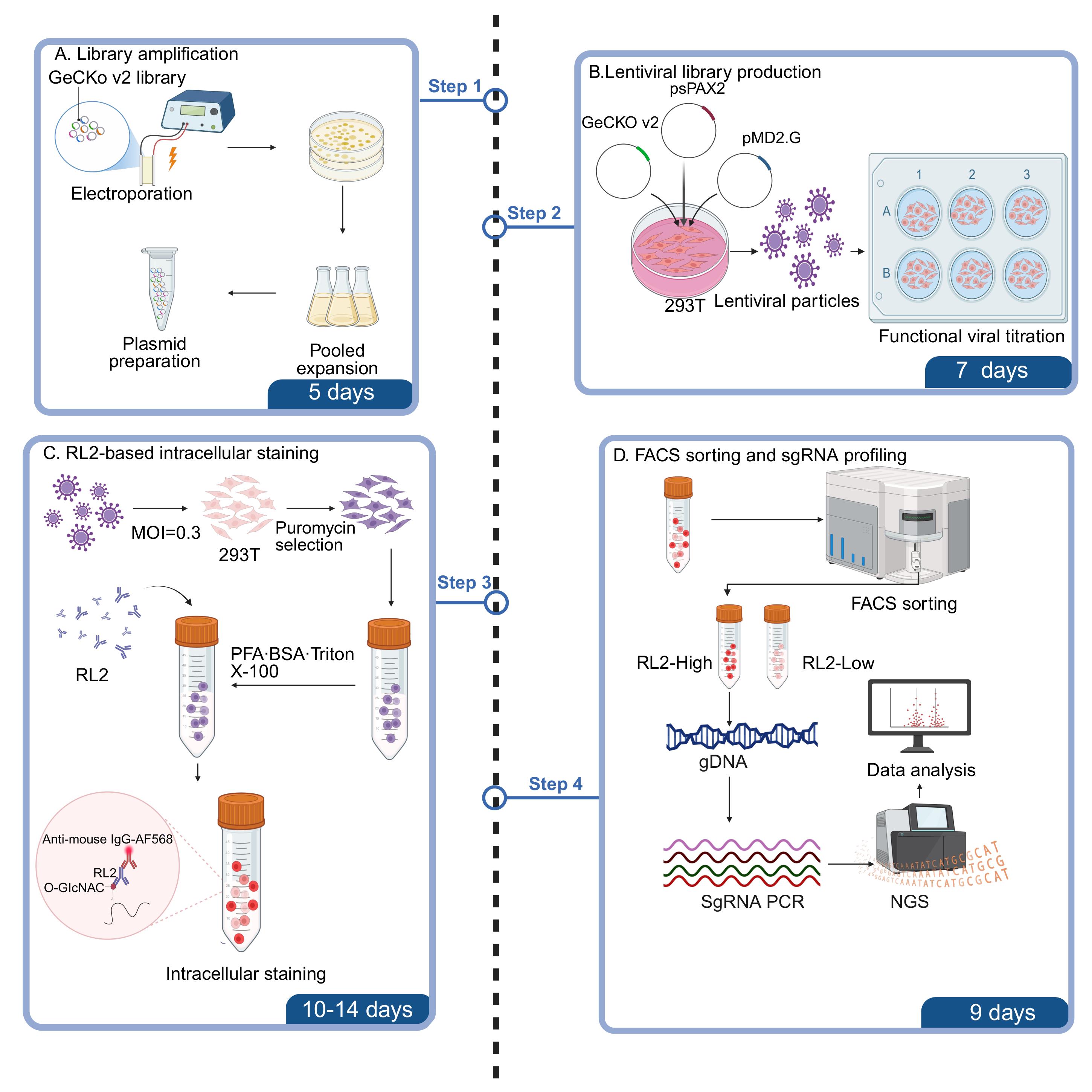
Workflow of the GeCKO v2–RL2 pooled CRISPR screen. The human GeCKO v2 library is amplified, packaged into lentivirus, and transduced into HEK293T cells at a low multiplicity of infection (MOI). Following puromycin selection, cells are stained with RL2. The top 5% RL2-high population and a corresponding unsorted/input reference are sequenced and compared using MAGeCK. The RL2-low population shown in the sorting schematic is a fluorescence and sorting control only and is not sequenced.
Background
Protein O-GlcNAcylation is a dynamic and reversible intracellular post-translational modification in which a single O-linked β-N-acetylglucosamine moiety is added to serine or threonine residues of nuclear and cytoplasmic proteins by O-GlcNAc transferase (OGT) and removed by O-GlcNAcase (OGA) [1]. Because UDP-GlcNAc, the donor substrate for OGT, is generated through the hexosamine biosynthetic pathway, cellular O-GlcNAcylation is closely coupled to nutrient availability, metabolic state, and stress responses. O-GlcNAcylation regulates multiple biological processes, including transcription, chromatin regulation, signal transduction, metabolism, proteostasis, and cell fate decisions. Dysregulation of O-GlcNAc homeostasis has been associated with pathological states, including cancer and metabolic disorders [1,2].
Existing approaches for investigating O-GlcNAc regulation mainly rely on targeted perturbation and detection strategies, including pharmacological perturbation of O-GlcNAc cycling, antibody-based immunodetection, chemoenzymatic labeling, and mass spectrometry–based profiling [2−4]. These approaches are useful for measuring global or protein-specific O-GlcNAcylation and for validating candidate regulators. However, they are not designed to identify, in an unbiased genome-scale manner, the broader genetic network that controls intracellular O-GlcNAc homeostasis. Therefore, a scalable functional-genomics strategy is needed to systematically discover genes whose loss increases or decreases global O-GlcNAcylation.
Pooled CRISPR-Cas9 screening provides a powerful strategy for connecting genetic perturbations with cellular phenotypes. The human GeCKO library established lentiviral genome-scale knockout screening in human cells, and the GeCKO v2 system further improved the vectors and genome-wide libraries for CRISPR screening [5,6]. Subsequent genome-scale CRISPR screening protocols have defined essential experimental principles, including low multiplicity of infection (MOI), antibiotic selection, maintenance of sufficient sgRNA library coverage, and sequencing-based guide recovery [7]. In parallel, phenotype-based and high-content CRISPR screening strategies, together with diverse applications in cancer and immune-cell systems, have expanded pooled screens beyond simple viability readouts to more complex cellular and molecular phenotypes [8−10]. For sortable phenotypes, fluorescence-activated cell sorting (FACS)-based pooled CRISPR screening enables the enrichment of cell populations with high or low levels of a defined fluorescence signal, including antibody-based intracellular molecular readouts [11,12].
Here, we describe a GeCKO v2–RL2 pooled CRISPR screening protocol that converts global O-GlcNAcylation into a sortable fluorescence phenotype. HEK293T cells are transduced with the GeCKO v2 lentiviral library at low MOI, selected with puromycin, expanded while maintaining sgRNA representation, fixed and permeabilized in suspension, and stained with the anti-O-GlcNAc antibody RL2 followed by an Alexa Fluor 568–conjugated secondary antibody and DAPI. The top 5% of cells with the highest intracellular RL2 fluorescence are collected as the RL2-high population, while a corresponding unsorted/input sample is retained as the reference for downstream sgRNA enrichment analysis. Integrated sgRNA cassettes are recovered from genomic DNA from the RL2-high and unsorted/input samples for next-generation sequencing and MAGeCK-based enrichment analysis [2,13].
By integrating genome-scale perturbation, suspension intracellular immunofluorescence staining, quantitative FACS enrichment, and sgRNA-level computational analysis, this protocol enables unbiased identification of candidate negative regulators whose knockout is associated with increased intracellular RL2 fluorescence. More broadly, the same framework can be adapted to other antibody-defined intracellular phenotypes, including phosphorylation, acetylation, ubiquitination, and chromatin-associated epitopes, providing a general strategy for dissecting intracellular signaling and regulatory networks.
Materials and reagents
Biological materials
1. HEK293T cells (ATCC, catalog number: CRL-3216); cells should be authenticated and routinely tested for mycoplasma contamination
2. Endura electrocompetent Escherichia coli cells (Lucigen, catalog number: 60242-2)
Plasmids and pooled libraries
1. Human GeCKO v2 CRISPR knockout pooled library in the lentiCRISPR v2 backbone (Addgene pooled library #1000000048); the complete library comprises library A and library B and contains 123,411 sgRNAs targeting 19,050 protein-coding genes, with six sgRNAs per gene and 1,000 non-targeting control sgRNAs
2. pMD2.G lentiviral envelope plasmid (Addgene, plasmid #12259)
3. psPAX2 lentiviral packaging plasmid (Addgene, plasmid #12260)
Reagents
1. Dulbecco’s modified Eagle medium (DMEM) (Vivocell, catalog number: 06-1055-57-1 ACS)
2. Fetal bovine serum (FBS) (Meisen, catalog number: CTCC-002-071)
3. 0.25% Trypsin-EDTA (Thermo Fisher Scientific, catalog number: 25200072)
4. Dulbecco’s phosphate-buffered saline (DPBS) without Ca2+ and Mg2+ (Thermo Fisher Scientific, catalog number: 14190144); critical for minimizing cell aggregation during intracellular staining
5. Accutase cell detachment solution (Innovative Cell Technologies, catalog number: AT-104)
6. Puromycin (Selleck, catalog number: S7417)
7. Hexadimethrine bromide (Polybrene) (Sigma, catalog number: H9268)
8. Opti-MEM I reduced-serum medium (Thermo Fisher Scientific, catalog number: 31985070)
9. Formaldehyde solution, 37% (Sigma, catalog number: 252549)
10. Bovine serum albumin (BSA) (BioFroxx, catalog number: 4240GR005); use ultrapure BSA for reduced flow cytometry background
11. Triton X-100 (Solarbio, catalog number: T8200)
12. Phosphate-buffered saline (PBS) (20× stock) (Servicebio, catalog number: G4202)
13. Anti-O-GlcNAc antibody (RL2), mouse monoclonal (Thermo Fisher Scientific, catalog number: MA1-072)
14. Goat anti-mouse immunoglobulin G (IgG) (H+L), Alexa Fluor 568–conjugated (Thermo Fisher Scientific, catalog number: A11004)
15. DAPI (Sigma, catalog number: D9542)
16. Proteinase K (Qiagen, catalog number: 19131)
17. RNase A (Qiagen, catalog number: 19101)
18. Phenol:chloroform:isoamyl alcohol (25:24:1) (Sigma, catalog number: P3803)
19. Chloroform (Sigma, catalog number: C2432)
20. Glycogen (Thermo Fisher Scientific, catalog number: 10814010)
21. KOD DNA polymerase (Merck, catalog number: 71086)
22. dNTP mix (Thermo Fisher Scientific, catalog number: R0192)
23. Polyethylene glycol 8000 (PEG8000) (Solarbio, catalog number: P8260)
24. LB broth (Sigma, catalog number: L3022)
25. Ampicillin sodium salt (Sigma, catalog number: A5354)
26. SOC medium (New England Biolabs, catalog number: B9020S)
27. QIAquick PCR Purification kit (QIAGEN, catalog number: 28104)
28. QubitTM dsDNA HS Assay kit, 500 assays (Invitrogen, Thermo Fisher Scientific, catalog number: Q32854)
29. NEBNext® UltraTM II DNA Library Prep Kit for Illumina® (New England Biolabs, catalog number: E7645S)
30. NEBNext® Multiplex Oligos for Illumina® (Index Primers Set 2) (New England Biolabs, catalog number: E7500S)
Critical: This product has since been discontinued. According to the manufacturer’s recommendation, the NEBNext® 96 Unique Dual Index Primer Pairs (NEB #E6440S, #E6442S, #E6444S, #E6446S, or #E6448S) can be used as a replacement.
31. Agencourt AMPure XP beads (Beckman Coulter, catalog number: A63881)
32. Penicillin-streptomycin solution (Vivacell, catalog number: C3420-0100)
33. Sodium chloride (NaCl) (Sinopharm, catalog number: 10019318-500g)
Solutions
1. Complete growth medium for HEK293T cells (see Recipes)
2. 4% PFA fixative (see Recipes)
3. Blocking/permeabilization buffer (see Recipes)
4. Primary antibody dilution buffer (see Recipes)
5. FACS sorting buffer (see Recipes)
6. Lentivirus storage buffer (see Recipes)
7. 5× PEG8000 virus concentration solution (see Recipes)
Recipes
1. Complete growth medium for HEK293T cells
Add 50 mL of FBS and 5 mL of 100× penicillin-streptomycin to 445 mL of high-glucose DMEM. Mix thoroughly and store at 4 °C. Use within 1 month.
2. 4% PFA fixative
Add 10 mL of 10× PBS to 10.8 mL of 37% formaldehyde. Bring the volume to 100 mL with ddH2O. Mix thoroughly. Prepare fresh on the day of use; the working solution is stable for up to 1 week at 4 °C.
3. Blocking/permeabilization buffer
Dissolve 2 g of BSA in 5 mL of 20× PBS. Add 100 μL of Triton X-100 and bring the volume to 100 mL with ddH2O. Mix thoroughly and filter through a 0.22 μm sterile filter. Store at 4 °C for up to 1 week.
4. Primary antibody dilution buffer
Dissolve 0.1 g of BSA in 5 mL of 20× PBS. Add 50 μL of Triton X-100 and bring the volume to 100 mL with ddH2O. Mix thoroughly and filter through a 0.22 μm sterile filter. Prepare fresh each time before use.
5. FACS sorting buffer
Dissolve 1 g of BSA and 200 μL of 0.5 M EDTA (pH 8.0) in DPBS (Ca2+/Mg2+-free) and bring the volume to 100 mL with DPBS. Mix thoroughly and filter through a 0.22 μm sterile filter. Keep the solution ice-cold until use.
6. Lentivirus storage buffer
Dissolve 10 g of BSA and add 10 mL of FBS to high-glucose DMEM to a final volume of 100 mL. Mix thoroughly and filter through a 0.22 μm sterile filter. Store at -80 °C.
7. 5× PEG8000 virus concentration solution
Dissolve 40 g of PEG8000 and 8.77 g of NaCl in ddH2O to a final volume of 100 mL. Autoclave to sterilize. Warm gently to dissolve before use and store at 4 °C.
Equipment
1. Flow cytometer (BD Biosciences, model: FACSAria III)
2. CO2 cell culture incubator (Thermo Fisher Scientific, model: 3111)
3. Confocal laser scanning microscope (Zeiss, model: LSM880)
4. Fluorescence stereomicroscope (Leica, model: M205 FCA)
5. Class II A2 biosafety cabinet (Thermo Fisher Scientific)
6. High-speed refrigerated centrifuge (Thermo Fisher Scientific, model: 75004530)
7. Microcentrifuge (Eppendorf, model: 5424R)
8. Electroporator (Bio-Rad, model: Gene Pulser Xcell)
9. NanoDrop spectrophotometer (Thermo Fisher Scientific, model: 2000C)
10. Electroporation cuvettes, 0.1 cm (Bio-Rad, model: 1652089)
Software and datasets
1. MAGeCK, version 0.5.9
2. R, version 4.0.3
3. FlowJo or equivalent flow cytometry analysis software, version 10.7.1
4. GraphPad Prism, version 8.0
5. BioRender
Procedure
登录/注册后免费查看全文
文章信息
稿件历史记录
提交日期: Aug 7, 2026
接收日期: Sep 16, 2026
在线发布日期: Oct 8, 2026
出版日期: Nov 5, 2026
版权信息
© 2026 The Author(s); This is an open access article under the CC BY-NC license (https://creativecommons.org/licenses/by-nc/4.0/).
如何引用
Liu, P., Lei, Q., Zhang, N., Yu, H., Chen, F. and Yuan, K. (2026). Suspension-Based Intracellular Immunofluorescence Staining Coupled With Genome-Wide CRISPR-Cas9 Screening to Identify Regulators of O-GlcNAcylation. Bio-protocol 16(21): e5858. DOI: 10.21769/BioProtoc.5858.

 Download Citation in RIS Format
您对这篇实验方案有问题吗?
在此处发布您的问题,我们将邀请本文作者来回答。同时,我们会将您的问题发布到Bio-protocol Exchange,以便寻求社区成员的帮助。
 发布问题
发布问题


