


Preparation of Phosphoinositide-Containing Supported Lipid Bilayers and Quantitative Assessment of Protein Recruitment to Membrane Surfaces

发布: 2026年10月20日第16卷第20期 DOI: 10.21769/BioProtoc.5853 浏览次数: 33
评审: Devika AndhareSrajan KapoorGundeep Kaur
 PDF
PDF  Q&A
Q&A





Abstract
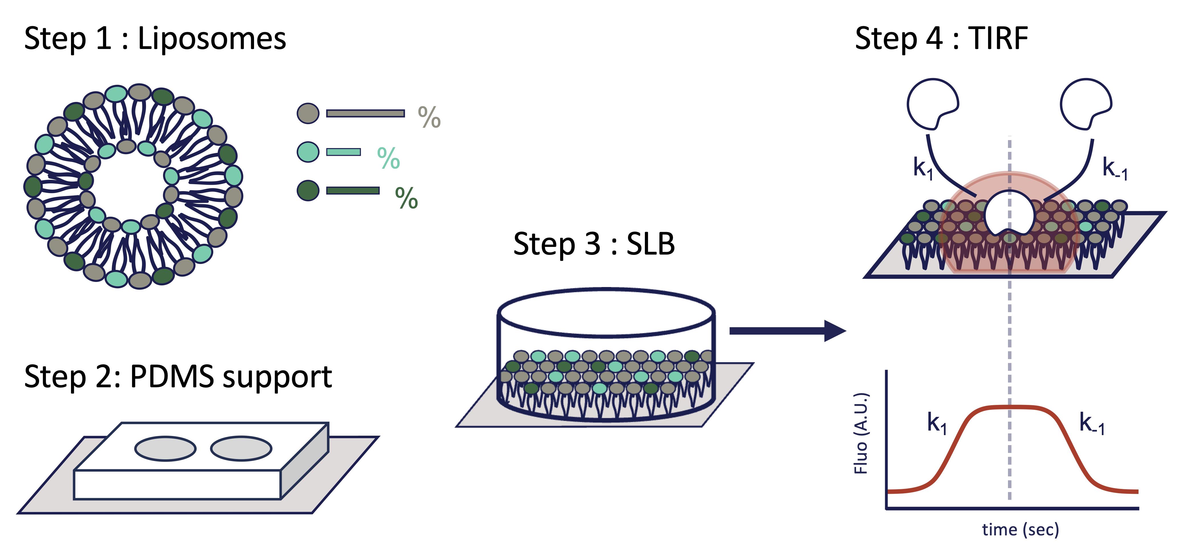
Small GTPases and many other peripheral membrane proteins regulate essential cellular processes through dynamic interactions with cellular membranes. These interactions are often controlled by membrane composition, particularly phosphoinositides (PIPs), which modulate the recruitment of GTPases, their regulators, and their effectors. Quantitative characterization of protein recruitment to defined membrane environments remains technically challenging. Here, we describe a protocol for preparing supported lipid bilayers (SLBs) containing defined phosphoinositide compositions in polydimethylsiloxane (PDMS) chambers and monitoring protein recruitment by total internal reflection fluorescence (TIRF) microscopy. Purified fluorescently labeled proteins are incubated with SLBs to measure membrane association in real time under controlled biochemical conditions. The method enables quantitative analysis of membrane recruitment kinetics and comparison of protein binding across different lipid compositions or in the presence of partner proteins. Combining the biochemical precision of a reconstituted membrane system with the sensitivity of TIRF imaging, this protocol provides a robust and versatile platform for studying lipid-dependent membrane recruitment. Although developed to investigate small GTPase signaling, it is readily applicable to a wide range of peripheral membrane proteins and membrane-associated signaling mechanisms.
Key features
• Reconstitutes small GTPase membrane recruitment on PIP-containing supported lipid bilayers compatible with TIRF microscopy.
• Enables quantitative comparison of lipid-dependent binding across defined membrane compositions, including phosphoinositide species, phosphatidylserine content, and additional membrane components.
• Suitable for purified small GTPases, GEFs, GAPs, and effectors, allowing direct testing of multicomponent membrane recruitment under controlled biochemical conditions.
• Can be adapted for steady-state measurements, recruitment kinetics, and protein competition assays to dissect any membrane-dependent signaling mechanism.
Keywords: Supported lipid bilayers Phosphoinositides
Small GTPases
TIRF microscopy
Membrane binding
Phosphoinositides
Small GTPases
TIRF microscopy
Membrane binding
Graphical overview
Background
Membrane interfaces provide dynamic platforms for numerous peripheral proteins that reversibly associate with lipid bilayers. The thermodynamic (binding avidity) and kinetic parameters governing these interactions—including the apparent recruitment (kobs) and dissociation (koff) rate constants—determine their specificity, efficiency, and temporal regulation. This is particularly important for small GTPases of the Ras superfamily.
Small GTPases function as molecular switches that regulate cell polarity, migration, membrane trafficking, cytoskeletal remodeling, and immune signaling [1]. Small GTPases constitute a paradigm for peripheral membrane proteins that dynamically associate with the cytosolic leaflet of the plasma membrane [2]. Their activation is tightly regulated by guanine nucleotide exchange factors (GEFs), which catalyze GDP-to-GTP exchange [3]; their inactivation and membrane dissociation are promoted by GTPase-activating proteins (GAPs), which stimulate GTP hydrolysis. Increasing evidence indicates that these regulatory reactions are not solely determined by protein–protein interactions but are strongly influenced by the membrane environment itself, including curvature, phosphoinositide composition, membrane charge, and lipid packing (reviewed in [4]). In addition, nanoscale membrane heterogeneities generated by phase separation create microenvironments with distinct biochemical and biophysical properties [5–7].
Reconstituting these processes in defined biomimetic systems has emerged as an important strategy to dissect the molecular principles governing spatiotemporal signal transduction, i.e., both their kinetic and thermodynamic features (reviewed in [8–10]). Several methodologies have been developed to investigate protein–membrane interactions in small GTPase signaling. Classical liposome co-sedimentation or flotation assays provide robust biochemical measurements of membrane association but lack spatial and temporal resolution [11]. Surface plasmon resonance and related biophysical approaches enable quantitative affinity measurements but do not reproduce the dynamic organization of signaling complexes on membrane surfaces. In contrast, live-cell imaging captures signaling in physiological contexts but often complicates mechanistic interpretation because of the complexity and heterogeneity of cellular environments.
Supported lipid bilayers (SLBs) combined with total internal reflection fluorescence (TIRF) microscopy provide a powerful intermediate approach by enabling direct visualization of membrane recruitment, diffusion, clustering, and signaling reactions in a controlled yet biomimetic environment. Previous work demonstrated that membrane confinement and local organization strongly influence small GTPase signaling outputs such as effector engagement [6,12–14]. Recent studies further highlighted how membrane composition and phosphoinositide identity modulate GEF activity and downstream signaling transmission [15].
The protocol presented here describes the preparation of supported lipid bilayers and their use in quantitative TIRF microscopy assays to monitor protein recruitment to membrane surfaces [13,16–18]. Compared with bulk biochemical approaches, this method provides direct access to membrane binding kinetics, residence times, lateral diffusion, nanoscale organization, and dynamic protein exchange under defined experimental conditions. The protocol is readily adaptable to systematic variations in lipid composition, including phosphoinositide content, cholesterol levels, membrane charge, and protein density. It also allows simultaneous visualization in real time of multiple fluorescently labeled proteins. Throughout this protocol, membrane recruitment refers to the accumulation of fluorescent proteins at the supported lipid bilayer resulting from direct lipid binding, interactions with membrane-associated partners, or both.
A major advantage of this approach is its versatility and quantitative nature. Beyond small GTPase signaling, the protocol can be adapted to study membrane-associated enzymatic reactions, scaffold assembly, cytoskeletal coupling, receptor signaling, protein phase separation, and dynamic signaling networks. The use of purified components additionally facilitates mechanistic interpretation and quantitative modeling of signaling reactions.
Several limitations should nevertheless be considered. Supported lipid bilayers do not fully reproduce the complexity, asymmetry, curvature, and active remodeling of cellular membranes. Certain curvature-sensitive or transmembrane systems may therefore require complementary approaches such as giant unilamellar vesicles or proteoliposomes. Despite these limitations, SLB-based TIRF assays provide a robust and accessible platform for dissecting membrane-associated signaling mechanisms with high spatiotemporal resolution.
Materials and reagents
Reagents
Liposomes
1. Chloroform (Sigma-Aldrich, catalog number: 366927-1L)
2. Argon gas
3. HEPES [4-(2-hydroxyethyl)-1-piperazineethanesulfonic acid], 1 M, pH 7.8 (Sigma-Aldrich, catalog number: H4034)
4. NaCl, 5 M (Euromedex, catalog number: 1112-A)
5. Phosphatidylcholine (Avanti, catalog number: 840054P-200mg, soy)
6. Phosphatidylethanolamine (Avanti, catalog number: 840022P-25mg, brain, porcine)
7. Phosphatidylserine (Avanti, catalog number: 840032P-25mg, brain, porcine)
8. Cholesterol (Avanti, catalog number: 700000-100mg, ovine)
9. NBD-PE [N-(7-nitrobenz-2-oxa-1,3-diazol-4-yl) phosphoethanolamine] (Avanti, catalog number: 840145P-1mg, 18:1)
10. Phosphatidylinositol-3,4-bisphosphate (Avanti, catalog number: 850153P-100μg, 18:1)
11. DGS-NiNTA (1,2-dioleoyl-sn-glycero-3-[(N-(5-amino-1-carboxypentyl)iminodiacetic acid)succinyl] (nickel salt) (Avanti, catalog number: A89404, 5mg, 18:1)
PDMS support
1. Sylgard 184 silicone elastomer (Sigma-Aldrich, catalog number: 761036-5EA)
2. Ethanol (≥99.8%) (VWR Chemicals, PROLABO, catalog number: 20821296-1L)
3. Isopropanol (≥99.8%) (Fluka, catalog number: 34965-1L)
4. Deionized water and MilliQ water
5. Absolute acetone (≥99.8%) (Merck, catalog number: 100014)
6. HMDS (hexamethyldisilazane) (Sigma-Aldrich, catalog number: 440191-100ML)
SLB
1. CaCl2, 1 M (Merck, catalog number: 10035-04-8)
TIRF imaging
1. Immersion oil (e.g., ZEISS, catalog number: 444960 518F)
Solutions
1. Lipid stock solutions in chloroform (see Recipes)
2. HN buffer (see Recipes)
3. SLB-FB buffer (see Recipes)
4. HEPES 20 mM (pH 7.5) (see Recipes)
Recipes
1. Lipid stock solutions in chloroform
| Lipids | Mass (mg) | Volume (mL) | Concentration (mM) |
|---|---|---|---|
| Phosphatidylcholine (PC) | 100 | 4.0 | 32.2 |
| Phosphatidylethanolamine (PE) | 25 | 2.5 | 13.0 |
| Phosphatidylserine (PS) | 25 | 2.5 | 12.3 |
| Cholesterol (cholesterol) | 100 | 13.3 | 19.4 |
| N-(7-nitrobenz-2-oxa-1,3-diazol-4-yl) phosphoethanolamine (NBD-PE) | 1 | 1.0 | 1.0 |
| Phosphatidylinositol-3,4-bisphosphate (PI(3,4)P2) | 0.1 | 0.1 | 0.9 |
| 1,2-dipalmitoyl-sn-glycero-3-[(N-(5-amino-1-carboxypentyl)iminodiacetic acid)succinyl] (DGS-NiNTA) | 10 | 2.0 | 4.8 |
We provide an example of the lipid composition used in the experiments presented in this protocol.
See the table below to prepare 0.5 mL of lipid stock solutions at a final lipid concentration of 2.0 mM (thereafter, “2 mM liposome”).
Lipid stock solutions to prepare 0.5 mL at 2 mM liposome
| Reagent | Target composition (%) | Stock volume required (μL) |
|---|---|---|
| PC | 31 | 10 |
| PE | 20 | 15 |
| PS | 20 | 16 |
| Cholesterol | 20 | 10 |
| NBD-PE | 1 | 10 |
| PI(3,4)P2 | 4 | 44 |
| DGS-NiNTA | 4 | 8 |
2. HN buffer
HEPES 20 mM (pH 7.5), NaCl 100 mM
3. SLB-FB buffer
HEPES 20 mM (pH 7.5), NaCl 120 mM, DTT 1 mM
4. HEPES 20 mM (pH 7.5)
NaCl 120 mM, DTT 1 mM, MgCl2 1 mM
Laboratory supplies
Liposomes
1. 1.5 mL opaque tubes (e.g., Eppendorf, catalog number: 0030120191) and Amber tubes (Eppendorf, catalog number: 10458312)
2. Glass vials (e.g., WHEATON®, catalog number: DWKW224681) (2 mL)
3. Powder-free nitrile gloves (e.g., SHIELD Scientific, catalog number: 625124)
4. Quickfit® round-bottom flasks (e.g., Sigma-Aldrich, catalog number: Z302732-1EA)
PDMS support
1. Silicon master molds (photolithography generated) (homemade)
2. Aluminum foil (e.g., Sigma-Aldrich, catalog number: Z691577)
3. Adhesive tape (e.g., Scotch 3M)
4. Glass coverslips, e.g., 24 × 60 mm with 0.13–0.17 mm thickness (VWR, catalog number: 48404-133)
5. 6-mm biopsy punch (e.g., Dutscher, catalog number: 030740)
SLB
1. Glass coverslip holder
Equipment
Liposomes
1. Rotavapor (e.g., Labortechnik AG, model: eR-II BÜCHI)
2. Vacuum pump (e.g., Büchi Labortechnik AG, model: V-700 with vacuum controller V-850)
3. Liquid cooling system (e.g., Fisher Bioblock Scientific)
4. Vortex mixer (e.g., Sigma-Aldrich, catalog number: Z258423-1EA)
5. Hamilton® syringes [e.g., Sigma-Aldrich, catalog numbers: HAM80075-1EA (10 μL); HAM7656-01-1EA (100 μL); 20739 (500 μL)]
PDMS support
1. Ultrasonic bath (e.g., Bandelin SONOREX, Merck, catalog number: Z659460)
2. Vacuum desiccator or vacuum chamber
3. Oven (75 °C)
4. Plasma cleaner (e.g., Harrick, catalog number: PDC-32G-2, 230V)
5. Closed chamber/container for HMDS vapor silanization
6. Chemical hood
TIRF imaging
1. Cyanine5.5 succinimidyl ester (CY5.5-SE) (e.g., Cytiva, catalog number: 10461835)
2. TIRF microscope, e.g., Nikon Ti-E inverted equipped with a 100× TIRF Nikon objective (NA 1.49, oil immersion) and a 1.5× Nikon magnification lens. Fluorescence excitation is achieved using 488, 561, and 640 nm lasers: 488 nm Coherent OBIS laser 150 mW for NBD-PE and GFP-derived probes; 561 nm Coherent OBIS laser 100 mW Cy3 protein fluorophores; 640 nm Coherent OBIS laser 150 mW for Cy5 or Cy5.5-labeled proteins. Excitation light and fluorescence emission are separated using a quad-band dichroic filter (TRF89901v2-NK, Chroma), and the fluorescence signal will be detected on an EMCCD camera (iXion Ultra 897, Andor, pixel size = 107 nm). Hardware control and image acquisition are performed using MetaMorph (Molecular Devices, v7.8.13.0)
3. Centrifuge (e.g., Sigma-Aldrich, model: Eppendorf EP5810000010-1EA)
4. Vacuum pump (e.g., Lagerwerk, model: KNF N86 KN.18)
Software and datasets
Image acquisition and TIRF illumination were controlled using MetaMorph Microscopy Automation and Image Analysis Software (Molecular Devices, San Jose, CA, USA; RRID: SCR_002368) and iLAS2/Modular TIRF control software (Gataca Systems, Massy, France). Quantification of membrane-associated fluorescence was performed with Fiji software [19] and fitting of raw curves and statistical analysis with PRISM 10 (GraphPad).
Procedure
登录/注册后免费查看全文
文章信息
稿件历史记录
提交日期: Jul 5, 2026
接收日期: Aug 27, 2026
在线发布日期: Oct 8, 2026
出版日期: Oct 20, 2026
版权信息
© 2026 The Author(s); This is an open access article under the CC BY-NC license (https://creativecommons.org/licenses/by-nc/4.0/).
如何引用
Pagès, C., Ghasemi, R., Zuber, M., Coelho, T., Peyroche, G. and Nawrotek, A. (2026). Preparation of Phosphoinositide-Containing Supported Lipid Bilayers and Quantitative Assessment of Protein Recruitment to Membrane Surfaces. Bio-protocol 16(20): e5853. DOI: 10.21769/BioProtoc.5853.

 Download Citation in RIS Format
您对这篇实验方案有问题吗?
在此处发布您的问题,我们将邀请本文作者来回答。同时,我们会将您的问题发布到Bio-protocol Exchange,以便寻求社区成员的帮助。
 发布问题
发布问题


