


An Optimized Protocol for TurboID-Based Proximity Labeling and Sample Preparation for Mass Spectrometry in Arabidopsis thaliana


(*contributed equally to this work) 发布: 2026年11月05日第16卷第21期 DOI: 10.21769/BioProtoc.5852 浏览次数: 23
评审: Deepti M NambiarTasleem JavaidAnonymous reviewer(s)
 PDF
PDF  Q&A
Q&A





Abstract
Plant proximity labeling proteomics enables the identification of transient and weak intracellular protein interactions that are undetectable via traditional biochemical assays. Conventional enrichment pipelines suffer severe quantitative bias caused by urea-derived chemical artifacts and high mass spectrometry background signals from sample- and carrier-derived impurities. This protocol describes a complete standardized workflow for biotinylated protein extraction, enrichment, and LC-MS/MS sample preparation optimized for Arabidopsis seedlings. The procedure integrates controlled urea dilution and pre-desalting to suppress non-enzymatic protein modifications, introduces acetylation blocking of streptavidin magnetic beads to mitigate streptavidin degradation during on-bead digestion, and applies two-step on-bead trypsin digestion to improve peptide coverage. Multi-point sample retention and dual protein quantification are implemented throughout all experimental stages to ensure full-process quality control. Subsequent data processing pipelines using Spectronaut for data-independent acquisition (DIA) data and MaxQuant for data-dependent acquisition (DDA) data are also detailed for consistent proteome quantification. This workflow delivers higher protein recovery and better signal-to-noise ratios than standard protocols while offering flexible adaptation for various crop proximity labeling and affinity enrichment proteomic research.
Key features
• Complete standardized preprocessing pipeline specially optimized for Arabidopsis seedlings undergoing TurboID-based proximity labeling proteomics.
• Urea dilution and pre-desalting to suppress artificial protein carbamylation.
• Magnetic bead acetylation blocking to mitigate streptavidin degradation during on-bead digestion, coupled with two-step tryptic digestion for higher peptide coverage.
• Full-process multi-point quality control, compatible with DIA (Spectronaut) and DDA (MaxQuant) mass spectrometry analysis.
Keywords: Arabidopsis Proximity labeling
TurboID
Streptavidin magnetic bead
Biotinylated protein
Quantitative proteomics
On-bead digestion
Proximity labeling
TurboID
Streptavidin magnetic bead
Biotinylated protein
Quantitative proteomics
On-bead digestion
Graphical overview
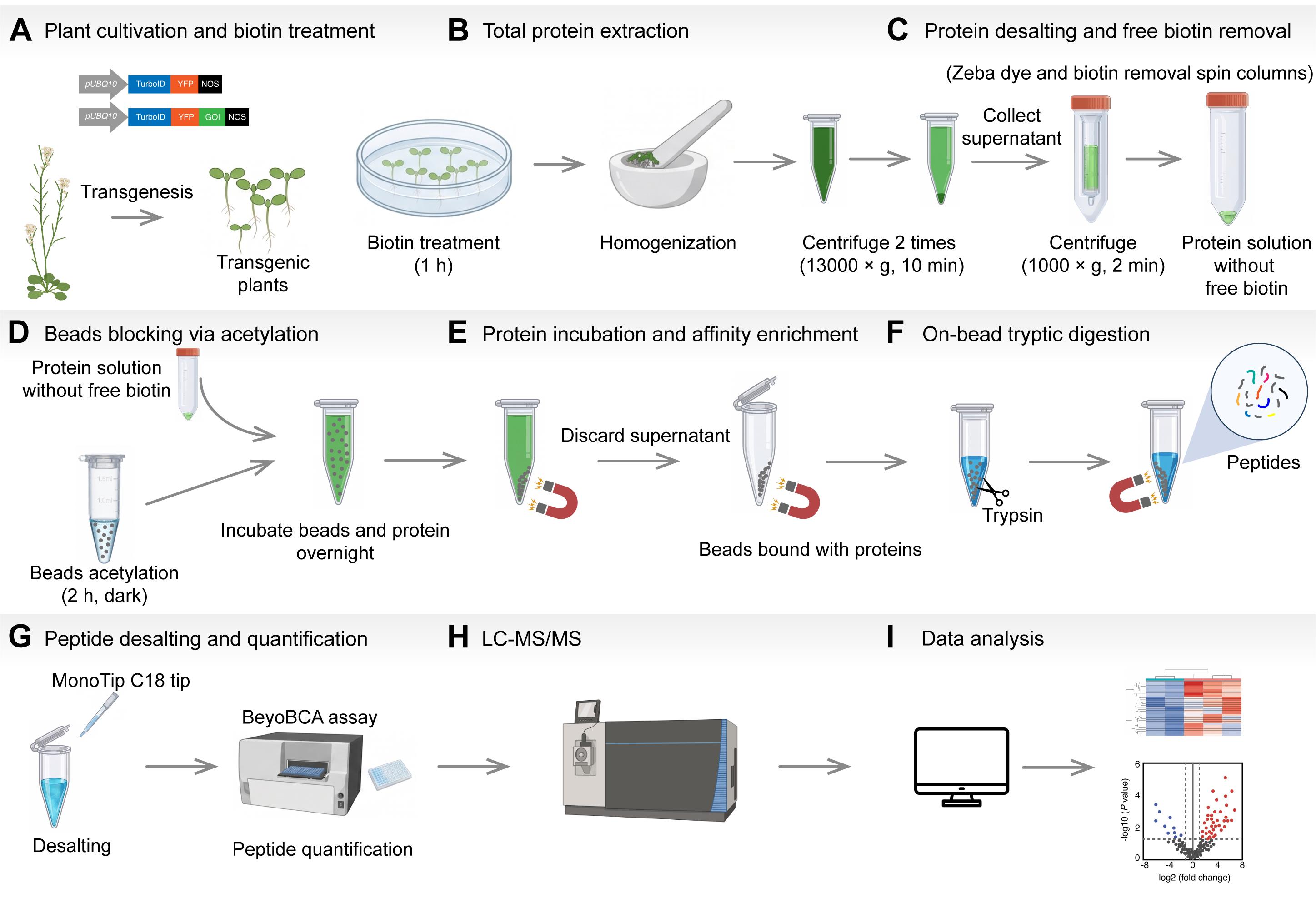
Workflow of TurboID biotinylated protein enrichment and mass spectrometry (MS) preprocessing in Arabidopsis thaliana. Complete experimental workflow for enrichment and MS preprocessing of biotinylated proteins via TurboID proximity labeling in Arabidopsis thaliana. The workflow consists of nine core steps: (A) plant cultivation and biotin treatment, (B) total protein extraction, (C) protein desalting and free biotin removal, (D) beads blocking via acetylation, (E) protein incubation and affinity enrichment of biotinylated proteins, (F) on-bead tryptic digestion, (G) peptide desalting and quantification, (H) mass spectrometry, and (I) proteomic data analysis. Parallel protein aliquots are reserved at each critical stage to perform quality control for protein loss throughout the entire pipeline.
Background
Protein–protein interactions (PPIs) govern most cellular biological processes. Nevertheless, conventional biochemical approaches such as co-immunoprecipitation show clear limitations in capturing weak and transient protein interactions. As a technique developed to address these drawbacks, proximity labeling (PL) works by fusing a labeling enzyme to a protein of interest to covalently tag its spatially neighboring proteins in living cells [1]. Two major classes of this technique are available: biotin-ligase-based systems represented by BioID, and peroxidase-based systems represented by APEX. Owing to the low signal-to-noise ratio (SNR) stemming from the high background activity of endogenous plant peroxidases, as well as cytotoxicity and stress effects caused by the substrate H2O2 in peroxidase-dependent approaches, BioID-based PL is therefore preferred for plant research [2]. Engineered from BioID, TurboID has been widely adopted in plant studies for its high catalytic activity, low cytotoxicity, and tolerance to a broad range of working temperatures [3]. Upon exogenous biotin supplementation, engineered TurboID produces short-lived, diffusible biotin-AMP intermediates that covalently modify the ε-amino groups of exposed lysine residues on proteins within an approximate 10 nm labeling radius. The biotin-tagged protein complexes or organellar components are affinity-enriched using streptavidin-coated magnetic beads and subsequently identified by mass spectrometry (MS)-based analysis [2].
Combined with quantitative proteomics, proximity-dependent biotinylation provides a powerful route to dissect plant subcellular protein interaction networks and identify key regulatory molecules within signaling pathways [4]. Capitalizing on its distinctive in vivo labeling capability, it broadens the scope of conventional interaction assays and supports mechanistic studies covering plant membrane trafficking, immune responses, and stress physiology [1,5–10].
Conventional preprocessing workflows for enriching biotinylated proteins from plant tissues suffer from multiple inherent technical limitations. The TurboID technique was originally developed using mammalian cell models. In animal systems, repeated PBS washing alone suffices to remove free biotin, yet this approach is entirely incompatible with plant tissues due to their complex endogenous biotin background. To address the technical bottleneck of high endogenous free biotin levels in plant tissues that cannot be eliminated via simple aqueous washing, Mair et al. (2019) standardized a PD-10 gel filtration desalting column procedure tailored for plant TurboID assays, which has been widely adopted as a core desalting step in subsequent plant TurboID experimental protocols [5].
Plant TurboID assays commonly employ lysis buffers containing high-concentration urea. When stored at low temperatures for extended periods, urea undergoes spontaneous decomposition to produce isocyanate, which induces artificial carbamylation modifications at the N-termini and lysine residues of proteins. Such modifications block tryptic cleavage sites, resulting in incomplete digestion and missed peptides; they additionally alter peptide physicochemical properties to reduce the number of proteins identified by MS. Furthermore, carbamylation interferes with tandem mass tags (TMT) and label-free quantification, amplifying quantitative errors and weakening experimental reproducibility [11]. Most published protocols omit acetylation blocking of streptavidin magnetic beads. Unblocked exposed lysine residues on streptavidin are simultaneously degraded by Lys-C and trypsin during digestion, generating abundant carrier-derived contaminant peptides that suppress signals from low-abundance interacting peptides. This limits the maximum loading volume for MS, impairs the efficiency of two-stage protease digestion, distorts quantitative results, and ultimately leads to the loss of low-abundance target proteins and poor experimental repeatability [12,13]. Moreover, nearly all universal workflows adopt a fixed bead dosage regardless of the gradient abundance of biotinylated proteins within samples. Fixed bead quantities easily saturate binding sites and fail to fully recover all biotin-labeled target proteins [14]. In addition, the vast majority of published protocols lack quality control (QC) designs involving parallel sample preservation at multiple experimental stages, making it impossible to track protein loss across the full workflow of lysis, desalting, bead enrichment, and protease digestion, which further compromises the reproducibility of experimental outcomes.
To address the above series of technical defects, this study establishes a standardized, complete preprocessing workflow tailored for Arabidopsis seedlings, covering the full pipeline from biotinylated protein enrichment to MS sample preparation. In this protocol, high-concentration 8 M urea lysis buffer is diluted to a final concentration of 2 M prior to desalting, which reduces urea content upfront and minimizes the risk of artificial carbamylation modifications originating from urea decomposition [15]. Meanwhile, Sulfo-NHS-acetate is introduced for magnetic bead acetylation blocking to shield free amino groups on bead surfaces and eliminate MS background interference caused by co-digestion of bead carriers [13,16]. Sequencing-grade modified trypsin is utilized for on-bead digestion with an optimized protein-to-protease mass ratio of 50:1, which is adjusted in accordance with commercial protease specifications to alleviate steric hindrance arising from protein immobilization on beads [7]. To mitigate incomplete digestion and peptide loss caused by bead-immobilized proteins, a two-step supplementary digestion strategy is implemented: primary overnight incubation with trypsin is followed by supplementation with half of the initial trypsin dosage for an additional 4 h, which markedly improves peptide coverage of target proteins [3,12,17]. At critical steps, including protein lysis, desalting purification, flowthrough fraction collection, and multiple rounds of bead washing, parallel protein aliquots are reserved separately for each sample. A dual quantitative system combining Coomassie Brilliant Blue semi-quantification and BeyoBCA peptide colorimetric quantification is integrated to enable comprehensive quality control over protein loss throughout all preprocessing and digestion steps. Notably, this full-process quality control framework has been robustly validated and successfully applied in our laboratory's published plant signaling pathway studies [9,10].
Compared with previously reported conventional preprocessing methods, this optimized workflow effectively elevates the total recovery rate of biotinylated proteins and SNR in quantitative MS analysis. Nevertheless, certain limitations remain. The bead acetylation blocking step requires a 2 h incubation in darkness, which prolongs the storage duration of desalted protein samples at 4 °C. Diluted low-concentration urea buffers exhibit poor stability; prolonged static storage at 4 °C accelerates urea decomposition and the continuous generation of isocyanate, introducing persistent artificial carbamylation. Therefore, clarified protein supernatants obtained after dilution and desalting must be subjected to magnetic bead enrichment and protease digestion as promptly as possible [11,18].
All buffer formulations and magnetic bead operating parameters of this workflow are optimized specifically for Arabidopsis leaf tissues. When applied to mammalian cells, fungi, or other heterologous biological materials, the full lysis and enrichment procedures require re-optimization. Beyond in vivo BioID/TurboID quantitative interactome analysis in Arabidopsis, this standardized preprocessing pipeline can be adapted for in vivo proximity labeling interactome profiling, plasma membrane surface biotinylated membrane proteome identification, and affinity enrichment experiments for diverse post-translational modifications such as ubiquitination via appropriate buffer optimization, replacement of affinity enrichment media, and supplementation with modification-specific inhibitors. This standardized protocol reduces systematic experimental bias and improves reproducibility, thereby providing stable and reliable preprocessing technical support for diverse plant interactome and post-translational proteomics research.
Materials and reagents
Biological materials
1. Arabidopsis thaliana Columbia-0 (Col-0)
2. Agrobacterium tumefaciens strain GV3101
3. Plasmid: pCAMBIA1300-pUBQ10::TurboID-EYFP
4. Plasmid: pCAMBIA1300-pUBQ10::TurboID-EYFP-ATG8a
5. Arabidopsis thaliana transgenic line pUBQ10::TurboID-EYFP
6. Arabidopsis thaliana transgenic line pUBQ10::TurboID-EYFP-ATG8a
Reagents
1. Sodium chloride (NaCl) (VETEC, catalog number: V900058)
2. Tris (hydroxymethyl) aminomethane (Tris base) (Sigma-Aldrich, catalog number: T1503-1KG)
3. Sodium deoxycholate (Sangon Biotech, catalog number: A600150-0050)
4. Urea (Sigma-Aldrich, catalog number: U1250-1KG)
5. Ammonium bicarbonate (Sangon Biotech, catalog number: A610032-0500)
6. Sodium dodecyl sulfate (SDS) (Sangon Biotech, catalog number: A600485-0500)
7. Bromophenol Blue (BPB) (Sangon Biotech, catalog number: A500922-0025)
8. Glycine (Sangon Biotech, catalog number: A610235-0005)
9. Sodium dihydrogen phosphate dihydrate (NaH2PO4·2H2O) (Guangzhou Chemical Reagent Factory, catalog number: BE13-AR-500G)
10. Disodium hydrogen phosphate dodecahydrate (Na2HPO4·12H2O) (Sangon Biotech, catalog number: A607793-0500)
11. Potassium chloride (KCl) (Sigma-Aldrich, catalog number: V900068-500G)
12. Sodium carbonate (Na2CO3) (VETEC, catalog number: V900801-500G)
13. Sodium bicarbonate (NaHCO3) (Sigma-Aldrich, catalog number: V900182)
14. Concentrated hydrochloric acid (HCl) (Tianjin Chemical Reagent Factory No.1, catalog number: SXH005)
15. Sodium hydroxide (NaOH) (Guangzhou Chemical Reagent Factory, catalog number: DS018-1)
16. Ammonium persulfate (APS) (Sangon Biotech, catalog number: A100486-0025)
17. Tween-20 (VETEC, catalog number: V900548)
18. HEPES (VETEC, catalog number: V900477-100G)
19. Sulfo-NHS-acetate (Aladdin, catalog number: S333917)
20. Dithiothreitol (DTT) (Sangon Biotech, catalog number: A620058-0005)
21. Iodoacetamide (IAM) (Sangon Biotech, catalog number: A600539-0005)
22. Formic acid (FA) [Guangzhou Chemical Reagent Factory (GCRF), catalog number: CB05]
23. Acetonitrile (ACN) (Sigma-Aldrich, catalog number: 34851-1L)
24. β-Mercaptoethanol (BME) (Sigma-Aldrich, catalog number: M3148-100ML)
25. Glycerol (Sangon Biotech, catalog number: A501745-0500)
26. 30% acrylamide-bisacrylamide mixed solution (Acry-bis) (Sangon Biotech, catalog number: B546017-0500)
27. Tetramethylethylenediamine (TEMED) (Sangon Biotech, catalog number: A610508-0100)
28. Dimethyl sulfoxide (DMSO) (VETEC, catalog number: V900090-500ML)
29. Protease inhibitor cocktail (PI) (GlpBio, catalog number: GK10014)
30. Sequencing-grade modified trypsin, porcine (Promega, catalog number: V511A)
31. BeyoBCA Peptide Quantitation Assay kit (colorimetric) (Beyotime Biotechnology, catalog number: P0397S)
32. BSA standard protein solution (2 mg/mL) (Sangon Biotech, catalog number: C900172-0010)
33. Coomassie Blue Fast Staining Solution (Zylome, catalog number: CS021-500 mL)
34. MS modified base salts with vitamins (Coolaber, catalog number: PM10111-100L)
35. Sucrose (Sigma-Aldrich VETEC, catalog number: V900116-500G)
36. Biotin (Sigma-Aldrich, catalog number: B4501-100MG)
37. Agar (Coolaber, catalog number: CA1331-1kg)
38. 1 mg/mL peptide standard (see Beyotime Biotechnology’s official website instructions)
39. BeyoBCA reagent A (see Beyotime Biotechnology’s official website instructions)
40. BeyoBCA reagent B (see Beyotime Biotechnology’s official website instructions)
41. BeyoBCA reagent C (see Beyotime Biotechnology’s official website instructions)
42. Clorox™ disinfecting bleach (Clorox, catalog number: CLO32251)
Solutions
1. 0 M urea dilution buffer (see Recipes)
2. 2 M urea washing buffer (see Recipes)
3. 4 M urea ammonium bicarbonate resuspension buffer (see Recipes)
4. 8 M urea denaturation buffer (see Recipes)
5. 20% Tween-20 stock solution (see Recipes)
6. HEPES-tween washing buffer 1 (buffer 1) (see Recipes)
7. 100 mM Sulfo-NHS-acetate stock solution (see Recipes)
8. Acetylation reaction mix (see Recipes)
9. Ammonium bicarbonate-tween washing buffer 2 (buffer 2) (see Recipes)
10. 100 mM DTT (see Recipes)
11. 400 mM IAM (see Recipes)
12. 25 mM ammonium bicarbonate stock solution (see Recipes)
13. 0.1 μg/μL sequencing-grade trypsin stock solution (see Recipes)
14. 20% formic acid (FA) (see Recipes)
15. 50% acetonitrile with 0.1% FA (see Recipes)
16. 0.1% FA (see Recipes)
17. 5× SDS-PAGE protein loading buffer (5× SDS) (see Recipes)
18. 5× SDS-PAGE electrophoresis buffer (see Recipes)
19. 3× separating buffer (see Recipes)
20. 5× stacking gel buffer (see Recipes)
21. Stacking gel mixture (see Recipes)
22. Separating gel mixture (see Recipes)
23. 1/2 MS culture medium formula (see Recipes)
24. 50 mM biotin stock solution (see Recipes)
25. 50 μM biotin stock solution (see Recipes)
Recipes
1. 0 M urea dilution buffer
| Reagent | Final concentration | Quantity or volume |
|---|---|---|
| NaCl | 150 mM | 0.1753 g |
| Tris base (MW = 121.14 g/mol) | 50 mM | 0.1211 g |
| Sodium deoxycholate | 0.5% (w/v) | 0.1 g |
| PI cocktail (add fresh right before use) | n/a | 200 μL |
| Milli-Q water | n/a | Add to a final volume of 20 mL after adjusting pH to 8.0 with HCl |
| Total | n/a | 20 mL |
2. 2 M urea washing buffer
| Reagent | Final concentration | Quantity or volume |
|---|---|---|
| Urea | 2 M | 12.012 g |
| NaCl | 150 mM | 0.8766 g |
| Tris base (MW = 121.14 g/mol) | 50 mM | 0.6057 g |
| Sodium deoxycholate | 0.5% (w/v) | 0.5 g |
| PI cocktail (add fresh right before use) | n/a | 1 mL |
| Milli-Q water | n/a | Add to a final volume of 100 mL after adjusting pH to 8.0 with HCl |
| Total | n/a | 100 mL |
3. 4 M urea ammonium bicarbonate resuspension buffer
| Reagent | Final concentration | Quantity or volume |
|---|---|---|
| Urea | 4 M | 1.2012 g |
| Ammonium bicarbonate | 25 mM | 0.0099 g |
| Milli-Q water | n/a | Add to final volume 5 mL |
| Total | n/a | 5 mL |
4. 8 M urea denaturation buffer
| Reagent | Final concentration | Quantity or volume |
|---|---|---|
| Urea | 8 M | 4.8048 g |
| NaCl | 150 mM | 0.0877 g |
| Tris base (MW = 121.14 g/mol) | 50 mM | 0.0606 g |
| Sodium deoxycholate | 0.5% (w/v) | 0.05 g |
| PI cocktail (add fresh right before use) | n/a | 100 μL |
| Milli-Q water | n/a | Add to a final volume of 10 mL after adjusting pH to 8.0 with HCl |
| Total | n/a | 10 mL |
5. 20% Tween-20 stock solution
| Reagent | Final concentration | Quantity or volume |
|---|---|---|
| 100% Tween-20 | 20% (v/v) | 10 mL |
| Milli-Q water | n/a | Add to a final volume of 50 mL |
| Total | n/a | 50 mL |
6. Buffer 1
| Reagent | Final concentration | Quantity or volume |
|---|---|---|
| HEPES | 50 mM | 0.1192 g |
| 20% (v/v) Tween-20 stock | 0.2% (v/v) | 100 μL (equivalent to 20 μL of pure undiluted 100% Tween-20) |
| Milli-Q water | n/a | Add to a final volume of 10 mL after adjusting pH to 7.8 with NaOH |
| Total | n/a | 10 mL |
7. 100 mM Sulfo-NHS-acetate stock solution
| Reagent | Final concentration | Quantity or volume |
|---|---|---|
| Sulfo-NHS-acetate | 100 mM | 0.00259 g |
| DMSO | n/a | Add to final volume 100 μL |
| Total | n/a | 100 μL |
8. Acetylation reaction mix
| Reagent | Final concentration | Quantity or volume |
|---|---|---|
| Buffer 1 | n/a | 380 μL |
| 100 mM Sulfo-NHS-acetate stock solution | 5 mM | 20 μL |
| Total | n/a | 400 μL |
9. Buffer 2
| Reagent | Final concentration | Quantity or volume |
|---|---|---|
| Ammonium bicarbonate | 50 mM | 0.04 g |
| 20% (v/v) Tween-20 stock | 0.2% (v/v) | 100 μL |
| Milli-Q water | n/a | Add to a final volume of 10 mL |
| Total | n/a | 10 mL |
10. 100 mM DTT
| Reagent | Final concentration | Quantity or volume |
|---|---|---|
| Dithiothreitol (DTT, MW = 154.25 g/mol) | 100 mM | 0.0154 g |
| Milli-Q water | n/a | Add to final volume 1 mL |
| Total | n/a | 1 mL |
11. 400 mM IAM
| Reagent | Final concentration | Quantity or volume |
|---|---|---|
| IAM, MW = 185.0 g/mol | 400 mM | 0.074 g |
| Milli-Q water | n/a | Add to final volume 1 mL |
| Total | n/a | 1 mL |
12. 25 mM ammonium bicarbonate stock solution
| Reagent | Final concentration | Quantity or volume |
|---|---|---|
| Ammonium bicarbonate | 25 mM | 0.0198 g |
| Milli-Q water | n/a | Add to final volume 10 mL |
| Total | n/a | 10 mL |
13. 0.1 μg/μL sequencing-grade trypsin stock solution
| Reagent | Final concentration | Quantity or volume |
|---|---|---|
| Sequencing-grade trypsin dry powder | 0.1 μg/μL | Appropriate amount of trypsin powder |
| Manufacturer-supplied reconstitution buffer | n/a | Add to fix the final volume to reach 0.1 μg/μL |
Dissolve trypsin dry powder exclusively with the reconstitution buffer provided by the manufacturer; do not use other solvents.
14. 20% FA
| Reagent | Final concentration | Quantity or volume |
|---|---|---|
| Pure FA | 20% (v/v) | 20 μL |
| Milli-Q water | n/a | 80 μL |
| Total | n/a | 100 μL |
15. 50% acetonitrile with 0.1% FA
| Reagent | Final concentration | Quantity or volume |
|---|---|---|
| Pure ACN | 50% (v/v) | 5 mL |
| Pure FA | 0.1% (v/v) | 10 μL |
| Milli-Q water | n/a | Add to a final volume of 10 mL |
| Total | n/a | 10 mL |
16. 0.1% FA
| Reagent | Final concentration | Quantity or volume |
|---|---|---|
| Pure FA | 0.1% (v/v) | 10 μL |
| Milli-Q water | n/a | Add to a final volume of 10 mL |
| Total | n/a | 10 mL |
17. 5× SDS-PAGE protein loading buffer (5× SDS)
| Reagent | Final concentration | Quantity or volume |
|---|---|---|
| Tris base (MW = 121.14 g/mol) | 50 mM | 0.3029 g |
| SDS | 2% (w/v) | 1 g |
| BPB | 0.1% (w/v) | 50 mg |
| Glycerol | 10% (v/v) | 5 mL |
| BME | 1% (v/v) | 500 μL |
| Milli-Q water | n/a | Add to a final volume of 50 mL after adjusting pH to 6.8 with HCl |
| Total | n/a | 50 mL |
Mix thoroughly, aliquot into 0.5 mL per tube, and store at 4 °C.
18. 5× SDS-PAGE electrophoresis buffer
| Reagent | Final concentration | Quantity or volume |
|---|---|---|
| Tris base (MW = 121.14 g/mol) | 125 mM | 15.14 g |
| Glycine | 960 mM | 72.05 g |
| SDS | 0.5% (w/v) | 5 g |
| Milli-Q water | n/a | Add to a final volume of 1 L after adjusting pH to 8.3 with HCl |
| Total | n/a | 1 L |
Note: Dilute the 5× stock buffer to 1× working concentration with Milli-Q water prior to each electrophoresis run.
19. 3× separating buffer
| Reagent | Final concentration | Quantity or volume |
|---|---|---|
| Tris base (MW = 121.14 g/mol) | 1.125 M | 27.24 g |
| SDS | 0.3% (w/v) | 0.6 g |
| Milli-Q water | n/a | Adjust volume to 200 mL after adjusting pH to 8.8 with HCl |
| Total | n/a | 200 mL |
20. 5× stacking gel buffer
| Reagent | Final concentration | Quantity or volume |
|---|---|---|
| Tris base (MW = 121.14 g/mol) | 0.5 M | 12.11 g |
| SDS | 0.4% (w/v) | 0.8 g |
| Milli-Q water | n/a | Adjust volume to 200 mL after adjusting pH to 6.8 with HCl |
| Total | n/a | 200 mL |
21. Stacking gel mixture
| Reagent | Final concentration | Quantity or volume |
|---|---|---|
| 30% Acry-bis | 5% | 833 μL |
| 5× stacking gel buffer | 1× | 1 mL |
| Milli-Q water | n/a | 3.167 mL |
| 30% APS | 0.12% (w/v) | 20 μL |
| TEMED | 0.1% (v/v) | 5 μL |
| Total | n/a | 5 mL |
22. Separating gel mixture
| Reagent | Final concentration | Quantity or volume |
|---|---|---|
| 30% Acry-bis | 10% | 3.33 mL |
| 3× separating gel buffer | 1× | 3.33 mL |
| Milli-Q water | n/a | 3.33 mL |
| 30% APS | 0.075% (w/v) | 25 μL |
| TEMED | 0.0625% (v/v) | 6.25 μL |
| Total | n/a | 10 mL |
23. 1/2 MS culture medium formula
| Reagent | Final concentration | Quantity or volume |
|---|---|---|
| MS modified base salts with vitamins | 0.5× | 2.2 g |
| Sucrose | 30 mM | 10 g |
| Milli-Q water | n/a | Adjust volume to 1 L after adjusting pH to 5.7 with HCl |
| Total | n/a | 1 L |
For solid 1/2 MS medium, add 7.5 g of agar per liter of liquid 1/2 MS medium.
24. 50 mM biotin stock solution
| Reagent | Final concentration | Quantity or volume |
|---|---|---|
| Biotin | 50 mM | 0.0122 g |
| DMSO | n/a | Adjust volume to 1 mL |
| Total | n/a | 1 mL |
25. 50 μM biotin stock solution
| Reagent | Final concentration | Quantity or volume |
|---|---|---|
| 50 mM biotin stock solution | 50 μM | 50 μL |
| 1/2 MS liquid medium | n/a | Adjust volume to 50 mL |
| Total | n/a | 50 mL |
Laboratory supplies
1. 1.5 mL polypropylene microcentrifuge tubes (Biosharp, catalog number: BS-15-M)
2. 2.0 mL polypropylene microcentrifuge tubes (Biosharp, catalog number: BS-20-M)
3. Low protein binding 1.5 mL microcentrifuge tubes (Thermo Scientific, catalog number: 90410)
4. 1,000 μL blue pipette tips (Biosharp, catalog number: BS-1000-T)
5. 200 μL yellow pipette tips (Biosharp, catalog number: BS-200-T)
6. 10 μL clear pipette tips (Biosharp, catalog number: BS-10-T)
7. 50 mL polypropylene centrifuge tubes (Corning, catalog number: 430829)
8. 15 mL polypropylene centrifuge tubes (Corning, catalog number: 430791)
9. 0.5–10 μL filter pipette tips, racked (Axygen, catalog number: TF-300-L-R-S)
10. 1–200 μL filter pipette tips, racked (Axygen, catalog number: TF-200-L-R-S)
11. 100–1,000 μL filter pipette tips, racked (Axygen, catalog number: TF-1000-L-R-S)
12. MonoTip C18 extraction tips, 200 μL (GL Sciences Inc., catalog number: 5010-21000)
13. ZebaTM dye and biotin removal columns, 5 mL (Thermo Scientific, catalog number: A44301)
14. Streptavidin magnetic beads, 5 mL (Smart-Lifesciences, catalog number: SM007005)
15. Sterile cell culture plates, 96-well flat bottom (Corning, catalog number: 3599)
16. Petri dish BS-90-D (Lablogic Technology, catalog number: BS-90-D)
17. 6-well cell culture plate (NEST, catalog number: 723321)
Equipment
1. Heraeus Pico 17 microcentrifuge (Thermo Fisher Scientific, catalog number: 75002410)
2. Bio-Rad Protein Electrophoresis System (Bio-Rad Laboratories, Inc.)
3. Dry bath incubator (Shanghai Baba Industrial Co., Ltd., model: GDC-100)
4. Rotational incubator (Haimen Kylin-Bell Instrument Manufacturing Co., Ltd., model: QB-128)
5. Reciprocating decolorizing shaker (Hangzhou Miulab Instrument Co., Ltd., model: TS-300S)
6. Water-jacketed incubator (Shanghai Xinmiao Medical Device Manufacturing Co., Ltd., model: GNP-9050BS-III)
7. Model 4-5 benchtop low-speed centrifuge (TOMOS Scientific Instruments, model: 4-5)
8. Heraeus Fresco 17 refrigerated microcentrifuge (Thermo Fisher Scientific, catalog number: 75002420)
9. Micro-Volume UV-Vis Spectrophotometer (Wuxi Bioteke Biotechnology Co., Ltd., model: ND5000)
10. Vacuum Concentrator Plus (Eppendorf, model: 5305)
11. EnSpire Multimode Plate Reader (PerkinElmer, model: 2300)
12. Orbitrap Astral Mass Spectrometer (Thermo Fisher Scientific)
13. Tissue culture incubator (Ruihua, model: TCC-15)
Software and datasets
1. Spectronaut (DIA proteomic data analysis); Spectronaut, Biognosys AG, Version 20.0; Source: https://biognosys.com/software/spectronaut/, accessed 04/27/2026
2. MaxQuant (DDA proteomic data analysis); MaxQuant, Version v2.8.1.0; Source: https://maxquant.org, accessed 07/19/2026
3. Reference Protein Databases for Arabidopsis thaliana: Dataset: Araport11_pep_20250411.fasta (48,354 protein entries); Source: https://www.arabidopsis.org/download/list?dir=Proteins%2FAraport11_protein_lists, accessed 04/27/2026; license: free public academic use
Procedure
登录/注册后免费查看全文
文章信息
稿件历史记录
提交日期: Jul 19, 2026
接收日期: Sep 19, 2026
在线发布日期: Oct 1, 2026
出版日期: Nov 5, 2026
版权信息
© 2026 The Author(s); This is an open access article under the CC BY-NC license (https://creativecommons.org/licenses/by-nc/4.0/).
如何引用
Zhang, L., Ye, Y., Zhuo, D., Yan, Q., Ye, Y., Li, H., Gao, C. and Feng, L. (2026). An Optimized Protocol for TurboID-Based Proximity Labeling and Sample Preparation for Mass Spectrometry in Arabidopsis thaliana. Bio-protocol 16(21): e5852. DOI: 10.21769/BioProtoc.5852.

 Download Citation in RIS Format
您对这篇实验方案有问题吗?
在此处发布您的问题,我们将邀请本文作者来回答。同时,我们会将您的问题发布到Bio-protocol Exchange,以便寻求社区成员的帮助。
 发布问题
发布问题


