


Microtube-Assisted Paraffin Embedding and Sectioning of Fragile 3D Tumor Spheroids for High-Quality Histology



发布: 2026年11月05日第16卷第21期 DOI: 10.21769/BioProtoc.5850 浏览次数: 25
评审: Hima Bindu DurumutlaAnonymous reviewer(s)
 PDF
PDF  Q&A
Q&A





Abstract
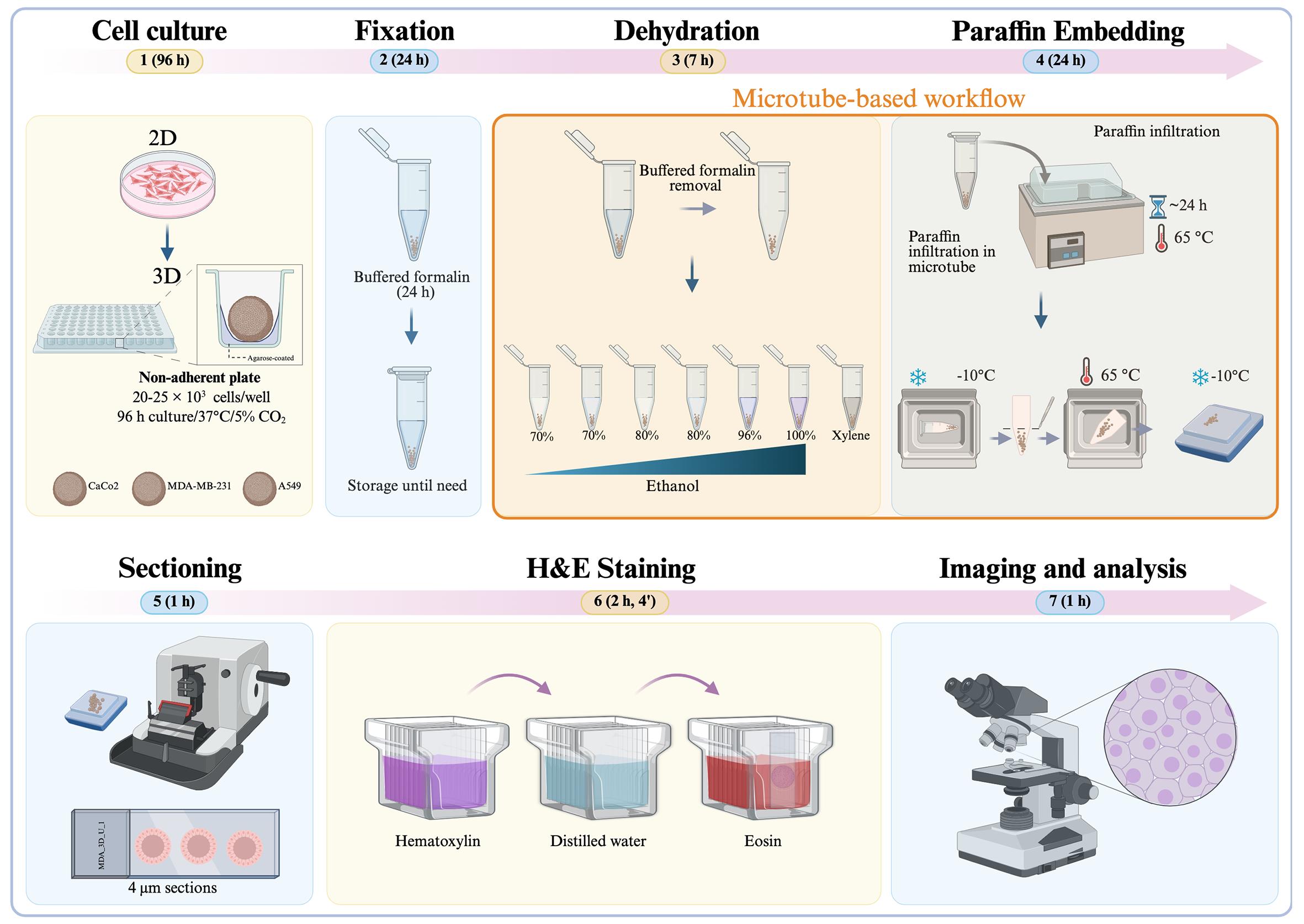
Three-dimensional (3D) spheroid models have become essential in cancer biology, drug screening, and tissue engineering. However, their small size, fragile structure, and tendency to disintegrate during routine histoprocessing present persistent technical challenges. Conventional paraffin embedding often results in tissue fragmentation, loss of spatial orientation, and poor section quality, whereas cryosectioning often compromises cellular morphology. Here, we present a robust, cost-effective protocol for preserving and sectioning fragile 3D spheroids, resulting in high-quality histological sections with intact architecture and excellent cellular detail. The method involves optimized handling and embedding procedures that stabilize spheroids during standard formalin fixation, paraffin infiltration, and microtomy, eliminating mechanical distortion and preserving spherical integrity for consistent sectioning. We demonstrate the successful application of this protocol across different tumor spheroids derived from distinct tissue types, including MDA-MB-231 (breast carcinoma), Caco-2 (colorectal adenocarcinoma), and A549 (lung carcinoma) cell lines, with subsequent compatibility with hematoxylin and eosin (H&E) staining protocols. This protocol is compatible with immunohistochemistry (IHC) and immunofluorescence (IF). In our qualitative comparison with conventional methods, we found that our approach effectively limits sample loss, promotes consistent inter-section reproducibility, and preserves fine structural features, such as necrotic cores, proliferative zones, and extracellular matrix components. This protocol provides a reliable, accessible solution for routine histological analysis of fragile 3D spheroids, facilitating more accurate morphological assessment in translational research settings.
Key features
• Cost-effective agarose-coated plates for reproducible spheroid formation.
• Complete in-tube processing to minimize sample loss and damage.
• Floating tube system for reliable paraffin infiltration without specialized equipment.
• Produces high-quality sections compatible with H&E.
Keywords: 3D spheroids Paraffin embedding
FFPE
Microtome sectioning
Histological analysis
Paraffin embedding
FFPE
Microtome sectioning
Histological analysis
Graphical overview
Background
Three-dimensional (3D) tumor models have advanced to become in vitro platforms that more accurately mimic the biological complexity of cancer than traditional two-dimensional (2D) monolayer cultures [1]. Cancer is a multifactorial and diverse disease characterized by uncontrolled cell growth, genomic instability, metabolic reprogramming, immune evasion, and the ability to invade and metastasize [2]. Notably, these processes are heavily influenced by the tumor microenvironment (TME), which includes oxygen and nutrient gradients, extracellular matrix (ECM) composition, biomechanical forces, and cell-to-cell communication [3]. While 2D systems are convenient for experiments, they do not replicate the spatial organization and microenvironmental conditions that control tumor behavior. Cells in 2D cultures assume an unnatural flat shape, with unlimited apical diffusion and limited basal attachment, lacking proper cell-cell contacts and a three-dimensional structure that regulates cell signaling, gene expression, and drug response. As a result, 2D models have limited relevance and predictive ability in preclinical drug testing [1].
3D tumor models include various platforms such as multicellular spheroids, organoids, scaffold-based cultures, and bioprinted constructs. Among these, tumor spheroids are particularly popular and cost-effective [3]. In non-adherent culture conditions, tumor cells spontaneously form compact multicellular aggregates that develop inherent structural organization and physiologically relevant gradients of oxygen, nutrients, metabolites, and signaling molecules. These gradients create spatially distinct cellular zones, including an outer proliferative layer, intermediate quiescent regions, and a central hypoxic or necrotic core, closely mimicking avascular tumor regions in vivo [4]. The radial structure of spheroids, with a high-oxygen, nutrient-rich outer layer and a progressively hypoxic, nutrient-deprived center, reflects the key mass-transfer limitations seen in solid tumors. This stratification enables researchers to investigate pathophysiological processes such as oxygen- and nutrient-driven metabolic adaptation, hypoxia-inducible factor (HIF)-mediated signaling, and the transition from quiescence to necrosis in a precisely controlled environment [5].
Establishing spatial heterogeneity within spheroids has significant implications for cancer research. Cells in different regions of the spheroid exhibit distinct transcriptional programs, metabolic states, proliferative capacities, and responses to stress. Hypoxic cores activate pathways such as HIF-mediated signaling, which promote metabolic adaptation and survival under nutrient deprivation [5]. Additionally, increased cell-cell adhesion, changes in polarity, and endogenous ECM deposition create diffusion barriers that affect drug penetration and therapeutic response. Consequently, spheroids model clinically relevant phenomena such as chemoresistance, microenvironment-driven plasticity, and treatment-induced selective pressures more effectively than 2D cultures [6]. Recent studies have demonstrated that including stromal or immune cell, such as co-culturing cancer cells with macrophages, can be integrated into spheroid models to imitate the complex cellular interactions of the TME, allowing tracking of region-specific changes in hypoxia, apoptosis, and metabolic profiles across the radial axis [3,7].
The use of 3D models greatly influences the study of cancer progression and drug development. Drug screening in spheroids often shows lower drug sensitivity compared to 2D cultures, emphasizing the importance of the microenvironment in treatment response [1]. Additionally, 3D systems allow investigation of tumor-matrix interactions, invasive behavior, and ECM remodeling when combined with biomimetic matrices such as collagen, matrigel, or synthetic hydrogels. These matrix-embedded systems mimic biomechanical constraints and stromal interactions found in vivo, offering a more realistic framework for studying tumor adaptation and growth [7]. For example, embedding spheroids in collagen I hydrogels enables direct observation of cells escaping from the tumor mass and invading surrounding tissue, a process key to metastasis and drug resistance [7]. The ability to observe such events in both space and time makes 3D spheroids especially useful for mechanistic research and developing anti-invasive treatments [3].
Despite these advantages, 3D spheroid models pose technical challenges, particularly during downstream histological processing. Because they are small, have compact cellular structures, and lack intrinsic support, spheroids are very prone to deformation, fragmentation, or loss during fixation, dehydration, paraffin embedding, and microtome sectioning [4,8]. Matrix-embedded spheroids face extra difficulties, as mechanical mismatch or uneven shrinkage between the spheroid and the surrounding hydrogel can weaken their structure [7]. Their small size also makes spheroids difficult to locate and position within a paraffin block, and incomplete dehydration or poor paraffin infiltration can lead to voids, tearing, or complete loss during sectioning [9,10]. As a result, many researchers choose low-resolution whole-mount imaging or cryosectioning, both of which trade off either spatial detail or molecular preservation [8].
Histological sectioning is crucial for thoroughly characterizing spheroids. While live imaging and viability assays offer overall functional insights, they cannot reveal the internal structure or localized treatment effects [11]. Histological analysis allows direct visualization of proliferative zones, hypoxic regions, necrotic cores, and area-specific therapeutic damage at a microscopic level [12]. Additionally, formalin-fixed, paraffin-embedded (FFPE) sections are preferable to whole-mount preparations for many applications, such as immunohistochemistry, because they prevent the depth-related antibody penetration issues seen in thick samples and enable long-term storage in biobanks [8]. Consequently, developing optimized and consistent histological workflows is essential for unlocking the full analytical potential of 3D tumor spheroid systems [9,10].
In this context, developing tailored, step-by-step histological protocols specifically adapted for 3D spheroid models is necessary to ensure structural preservation, reproducibility, and reliable spatial analysis [4,13]. These methodological improvements support the wider adoption of 3D tumor models in cancer biology research, drug discovery, and preclinical evaluation [1,3].
Materials and reagents
Biological materials
1. MDA-MB-231 cell line (ATCC, catalog number: CRM-HTB-26)
2. A549 cell line (ATCC, catalog number: CRM-CCL-185)
3. Caco-2 cell line (ATCC, catalog number: HTB-37)
Reagents
1. 10% neutral buffered formalin (J.T. Baker, catalog number: 2106-03)
2. Phosphate-buffered saline (1× PBS) (commercial or lab-made)
3. Ethanol (Meyer, catalog number: 0390)
4. Xylene (J.T. Baker, catalog number: 9490-03)
5. Paraffin wax, histology grade (melting point 56–60 °C) (Sigma-Aldrich, catalog number: 1.07164.2504)
6. Hematoxylin solution (Hycel, catalog number: 738)
7. Eosin Y solution (Sigma-Aldrich, catalog number: 45380)
8. Lithium carbonate (Fagan Lab, catalog number: 2518)
9. Synthetic resin for mounting (Sigma-Aldrich, catalog number: 1.07960.0500)
10. Methanol (J.T. Baker, catalog number: 9070-03)
11. Dulbecco’s modified Eagle medium (DMEM) (Sigma Merck, catalog number: D5648) or RPMI media
12. Sodium bicarbonate (NaHCO3) (J.T. Baker, catalog number: 3506-01)
13. L-glutamine (Gibco, catalog number: 20530-081)
14. Amphotericin B (Sigma-Aldrich, catalog number: A2942)
15. Fetal bovine serum (FBS) (BioWest, catalog number: BIO-S1400)
16. Bovine calf serum (BCS) (BioWest, catalog number: S0400-500)
17. Penicillin-streptomycin (Gibco, catalog number: 15140-122)
18. Trypsin (Sigma-Aldrich, catalog number: T4799-5G)
19. Ethylenediaminetetraacetic acid (EDTA) (J.T. Baker, catalog number: 8993-01)
20. Dimethyl sulfoxide (DMSO) (Sigma-Aldrich, catalog number: D2650)
21. Actinomycin D (Sigma-Aldrich, catalog number: A9415)
22. Potassium chloride (J.T. Baker, catalog number: 3040-01)
23. Sodium chloride (J.T. Baker, catalog number: 3624-01)
24. Disodium phosphate (Na2HPO4) (J.T. Baker, catalog number: 3828-01)
25. Monobasic potassium phosphate (KH2PO4) (J.T. Baker, catalog number: 3246-01)
25. Agarose (Invitrogen, catalog number: 16500-100)
27. MilliQ water, sterile (J.T. Baker, catalog number: 4220-20)
28. Hydrochloric acid (HCl) (J.T. Baker, catalog number: 9535-05)
29. Sodium hydroxide (NaOH) (Macron, Fine Chemicals, catalog number: 7708-10)
30. Sodium dihydrogen phosphate (NaH2PO4) (J.T. Baker, catalog number: 10049-21-5)
Laboratory supplies
1. 96-well cell culture plate (NEST, catalog number: 701002)
2. Cell culture dish (60 mm × 15 mm) (NEST, catalog number: 705007)
3. Conical-bottom centrifuge tube, 50 mL (Uniparts, catalog number: 32117F)
4. Conical-bottom centrifuge tube, 15 mL (Uniparts, catalog number: 34117F)
5. Microcentrifuge tubes 1.5 mL (Axygen, catalog number: MCT-150-C)
6. Pipette tip 100–1,000 μL (Uniparts, catalog number: 51131)
7. Pipette tip 1–200 μL (Uniparts, catalog number: 51121Y)
8. Pipette tip 0.5–10 μL (Axygen, catalog number: T-300)
9. Embedding molds (CR Globe, catalog number: P2030)
10. Microscope slide (BRAND, catalog number: BR474701)
11. Cover glasses for microscope slides (BRAND, catalog number: 470045)
12. Surgical design disposable scalpels (Fisher Scientific, catalog number: 22-079-707)
Solutions
1. Culture medium for MDA-MB-231, Caco-2, and A549 cell lines (supplemented DMEM) (see Recipes)
2. 1.5% agarose solution (see Recipes)
3. 1× PBS (see Recipes)
4. 0.04% trypsin-EDTA (see Recipes)
5. 500 μM Actinomycin D (see Recipes)
6. Ethanol solutions (see Recipes)
7. Eosin solution (see Recipes)
8. 10% neutral buffered formalin (see Recipes)
9. Lithium carbonate bluing solution (see Recipes)
10. 1 M HCl (see Recipes)
11. 1 M NaOH (see Recipes)
Recipes
1. Supplemented DMEM
| Reagent | Final concentration | Quantity |
| DMEM powder | - | 15.6 g |
| NaHCO3 | 3 g/L | 3 g |
| FBS | 5% (v/v) | 50 mL |
| BCS | 5% (v/v) | 50 mL |
| Penicillin-streptomycin | 1% (v/v) | 10 mL |
| Amphotericin B | 0.05% (v/v) | 500 μL |
| L-glutamine (200 mM) | 0.05% (v/v) | 500 μL |
| Sterile distilled water | - | To 1 L final volume |
1. Base medium preparation:
a. Combine the DMEM powder with sterile distilled water and stir continuously until fully dissolved.
b. Add 3 g of NaHCO3 to the solution.
c. Adjust the pH to 7.4 by carefully adding 1 M NaOH or 1 M HCl dropwise as required.
2. Supplementation (perform inside a laminar flow hood): Add the following components to the base medium:
a. 5% FBS → 50 mL
b. 5% BCS → 50 mL
c. 1% penicillin-streptomycin → 10 mL
d. 0.05% amphotericin B → 500 μL
e. 0.05% L-glutamine (200 mM stock) → 500 μL
3. Final sterilization and storage:
a. Filter-sterilize the complete medium using a vacuum filtration system equipped with a 0.22 μm MCE membrane.
b. Store the sterile medium at 4 °C and use within 4 weeks.
Notes:
1. This formulation is routinely used for culturing MDA-MB-231 cells; RPMI medium also works.
2. The formulation for Caco-2 and A549 is the same, but without 0.05% L-glutamine.
2. 1.5% agarose solution
| Reagent | Final concentration | Quantity |
| Agarose | 1.5% (w/v) | 1.5 g |
| Triple-distilled water | - | To 100 mL |
| Final volume | - | 100 mL |
a. In a glass flask, combine 1.5 g of agarose with 100 mL of triple-distilled water.
b. Autoclave the mixture for 15 min to achieve sterility.
Note: This solution is intended to create non-adherent surfaces in standard 96-well plates. After autoclaving, it should be used immediately or kept at approximately 60–70 °C until ready to dispense.
3. 1× PBS (pH 7.4)
| Reagent | Final concentration | Quantity |
| NaCl | 137 mM | 8.0 g |
| KCl | 2.7 mM | 0.2 g |
| Na2HPO4 | 10 mM | 1.44 g |
| KH2PO4 | 1.8 mM | 0.24 g |
| ddH2O | - | 1 L |
a. Dissolve NaCl (8.0 g), KCl (0.2 g), Na2HPO4 (1.44 g), and KH2PO4 (0.24 g) in approximately 800 mL of distilled water using a magnetic stirrer. Continue stirring until the solution is completely clear and free of any undissolved particulates.
b. Calibrate the pH meter using standard buffer solutions. Then, while continuously stirring, titrate the solution to pH 7.4 by adding 1 M HCl or 1 M NaOH drop by drop. Allow the pH reading to stabilize for 15–20 s after each addition before recording the value.
c. Transfer the solution to a 1 L volumetric flask and bring the total volume to exactly 1 L with distilled water. Mix thoroughly by inverting the capped flask at least five times to ensure homogeneity.
d. Pour the solution into an autoclave-safe bottle, loosen the cap slightly to permit pressure equilibration, and autoclave at 121 °C (15 psi) for 20–30 min.
e. After autoclaving, let the solution cool to room temperature before use. If not used immediately, store it under appropriate conditions (e.g., 4 °C for short-term storage) and label the bottle with the solution name, pH, and preparation date.
4. 0.04% trypsin-EDTA
| Reagent | Final concentration | Quantity |
| Trypsin | 0.04% (w/v) | 390 mg |
| EDTA | 0.037% (w/v) | 370 mg |
| NaCl | - | 85 mg |
| PBS 1× | - | To 1 L |
| Final volume | - | 1 L |
a. Use 1 L of 1× PBS (homemade or commercial).
b. In a separate sterile container, dissolve 390 mg of trypsin and 85 mg of NaCl in 10 mL of the prepared PBS. Mix gently until completely dissolved.
c. Add 370 mg of EDTA to the remaining PBS (approximately 990 mL) and stir until fully dissolved.
d. Add the trypsin-NaCl solution to the EDTA-containing PBS and stir thoroughly to ensure even mixing.
e. Under aseptic conditions, filter-sterilize the combined solution using a 0.22 μm vacuum filtration unit.
f. Divide the sterile solution into appropriate aliquots, clearly label each with the concentration and date, and store at -20 °C until use.
5. 500 μM actinomycin D
| Reagent | Final concentration | Quantity |
| Actinomycin D | 3,980 μM | 5 mg |
| DMSO | 100% | 1 mL |
Actinomycin D is a powerful cytotoxic and teratogenic agent. Always wear appropriate personal protective equipment (PPE), such as gloves and safety goggles, and handle all materials in a designated chemical fume hood or biological safety cabinet.
Prepare the actinomycin D stock at 3,980 μM in 100% DMSO.
a. Under sterile conditions in a cell culture hood, dilute the stock in sterile 1× PBS to a final concentration of 500 μM.
b. To prepare 1 mL of 500 μM actinomycin D, combine 874.38 μL of sterile 1× PBS with 125.62 μL of the 3,980 μM stock in a sterile 1.5 mL microtube.
c. Mix thoroughly by gentle pipetting or vortexing until fully dissolved.
d. Protect the solution from the light by transferring it to amber microcentrifuge tubes or wrapping standard tubes in aluminum foil.
e. Clearly label each tube with the compound name, concentration, and preparation date.
f. Store aliquots at -20 °C, protected from light. Avoid repeated freeze-thaw cycles.
6. Ethanol solutions
| Reagent | Final concentration | Quantity or volume |
| Absolute ethanol | 70/80/96% (v/v) | 70/80/96 mL |
| Distilled water | - | 30/20/4 mL |
| Final volume | - | 100 mL |
To prepare 100 mL of each concentration, mix absolute ethanol with distilled water as follows:
70% ethanol: 70 mL of absolute ethanol + 30 mL of distilled water.
80% ethanol: 80 mL of absolute ethanol + 20 mL of distilled water.
96% ethanol: 96 mL of absolute ethanol + 4 mL of distilled water.
Combine the two components in a suitable container and mix thoroughly.
7. Eosin solution
| Reagent | Final concentration | Quantity or volume |
| Eosin Y powder | 1% (w/v) | 1 g |
| Distilled water | - | 80 mL |
| Absolute ethanol | 20% (v/v) | 20 mL |
| Glacial acetic acid | 0.25% (v/v) | 0.25 mL |
| Final volume | - | 100 mL |
a. In a glass container, dissolve 1 g of eosin Y powder in 80 mL of distilled water.
b. Add 20 mL of absolute ethanol and mix until the powder is fully dissolved.
c. Add 0.25 mL of glacial acetic acid to enhance cytoplasmic staining.
d. Carefully stir the solution until it is uniform.
e. If necessary, filter the solution, then transfer it to an amber bottle and store it at room temperature.
8. 10% neutral buffered formalin
| Reagent | Final concentration | Quantity or volume |
| Commercial formaldehyde (37%–40%) | 10% (v/v) | 100 mL |
| Sodium dihydrogen phosphate (NaH2PO4) | - | 4 g |
| Disodium hydrogen phosphate (Na2HPO4) | - | 6.5 g |
| Distilled water | - | 900 mL |
| Final volume | - | 1 L |
a. Dissolve 4 g of sodium dihydrogen phosphate (NaH2PO4) and 6.5 g of disodium hydrogen phosphate (Na2HPO4) in 900 mL of distilled water.
b. Add 100 mL of commercial formaldehyde solution (37%–40%).
c. Mix thoroughly and confirm that the final pH is approximately 7.0.
d. Label the container and store at room temperature.
9. Lithium carbonate bluing solution
| Reagent | Final concentration | Quantity or volume |
| Lithium carbonate (Li2CO3) | 0.1% (w/v) | 0.1 g |
| Distilled water | - | 100 mL |
| Final volume | - | 100 mL |
a. Weigh 0.1 g of lithium carbonate and dissolve it in 100 mL of distilled water.
b. Mix thoroughly until fully dissolved.
c. Label the container.
d. Store at room temperature. For best results, prepare fresh when possible.
10. 1 M HCl
| Reagent | Final concentration | Quantity |
| 37% HCl | 1 M | 9.05 mL |
| MilliQ water | - | Up to 100 mL |
a. Put on gloves and safety goggles. Work inside a fume hood.
b. Measure 9.05 mL of 37% HCl using a graduated cylinder or pipette.
c. Add approximately 50–70 mL of Milli-Q water to a 100 mL volumetric flask.
d. Slowly pour the measured HCl into the water while gently swirling the flask.
e. Allow the solution to cool to room temperature (the flask will become warm).
f. Top up with Milli-Q water to the 100 mL mark (meniscus at eye level).
g. Mix thoroughly by inverting the capped flask at least five times.
h. Transfer to a labeled glass bottle and store at room temperature.
Caution: Always add acid to water, never water to acid.
11. 1 M NaOH
| Reagent | Final concentration | Quantity |
| NaOH | 1 M | 4 g |
| MilliQ water | - | Up to 100 mL |
a. Put on gloves and safety goggles. Work in a well-ventilated area or inside a fume hood.
b. Weigh 4.0 g of NaOH pellets using an analytical balance.
c. Add approximately 50–70 mL of Milli-Q water to a 100 mL volumetric flask (or a glass beaker for initial dissolution).
d. Add the NaOH pellets slowly to the water while gently stirring with a glass rod.
e. Allow the solution to cool to room temperature (the dissolution is exothermic).
f. If using a beaker, quantitatively transfer the solution to the volumetric flask. Bring the final volume to the 100 mL mark with Milli-Q water (meniscus at eye level).
g. Mix thoroughly by inverting the capped flask at least five times.
h. Transfer to a labeled plastic or glass bottle and store at room temperature.
Caution: NaOH is caustic. Always add NaOH to water, not water to NaOH, to avoid violent boiling and splashing.
Equipment
1. Centrifuge (PowerSpinTM, model: C856)
2. Micropipettes (Axygen, AP-10, AP-100 and AP-1000):
a. 0.5–10 μL single-channel pipettor (Axygen® Axypet®, catalog number: AP-10)
b. 10–100 μL single-channel pipettor (Axygen® Axypet®, catalog number: AP-100)
c. 100–1,000 μL single-channel pipettor (Axygen® Axypet®, catalog number: AP-1000)
3. Orbital shaker (Benchmark, model: BT302)
4. pH meter (Apera, model: PH700)
5. Digital stirring hot plate (Thermo Scientific, model: SP131015Q)
6. Inverted microscope (Carl Zeiss, model: 37081) equipped with a digital camera (Leica, model: DFC310 FX)
7. Tissue flotation bath (Chicago Surgical & Electrical Co., catalog number: 26103)
8. Tissue embedding center (Ecoshel, model: ECO-6L)
9. Rotary microtome (Microm International, model: HM325)
10. Hot plate (Thermolyne, model: type 2200)
11. Spin tissue processor (Microm International, model: STP-120)
12. Laminar flow cabinet (Thermo Fisher Scientific, model: 1340)
13. CO2 incubator for cell culture (Thermo Fisher Scientific, model: 3422)
14. Neubauer chamber (Marienfeld, catalog number: 0610010)
15. Dissecting dressing forceps (Fisherbrand, catalog number: 13-812-40)
16. Glass 20-slide staining dish with removable rack (DWK Life Sciences Wheaton, catalog number: 08-812)
Software and datasets
1. ImageJ (https://imagej.net/ij/download.html)
2. LAS v4.13 (Leica Application Suite) software (https://imillermicroscopes.com/pages/software-download)
Procedure
登录/注册后免费查看全文
文章信息
稿件历史记录
提交日期: Aug 6, 2026
接收日期: Sep 14, 2026
在线发布日期: Oct 10, 2026
出版日期: Nov 5, 2026
版权信息
© 2026 The Author(s); This is an open access article under the CC BY-NC license (https://creativecommons.org/licenses/by-nc/4.0/).
如何引用
Cervantes-Rivera, R., Ortíz, S. J. F., Rosas, A. Z. R., Orozco, A. S., Herrera-Vargas, M. A., Meléndez-Herrera, E., Rodríguez, M. L., Ochoa-Zarzosa, A. and López-Meza, J. E. (2026). Microtube-Assisted Paraffin Embedding and Sectioning of Fragile 3D Tumor Spheroids for High-Quality Histology. Bio-protocol 16(21): e5850. DOI: 10.21769/BioProtoc.5850.

 Download Citation in RIS Format
您对这篇实验方案有问题吗?
在此处发布您的问题,我们将邀请本文作者来回答。同时,我们会将您的问题发布到Bio-protocol Exchange,以便寻求社区成员的帮助。
 发布问题
发布问题


