


A Dissociation-Based Seahorse XF Workflow for Quantifying Averaged Bioenergetics in Anchorage-Independent Cancer Cell Clusters

发布: 2026年10月20日第16卷第20期 DOI: 10.21769/BioProtoc.5849 浏览次数: 37
评审: Elena A. OstrakhovitchSomia ShehzadiShrabastee Chakraborty
 PDF
PDF  Q&A
Q&A






Abstract
Cell clustering during anchorage-independent growth (AIG) is a key hallmark of malignant progression, supporting survival under detachment during metastatic dissemination. However, bioenergetic profiling of cell clusters grown under AIG conditions remains technically challenging, in part because of steep nutrient and oxygen gradients and irregular cell cluster shapes that complicate Seahorse XF assay normalization. Here, we present a dissociation-based Seahorse XF96/XF Pro method that enables high-throughput quantification of the average bioenergetic state of anchorage-independent cancer cell clusters. In this workflow, clusters generated in ultra-low adherent plates are enzymatically and mechanically dissociated into a single-cell suspension immediately prior to Seahorse XF Mito Stress Test analysis on poly-D-lysine-coated 2D microplates. This approach ensures that all viable cells throughout each cluster contribute equally to oxygen consumption rate (OCR) measurements and reduces the need for complex post hoc normalization. We further apply this workflow to an in vitro metastasis model in which A549 lung cancer cells are studied as attached, detached (anchorageindependent), and reattached populations, mimicking primary tumor cells, circulating/clustered cells, and secondary lesions, respectively. Detached cells exhibit reduced mitochondrial membrane potential and diminished basal and maximal OCR, whereas reattached cells largely restore membrane potential and respiratory capacity to levels comparable to attached cells, demonstrating that detachment-induced mitochondrial dysfunction is reversible upon reattachment. These findings align with previous work showing that anchorage-independent and clustered cancer cells undergo reversible metabolic reprogramming, including suppression of oxidative metabolism, engagement of reductive pathways to support redox homeostasis, and altered ROS handling to facilitate metastatic colonization. Collectively, this dissociation-based Seahorse workflow provides a practical and accessible platform to interrogate metabolic adaptations to anchorage-independent growth and cell clustering and identify bioenergetic vulnerabilities of metastasis-relevant cancer cell states.
Key features
• Dissociation-based Seahorse XF Mito Stress Test enabling per-cell bioenergetic profiling of anchorage-independent cancer cell clusters grown in ultra-low adherent plates.
• Overcomes structural heterogeneity by enzymatically dissociating clusters into single-cell suspensions immediately before analysis, ensuring each viable cell contributes equally to OCR measurements.
• Compatible with adherent monolayer, anchorage-independent, and reattached cell populations, enabling direct bioenergetic comparison across conditions that model primary tumor, circulating, and metastatic colonization states.
• Validated in the A549 lung adenocarcinoma cell line; includes PDL coating, in-house DMEM preparation, and BAM15 concentration optimization steps adaptable to other cancer cell lines.
Keywords: Anchorage-independent growth Seahorse XF Mito Stress Test
Mitochondrial respiration
Cancer cell clusters
Oxygen consumption rate (OCR)
Seahorse XF Mito Stress Test
Mitochondrial respiration
Cancer cell clusters
Oxygen consumption rate (OCR)
Graphical overview
Background
Anchorage-independent growth is a defining feature of malignant transformation and a prerequisite for cancer cell survival during dissemination [1]. Cancer cells that detach from the extracellular matrix encounter profound metabolic and redox stress, including mitochondrial dysfunction and elevated reactive oxygen species (ROS), and must accordingly rewire central carbon metabolism to persist in circulation and successfully colonize distant organs [2,3]. Detachment is frequently accompanied by cell clustering, which further shapes metabolic demands and constraints [4]. In particular, clustered cancer cells experience localized hypoxia that can drive hypoxia-inducible factor 1-alpha (HIF1α)-dependent mitophagy, reduce mitochondrial oxidative capacity, and increase reliance on glycolysis and cytosolic reductive pathways to sustain ATP levels and limit ROS accumulation, thereby enhancing metastatic fitness [3]. Furthermore, under detachment, oxidative metabolism of glucose and glutamine is suppressed, while reductive carboxylation of glutamine-derived α-ketoglutarate through cytosolic isocitrate dehydrogenase 1 (IDH1) increases, supporting mitochondrial reduced nicotinamide adenine dinucleotide phosphate (NADPH) production and buffering oxidative stress [2]. This metabolic adaptation maintains redox homeostasis and enables continued proliferation despite the absence of matrix-derived survival signals.
Despite this conceptual framework, experimental access to the bioenergetics of anchorage-independent cells and clusters remains limited. Conventional Seahorse XF assays typically profile adherent monolayers, while 3D models often rely on intact spheroids plated in specialized microplates and analyzed as functional units [5]. These intact 3D measurements are powerful when the research question centers on whole-structure behavior, but they can be less informative when the goal is to quantify the average bioenergetic state of individual cells within heterogeneous clusters. Anchorage-independent clusters inherently develop steep oxygen and nutrient gradients, with proliferative, well-oxygenated cells at the periphery and more quiescent, stressed, or necrotic cells in inner regions [6,7]. This structural heterogeneity can bias respiratory readouts toward outer layers and complicate normalization based on cluster size or morphology, particularly when aggregates are irregular rather than spherical.
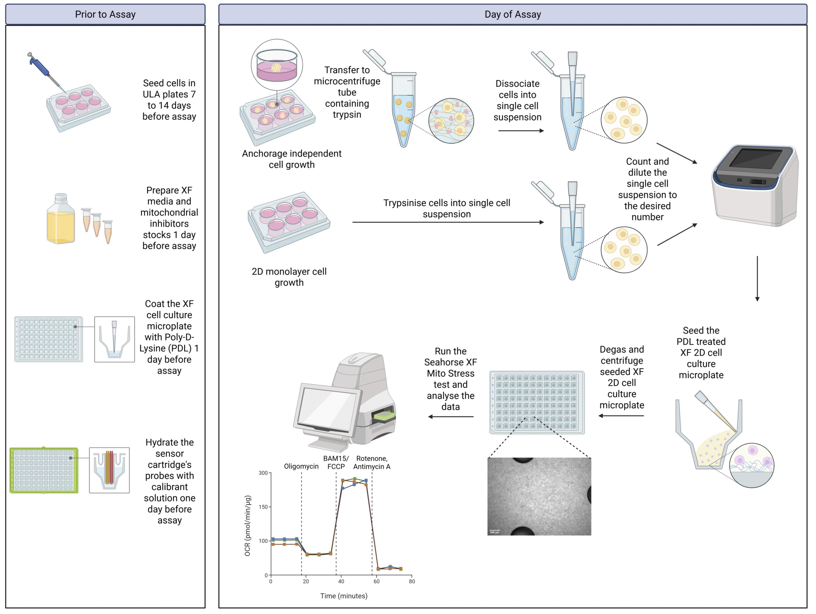
Here, we describe a dissociation-based Seahorse XF96/XF Pro workflow designed to overcome these limitations and enable robust quantification of the average bioenergetic state of anchorage-independent cancer cell clusters. In this approach, clusters generated in ultra-low adherent plates are enzymatically and mechanically dissociated into a single-cell suspension immediately before analysis, then seeded onto poly-D-lysine-coated 2D Seahorse microplates and profiled using the Mito Stress Test. By ensuring that each viable cell contributes equally to oxygen consumption rate (OCR) measurements under uniform acute conditions, the method reduces dependence on complex cluster-level normalization and directly reports per-cell respiratory capacity after growth in anchorage-independent conditions. This approach can also be applied to cells grown in 2D monolayers for direct comparison of bioenergetic profiles.
To demonstrate the utility of this workflow in a metastasis-relevant context, we applied it to an in vitro model in which A549 lung adenocarcinoma cells are examined as attached, detached, and reattached populations, modeling primary tumor cells (in terms of cell–extracellular matrix interactions), anchorage-independent circulating or clustered cells, and colonized secondary lesions, respectively. Using this dissociation-based Seahorse assay, we show that detachment induces a reversible state characterized by decreased oxidative capacity and that these features are largely restored upon reattachment. These proof-of-concept data illustrate how the workflow can capture dynamic, reversible bioenergetic remodeling associated with anchorage independence and reattachment.
Taken together, this work provides a practical and accessible platform to interrogate metabolic adaptations to anchorage-independent growth and cell clustering, complementing intact 3D assays and opening new opportunities to systematically explore bioenergetic vulnerabilities of metastasis-relevant cancer cell states. By enabling average per-cell bioenergetic profiling of cluster-derived cells with straightforward normalization, the described method supports more mechanistic studies of redox homeostasis, substrate usage, and mitochondrial function in models that better reflect the detachment and clustering events central to metastatic progression.
Materials and reagents
Biological materials
1. A549 lung adenocarcinoma cell line (Cellonex, catalog number: CA54-C)
Reagents
1. Dulbecco’s modified Eagle’s medium (DMEM), high glucose + L-glutamine (Thermo Fisher Scientific, catalog number: 11965092)
2. DMEM powder (Sigma-Aldrich, catalog number: D5030)
3. Fetal bovine serum (FBS) (Gibco, catalog number: A5256701)
4. Glucose, 200 g/L (Thermo Fisher Scientific, catalog number: A2494001)
5. L-Glutamine, 200 mM (Thermo Fisher Scientific, catalog number: 25030081)
6. Sodium pyruvate, 100 mM (Thermo Fisher Scientific, catalog number: 11360070)
7. Trypsin-EDTA, 0.5% (Thermo Fisher Scientific, catalog number: 15400054)
8. Cell-culture-grade water (Thermo Fisher Scientific, catalog number: A1287301)
9. Dimethyl sulfoxide (DMSO) (Sigma-Aldrich, catalog number: D2650)
10. Antimycin A (Sigma-Aldrich, catalog number: A8674)
11. BAM15 (5-N,6-N-bis(2-fluorophenyl)-1,2,5-oxadiazolo[3,4-b]pyrazine-5,6-diamine) (Sigma-Aldrich, catalog number: SML1760)
12. FCCP (Carbonyl cyanide-4-(trifluoromethoxy) phenylhydrazone) (Sigma-Aldrich, catalog number: C2920)
13. Oligomycin (Sigma-Aldrich, catalog number: O4876)
14. Rotenone (Sigma-Aldrich, catalog number: R8875)
15. HEPES (2-[4-(2-hydroxyethyl)piperazin-1-yl]ethanesulfonic acid) (Sigma-Aldrich, catalog number: H7523)
16. Phosphate-buffered saline (PBS) (Thermo Fisher Scientific, catalog number: 10010023)
17. Poly-D-Lysine (PDL), 0.1 mg/mL (Thermo Fisher Scientific, catalog number: A3890401)
18. Trypan Blue, 0.4% (Thermo Fisher Scientific, catalog number: T10282)
19. Sodium chloride (NaCl) (Sigma-Aldrich, catalog number: S9888)
Solutions
1. DMEM complete cell culture medium (see Recipes)
2. DMEM base medium (see Recipes)
3. XF cell assay medium (see Recipes)
4. Oligomycin, 2.5 mM in DMSO (see Recipes)
5. FCCP, 2.5 mM in DMSO (see Recipes)
6. BAM15, 3 mM in DMSO (see Recipes)
7. Rotenone and antimycin A mixture, 2.5 mM in DMSO (see Recipes)
Recipes
1. DMEM complete cell culture medium
| Reagent | Final concentration | Quantity or volume |
|---|---|---|
| DMEM, high glucose + L-glutamine | 90% | 500 mL |
| FBS | 10% | 50 mL |
| Total | 100% | 550 mL |
2. DMEM base medium
| Reagent | Final concentration | Quantity or volume |
|---|---|---|
| DMEM powder | 8.3 g/L | 8.3 g |
| Cell-culture-grade water | 99% | 990 mL |
| NaCl (3.2 M) | 32 mM | 10 mL |
| Total | n/a | 1000 mL |
Although it is recommended by Agilent to use the XF DMEM media for the Mito Stress Test, it is possible to prepare a similar base medium in-house.
3. XF cell assay medium
| Reagent | Final concentration | Quantity or volume |
|---|---|---|
| DMEM base medium | 92% | 46.05 g |
| HEPES (100 mM stock) | 5 mM | 2.5 mL |
| Glucose (200 g/L stock) | 10 mM | 450 μL |
| Sodium pyruvate (100 mM stock) | 1 mM | 500 μL |
| L-Glutamine (200 mM stock) | 2 mM | 500 μL |
| Total | n/a | 50 mL |
Prepare a total volume of 50 mL of XF cell assay medium no more than 4 h in advance to prevent glutamine degradation. Any XF cell assay medium can be used, preferably without phenol red.
4. Oligomycin, 2.5 mM in DMSO
| Reagent | Final concentration | Quantity or volume |
|---|---|---|
| Oligomycin | 2.5 mM | 39.6 mg |
| DMSO | 100% | 20 mL |
| Total | 20 mL |
5. FCCP, 2.5 mM in DMSO
| Reagent | Final concentration | Quantity or volume |
|---|---|---|
| FCCP | 2.5 mM | 24.5 mg |
| DMSO | 100% | 40 mL |
| Total | 40 mL |
6. BAM15, 3 mM in DMSO
| Reagent | Final concentration | Quantity or volume |
|---|---|---|
| BAM15 | 3 mM | 34 mg |
| DMSO | 100% | 40 mL |
| Total | 40 mL |
7. Rotenone and antimycin A mixture, 2.5 mM in DMSO
| Reagent | Final concentration | Quantity or volume |
|---|---|---|
| Rotenone | 2.5 mM | 39.4 mg |
| Antimycin A | 2.5 mM | 54.9 mg |
| DMSO | 100% | 40 mL |
| Total | 40 mL |
Note: Oligomycin, FCCP, BAM15, Rotenone, and Antimycin A are light-sensitive and should be stored appropriately. Stock solutions in DMSO should not be stored for longer than 1 month at -20 or -80 °C.
Laboratory supplies
1. T-75 tissue culture flask, vent cap, sterile (Celltreat, catalog number: 229341)
2. Ultra-low adherent plates, 6-well (Corning, catalog number: CLS3471)
3. Seahorse XFe96/XF Pro cell culture microplate (untreated) (Agilent, catalog number: 103794-100)
4. Seahorse XFe96/XF Pro FluxPak (includes sensor cartridge and calibrant solution) (Agilent, catalog number: 103793-100)
5. Cellulose acetate (CA) syringe filter, 0.22 μm (Bio-Smart Scientific, catalog number: SF-0.2-CA)
6. Countess cell counting chamber slides (Thermo Fisher Scientific, catalog number: C10228)
7. 5 mL serological pipettes (Whitehead Scientific, catalog number: 326001)
8. 10 mL serological pipettes (Whitehead Scientific, catalog number: 327001)
9. 25 mL serological pipettes (Whitehead Scientific, catalog number: 328001)
10. 10 μL pipette tips (Whitehead Scientific, catalog number: TFLR102-10)
11. 200 μL pipette tips (Whitehead Scientific, catalog number: 312012)
12. 1,000 μL pipette tips (Whitehead Scientific, catalog number: 313012)
Equipment
1. Agilent Seahorse XF Pro Analyzer (Agilent Technologies, model: S7850A/XF Pro)
2. Countess 3 Automated Cell Counter (Thermo Fisher Scientific, model: AMQAX2000)
3. EVOS M700 fluorescent cell imaging system (Thermo Fisher Scientific, model: AMF7000)
4. pH meter (Metrohm, catalog number: 2.913.0010, model: 913)
5. HeracellTM 150i CO2 incubator, 150 L (Thermo Fisher Scientific, catalog number: 51032871)
6. FILTA-MATIX Biological Safety Cabinet (FILTA-MATIX, model: BIO SAFE 6)
Note: Any Class II, Type A2 biosafety cabinet suitable for BSL-2 cell culture work may be used as an alternative, such as the Thermo ScientificTM 1500 Series A2 Class II, Type A2 Biological Safety Cabinet (Thermo Scientific, catalog number: 1598A2).
7. MyTempTM mini digital incubator (non-CO2) (Merck, catalog number: Z763330)
8. Eppendorf® Centrifuge 5810R (Merck, model: Eppendorf® Centrifuge 5810)
9. Water bath (Grant Instruments, catalog number: NC1808978)
10. Corning® StripettorTM Ultra Pipet Controller (Merck, catalog number: CLS4099)
11. FinnpipetteTM F2 GLP Kit (Thermo Fisher Scientific, catalog number, 4700870)
12. FinnpipetteTM F2 Multichannel Pipette (Thermo Fisher Scientific, catalog number: 4662020)
Software and datasets
1. Seahorse Pro Controller (Agilent, Version 10.2.1)
2. Seahorse Wave Pro (Agilent, Version 10.4.1)
3. Excel (Microsoft Office 2024)
4. GraphPad Prism 11
Procedure
登录/注册后免费查看全文
文章信息
稿件历史记录
提交日期: Aug 17, 2026
接收日期: Sep 17, 2026
在线发布日期: Oct 11, 2026
出版日期: Oct 20, 2026
版权信息
© 2026 The Author(s); This is an open access article under the CC BY-NC license (https://creativecommons.org/licenses/by-nc/4.0/).
如何引用
Lindeque, R., Visagie, J. L., Fouché, B. R. and Labuschagne, C. F. (2026). A Dissociation-Based Seahorse XF Workflow for Quantifying Averaged Bioenergetics in Anchorage-Independent Cancer Cell Clusters. Bio-protocol 16(20): e5849. DOI: 10.21769/BioProtoc.5849.

 Download Citation in RIS Format
您对这篇实验方案有问题吗?
在此处发布您的问题,我们将邀请本文作者来回答。同时,我们会将您的问题发布到Bio-protocol Exchange,以便寻求社区成员的帮助。
 发布问题
发布问题


