


An Improved Method for Rapid Unmarked Gene Deletion in Bacteria: Pseudomonas aeruginosa as an Example

发布: 2026年10月20日第16卷第20期 DOI: 10.21769/BioProtoc.5847 浏览次数: 53
评审: Alba BlesaVandana SinghAnonymous reviewer(s)
 PDF
PDF  Q&A
Q&A
参见作者原研究论文
The authors used this protocol in:
Sep 2012





Abstract
The release and updating of whole-genome sequences of several representatives of the human opportunistic pathogen Pseudomonas aeruginosa have laid the ground for investigating the mechanisms of antibiotic resistance, biofilm formation, and virulence, while also offering opportunities for researchers to find new therapeutic targets to control this bacterium using a functional genomics approach. However, there is still a lack of detailed protocols describing gene inactivation methods in P. aeruginosa, resulting in failures and extra time spent designing in-house protocols. Here, we introduce a rapid, efficient, and unmarked deletion mutagenesis method combining overlap extension PCR, efficient conjugation, and the traditionally used sacB-based counter-selection procedure. Efficient generation of deletion mutants using this detailed protocol can be easily completed in one week using standard lab reagents. Importantly, this method may be adaptable to other bacteria where the sacB-based counter-selection system works.
Key features
• Using modified overlap extension PCR (MOE-PCR), the deletion construct can be generated rapidly in a single fusion PCR without intermediate gel purification.
• Only one transformation step is involved in the whole protocol, thus saving time compared to marked mutagenesis involving Flp (flippase) recombinase recognition target (FRT)-mediated resolution.
• Pseudomonas agar P medium (PM) and casamino acid medium (CAA) reduce reactive oxygen species (ROS)-mediated toxicity, facilitating deletion of oxidative stress–sensitive genes.
• The sacB-based counter-selection works in many Gram-negative bacteria and even some Gram-positive actinobacteria, so the protocol may be adaptable to such species.
Keywords: P. aeruginosa Gene deletion
Unmarked mutagenesis
Overlap extension PCR
sacB counter-selection
Allelic exchange
MOE-PCR
Gene deletion
Unmarked mutagenesis
Overlap extension PCR
sacB counter-selection
Allelic exchange
MOE-PCR
Graphical overview
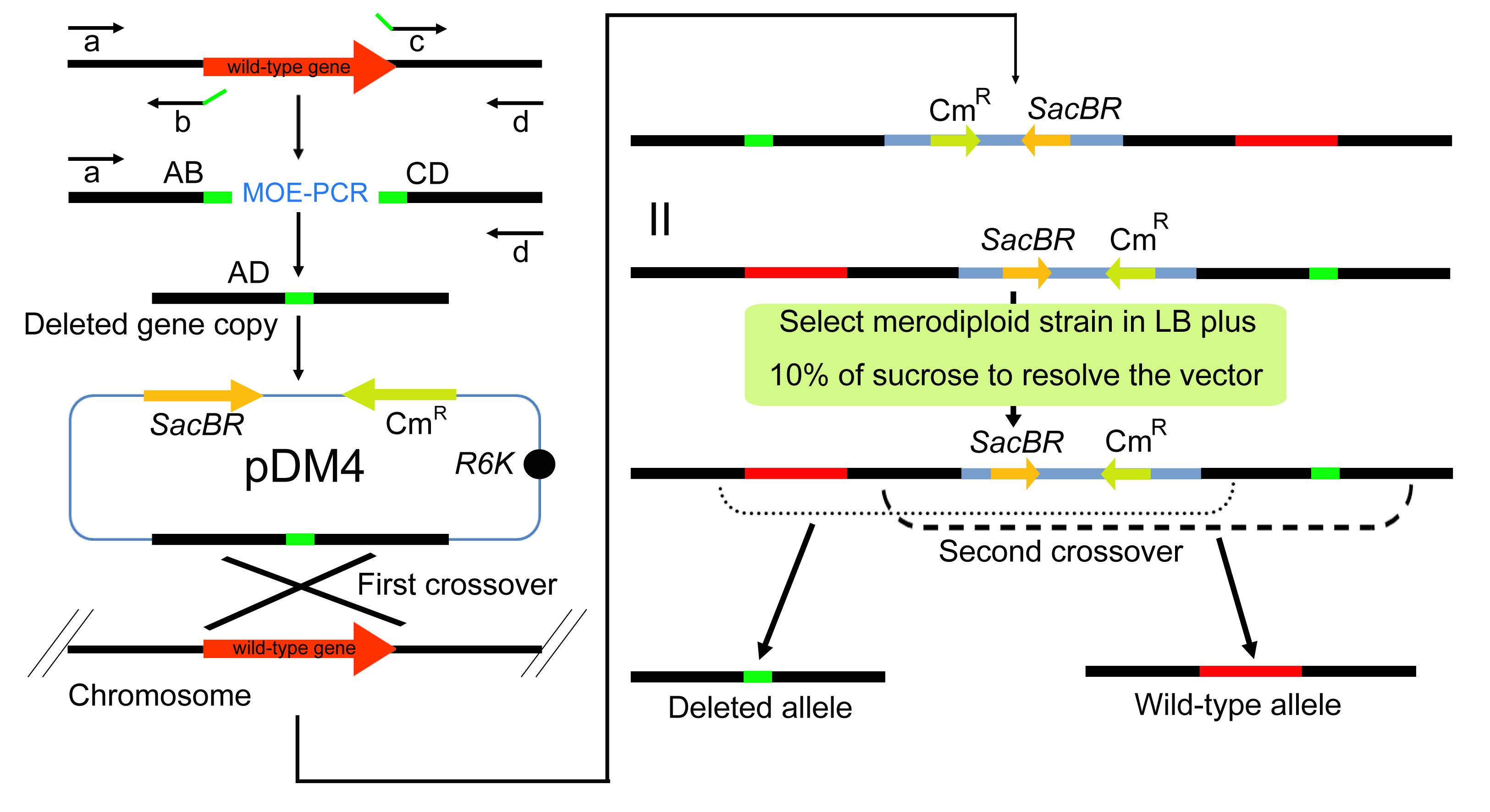
Workflow of deletion mutagenesis in P. aeruginosa. (A) The upstream (AB) and downstream (CD) homologous arms are amplified from the wild-type chromosome with primer pairs a/b and c/d. AB and CD share complementary sequences at their internal junction and are fused by MOE-PCR to generate the deletion cassette AD, in which the target gene is removed. The AD cassette is cloned into the sacB-containing suicide vector pDM4, and the recombinant plasmid is established in E. coli before its transfer into P. aeruginosa by conjugation. A single crossover between the cassette and the chromosomal target integrates pDM4 and creates a merodiploid carrying both the wild-type allele and the deleted allele. (B) The merodiploid is plated on Luria-Bertani (LB) agar without NaCl, containing 10% sucrose. Cells that still carry the integrated pDM4 vector express sacB and die from the accumulation of levan, so only cells in which a second crossover has excised the vector together with one duplicated allele survive. The chromosome of such cells retains either the wild-type allele or the deleted allele, and colony PCR with primers a and d identifies the markerless deletion mutants among the survivors. Note: The flanking primers (a and d) contain at least 17 nt perfectly matched to the target sequence plus a unique restriction site at their 5′ ends for cloning into pDM4. The internal primers (b and c) are 17–35 nt in length and share a 15–25 bp complementary overlap at their 5′ ends, and their Tm values are balanced against those of the flanking primers. The pDM4-AD construct is first established in E. coli strain S17-1 λpir and is then transferred into P. aeruginosa by conjugation.
Background
P. aeruginosa is a ubiquitous Gram-negative environmental bacterium that has remarkable metabolic diversity, resulting in the colonization of a multitude of ecological niches. This bacterium is noted as an important human opportunistic pathogen with the ability to cause acute and chronic infections. In 2000, Stover and his colleagues reported the whole genome sequence of P. aeruginosa type strain PAO1, providing insights into the basis of its pathogenesis, as well as multiple antibiotic resistance and versatile metabolism [1]. However, more than 2,000 genes (ca. 40% of the entire genome) of P. aeruginosa were predicted to encode (conserved) hypothetical proteins, with no clearly predicted functions. This blueprint provides potential opportunities for researchers to explore the functions of undefined genes of this bacterium, thus opening the post-genomic era for Pseudomonas.
Multiple genetic tools and methods have been generated and applied to study the biology of P. aeruginosa, including transposon (knockin) and deletion mutagenesis (knockout). Both have proved to be powerful techniques in the identification of functions of some of those uncharacterized genes, although the functions of certain essential genes (ca. 300–400 for P. aeruginosa) could not be resolved this way [2]. A variety of transposon libraries have been developed for P. aeruginosa strains based on different reporter genes, such as phoA [2–4], lacZ [2], gfp [5], and lux [6]. In addition, a plasposon and a mariner transposon library were also constructed to study the pathogenesis of P. aeruginosa [4,7,8]. While these mutant libraries have greatly facilitated high-throughput genomic studies, functional genomics involving specific non-polar gene knockouts necessitates an efficient deletion mutagenesis approach. Several methods based on allelic exchange have been developed, which combine the use of suicide vectors and counter-selectable markers [9–11]. Among these, Hmelo et al. (2015) [12] established a field-standard two-step allelic exchange protocol for P. aeruginosa, which has since served as the methodological benchmark for unmarked deletion methods in this organism. Unfortunately, although the previously described methods for rapid and efficient mutant generation have been useful in numerous studies, detailed protocols are often still lacking and require further modification to reduce the time and cost of mutant generation and improve its speed and reproducibility.
In this protocol, we describe an efficient approach to generate unmarked deletion mutants in P. aeruginosa based on the gene deletion method we used in previous studies [13–17]. Briefly, MOE-PCR was used to amplify a region containing the flanking sequences of the target gene to be deleted, which was subsequently ligated to a suicide vector (pDM4) [18–20] and introduced into P. aeruginosa by triparental mating with the mobilizable pDM4 derivative. The donor E. coli S17-1 λpir carries the recombinant suicide plasmid, the helper E. coli S17-1 λpir carrying pRK2013 supplies the RP4-derived Mpf machinery in trans, and the recipient is P. aeruginosa PAO1. Because pDM4 carries only the oriT and the TraJ relaxosome but lacks the Mpf machinery, it is mobilizable but not self-transmissible, and the helper strain is essential for plasmid transfer into PAO1. This configuration gives robust conjugation efficiencies sufficient for routine unmarked deletion mutagenesis and avoids the lower efficiencies typical of direct electroporation or chemical transformation of P. aeruginosa, which are limited by its restriction-modification barrier [21]. Second, merodiploids arising from chromosomal integration of the suicide vector through homologous recombination are resolved by the use of the counter-selection imposed by the sacB gene harbored on pDM4 [22]. Finally, gene disruption is confirmed by colony PCR (coPCR) and sequencing, if required. The entire procedure takes less than one week, is easy to perform, is not laborious, and is suitable for large-scale deletion analysis. In addition, the lethal effect of the sacB gene when cells are grown in the presence of sucrose is due to the accumulation of the levan polysaccharide in the periplasm [23,24], which means that this selection method works in most Gram-negative bacteria such as Acinetobacter baumannii, Erwinia chrysanthemi, Legionella pneumophila, Anabaena sp., Yersinia sp., Rhizobium sp., Xanthomonas sp., Pseudomonas sp., Klebsiella pneumoniae, Helicobacter pylori, Agrobacterium sp., and even in some Gram-positive actinobacteria, such as Corynebacterium glutamicum and Mycobacterium sp. [25]. Therefore, the method described here could serve as a general protocol for the genetic manipulation of those bacteria.
This proposed deletion mutagenesis approach has several advantages over other methods. First, MOE-PCR eliminates the intermediate gel-purification step required in conventional overlap extension PCR, saving approximately 4 h of hands-on time per target, reducing gel extraction reagent cost, and avoiding UV exposure of the amplicon, a known source of UV-induced mutations. Second, only one transformation step is involved, saving substantial time relative to marked mutagenesis with FRT-mediated resolution. Third, PM and CAA make it considerably easier to delete oxidative stress-response genes. Aerated LB auto-oxidizes during shaking culture and generates micromolar concentrations of H2O2 lethal to ROS-defective mutants [26]. In our hands, PM and CAA result in a higher recovery of oxidative stress–defective mutants than LB.
Materials and reagents
Biological materials
1. P. aeruginosa PAO1 (wild type; recipient strain) [1]
2. E. coli S17-1 λpir (thi pro hsdR hsdM+ recA RP4-2-Tc::Mu-Km::Tn7 λpir; cloning host and donor strain) [27]
3. E. coli S17-1 λpir (pRK2013) (helper strain for triparental conjugation) [28]
The strains and plasmids used in this protocol are listed in Table 1.
Table 1. Strains and plasmids used in this protocol
| Strain or plasmid | Relevant features | Use in this protocol | Antibiotic concentration |
|---|---|---|---|
| P. aeruginosa PAO1 | Wild type | Recipient strain for conjugation | None; transconjugants are selected on LB agar with Cm 300 μg/mL (section M) |
| E. coli S17-1 λpir | thi pro hsdR hsdM+ recA RP4-2-Tc::Mu-Km::Tn7 λpir | Cloning host. Donor strain carrying pDM-AD | Cm 50 μg/mL on LB agar (section J); Cm 25 μg/mL in LB broth for pDM-AD clones (section K) |
| E. coli S17-1 λpir (pRK2013) | Helper plasmid pRK2013 (ColE1 replicon, Tra+, KmR) | Helper strain for triparental conjugation (section L) | Km 50 μg/mL |
| pDM4 | Suicide vector. oriR6K (requires λpir), sacB, CmR, mob/oriT (RP4) | Suicide vector for allelic exchange | Cm as above |
| pDM-AD | pDM4 carrying the fused deletion construct AD | Recombinant suicide vector | Cm as above |
Reagents
1. Phusion High-Fidelity PCR kit (200 reactions) (New England BioLabs, catalog number: F553L) and Taq DNA polymerase (Qiagen, catalog number: 201203)
Critical: Use high-fidelity DNA polymerase to amplify the deleted gene copy, but use normal DNA polymerase (which is cheaper than the high-fidelity polymerase) to perform coPCRs when screening for positive clones containing the deletion. Phusion High-Fidelity DNA Polymerase is used in section B, and Taq DNA Polymerase is used in section N.
2. Gentra Puregene Yeast/Bact. kit (200 mL) (Qiagen, catalog number: 158567)
3. QIAprep Spin Miniprep kit (250) (Qiagen, catalog number: 27106)
4. Deionized H2O or Milli-Q ultrapure H2O or double-distilled H2O, autoclaved
5. 10 mM dNTP mix (10 mM each dNTP) (Invitrogen, catalog number: 18427-013)
6. Ethidium bromide (Sigma-Aldrich, catalog number: E1510)
7. DNA marker (Smart ladder 1000 lanes) (Eurogentec, catalog number: MW-1700-10)
8. Restriction enzymes: SalI (Thermo Fisher Scientific, Fermentas, catalog number: ER0641) and XbaI (Thermo Fisher Scientific, Fermentas, catalog number: ER0681)
9. Rapid DNA Ligation kit (Fermentas, catalog number: K1422)
10. GenElute Gel Extraction kit (50) (Sigma-Aldrich, catalog number: NA1111-1KT)
11. QIAquick PCR Purification kit (250) (Qiagen, catalog number: 28106)
12. UltraPure agarose (Invitrogen, catalog number: 16500500)
13. 6× DNA loading dye (Fermentas, catalog number: R0611)
14. Tris-HCl (1 M, pH 8.0) (Thermo Fisher, catalog number: 15567-027)
15. EDTA (0.5 M, pH 8.0) (Thermo Fisher, catalog number: 15575-038)
16. Sucrose (Merck, catalog number: 1.07687)
17. RbCl (Sigma-Aldrich, catalog number: R2252)
18. MnCl2·4H2O (Sigma-Aldrich, catalog number: M3634)
19. Potassium acetate (Sigma-Aldrich, catalog number: P1190)
20. CaCl2·2H2O (Sigma-Aldrich, catalog number: C5080)
21. Glycerol (Sigma-Aldrich, catalog number: G5516)
22. MOPS (Sigma-Aldrich, catalog number: M3183)
23. Tris base (Sigma-Aldrich, catalog number: T1503)
24. Glacial acetic acid (Sigma-Aldrich, catalog number: A6283)
25. Chloramphenicol (Sigma-Aldrich, catalog number: C0378)
26. Gelysate peptone (BD BBL, catalog number: 211870)
27. K2SO4 (Sigma-Aldrich, catalog number: P9458)
28. MgCl2 (Sigma-Aldrich, catalog number: M8266)
29. MgSO4·7H2O (Sigma-Aldrich, catalog number: M1880)
30. DMSO (Sigma-Aldrich, catalog number: D8418)
31. Agar (Sigma-Aldrich, catalog number: A1296)
32. Casamino acids (BD, Bacto, catalog number: 223050)
33. K2HPO4·3H2O (Sigma-Aldrich, catalog number: P9666)
34. GelRed nucleic acid gel stain (Biotium, catalog number: 41003) or equivalent safer DNA stain
35. Oligonucleotide primers a–d (17–35 nt each, designed as described in Primer design), HPLC-purified grade (Sangon Biotech, Shanghai, China; custom-synthesized)
Critical: All primers must be ordered as HPLC-purified grade to ensure high fidelity and avoid synthesis errors that could compromise the overlap extension PCR.
36. Absolute ethanol (Sinopharm Chemical Reagent Co., Ltd., Shanghai, China, catalog number: 10009218)
37. Tryptone (BD Bacto, catalog number: 211705)
38. Yeast extract (BD Bacto, catalog number: 212750)
39. Sodium chloride (NaCl) (Sigma-Aldrich, catalog number: S9888)
40. Potassium hydroxide (KOH) (Sigma-Aldrich, catalog number: 221473)
41. Sodium hydroxide (NaOH) (Sigma-Aldrich, catalog number: 221465)
42. Hydrochloric acid (HCl) (Sigma-Aldrich, catalog number: 258148)
43. Syringe filter (0.22 μm, PES) (Millipore, catalog number: SLGP033RS)
Solutions
1. Chemically competent E. coli S17-1 λpir cells (see Recipes)
a. RF1 buffer
b. RF2 buffer
2. Bacterial plates containing appropriate antibiotics (see Recipes)
a. LB broth
b. LB broth without NaCl containing 10% (w/v) sucrose
c. Cm stock solution
d. King's A medium (Pseudomonas agar P medium, PM)
3. 50× TAE stock buffer (see Recipes)
4. TE buffer (see Recipes)
5. 50% sucrose (see Recipes)
6. CAA medium (see Recipes)
Recipes
1. Chemically competent E. coli S17-1 λpir cells
For cloning of the deleted copy into the suicide vector pDM4 (Figure 1), E. coli strain S17-1 λpir (thi pro hsdR hsdM+ recA RP4-2-Tc::Mu-Km::Tn7 λpir) was used, since it produces the λpir protein to maintain the replication of pDM4 plasmid and its derivatives [27]. Chemically competent cells are used in our protocol and made according to the rubidium chloride (RbCl) method [29], which is supposed to increase competence compared to the calcium chloride (CaCl2) method. Alternatively [30], the CaCl2 method can be substituted. The relevant features and antibiotic concentrations of the strains and plasmids used in this protocol are summarized in Table 1 above.
Critical: Prepare RF1 and RF2 buffers freshly. Add metal salts (RbCl, MnCl2, CaCl2) after pH adjustment to avoid precipitation.
a. RF1 buffer (Buffer I for inducing chemical competence)
| Reagent | Final concentration | Quantity or volume |
|---|---|---|
| RbCl | 100 mM | 1.21 g |
| MnCl2·4H2O | 50 mM | 0.99 g |
| Potassium acetate | 30 mM | 0.294 g |
| CaCl2·2H2O | 10 mM | 0.148 g |
| Glycerol | 15% (w/v) | 15 g |
| Deionized H2O | n/a | Up to 100 mL |
Note: Dissolve potassium acetate and glycerol in ~80 mL of deionized H2O. Adjust pH to 5.8 with 0.2 M acetic acid. Do NOT overshoot (Mn2+ precipitates at alkaline pH). Then, dissolve metal salts. Sterilize by filtration (0.22 μm). Store at 4 °C.
b. RF2 buffer (Buffer II for inducing chemical competence)
| Reagent | Final concentration | Quantity or volume |
|---|---|---|
| 3-(N-morpholino) propane sulfonic acid (MOPS) | 10 mM | 0.209 g |
| RbCl | 10 mM | 0.121 g |
| CaCl2·2H2O | 75 mM | 1.10 g |
| Glycerol | 15% (w/v) | 15 g |
| Deionized H2O | n/a | Up to 100 mL |
Note: Dissolve MOPS and glycerol in ~80 mL of deionized H2O. Adjust pH to 6.5 with 1 M KOH. Do NOT overshoot. Then, dissolve metal salts. Sterilize by filtration (0.22 μm). Store at 4 °C.
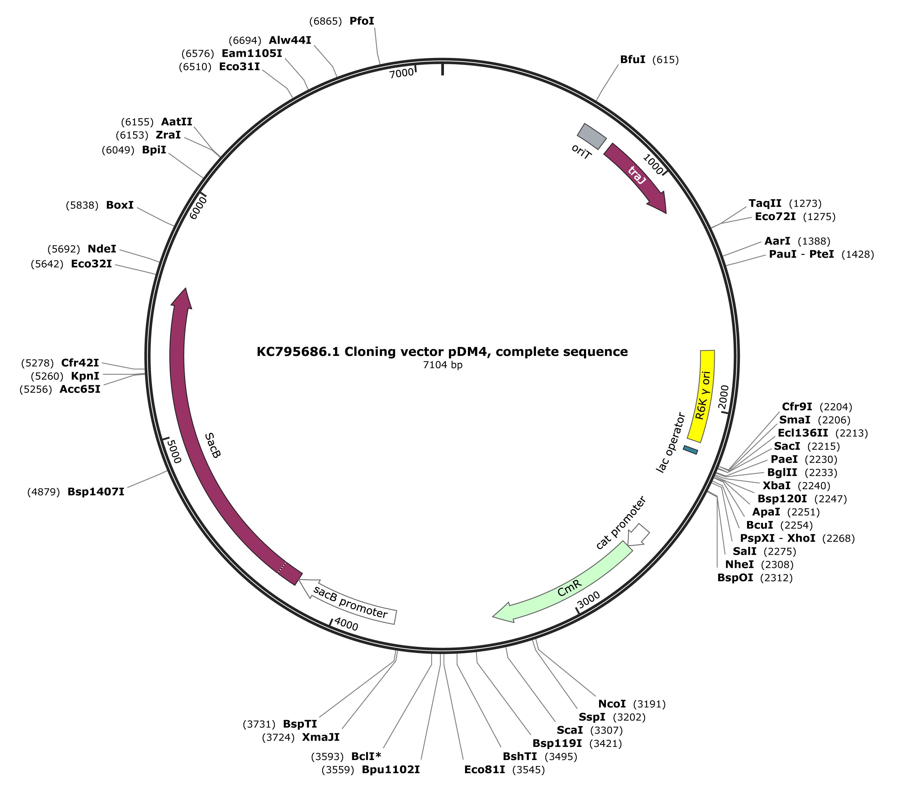
Figure 1. Map of the pDM4 suicide vector. The pDM4 vector (KC795686.1, 7104 bp) carries the R6K γ origin of replication, which requires the λpir protein and therefore restricts propagation to E. coli S17-1 λpir, the RP4 origin of transfer (oriT) and the traJ gene for conjugative mobilization, the sacB gene and its promoter for sucrose counter-selection, and the chloramphenicol resistance gene (cat) under the control of its own promoter. Restriction sites suitable for cloning the deletion cassette AD are indicated.
2. Bacterial plates containing appropriate antibiotics
For cloning of the deletion construct, LB agar plates containing 50 μg/mL chloramphenicol (Cm) are used. Cm serves as the selection marker, because the pDM4 suicide vector carries a chloramphenicol resistance gene. For the selection of P. aeruginosa transconjugants, LB agar plates containing 300 μg/mL Cm are used. For the counter-selection of double recombinants, LB medium without NaCl and containing 10% (w/v) sucrose is used. King's A medium (Pseudomonas agar P medium, PM) is suitable for the isolation of mutants, especially for strains with defects in oxidative stress defense. The recipes of these media are given below.
Critical: Sucrose must be filter-sterilized and added to the cooled autoclaved medium to avoid caramelization.
a. LB broth
| Reagent | Final concentration | Quantity or volume |
|---|---|---|
| Tryptone | 1% (w/v) | 10 g |
| Yeast extract | 0.5% (w/v) | 5 g |
| NaCl | 1% (w/v) | 10 g |
| Deionized H2O | n/a | Up to 1 L |
Note: Dissolve the components in about 900 mL of deionized water, adjust the pH to 7.0 with NaOH if necessary, and bring the volume to 1 L. Sterilize by autoclaving at 121 °C for 20 min. For LB agar plates, add 15 g of agar per liter before autoclaving. LB medium without NaCl is prepared by omitting NaCl from this recipe. Add the Cm stock solution only after the autoclaved medium has cooled to about 50 °C.
b. LB broth without NaCl containing 10% (w/v) sucrose (1 L, approx. 40 plates)
Critical: Sucrose must be filter-sterilized (0.22 μm) and added to the autoclaved medium after it has cooled to approximately 50 °C to avoid caramelization.
| Reagent | Final concentration | Quantity or volume |
|---|---|---|
| LB broth (prepare as above, but omit NaCl) | n/a | 800 mL |
| Sucrose stock [50% (w/v), filter-sterilized] | 10% (w/v) | 200 mL |
Note: The purpose of removing NaCl from the LB broth is to decrease the osmotic potential, which enhances the osmotic stress mediated by the levansucrase encoded by the sacB gene integrated into the chromosome of P. aeruginosa, leading to the rapid resolution of the plasmid from the genome. A sucrose concentration of 10% (w/v) is recommended as a starting concentration for counter-selection. The concentration can be increased up to 20% (w/v) if background growth or false-positive survivors indicate insufficient counter-selection, or decreased to 5% (w/v) if sucrose toxicity substantially impairs the recovery or growth of candidate recombinants. For LB agar plates, add 15 g of agar per liter before autoclaving.
c. Cm stock solution
| Reagent | Final concentration | Quantity or volume |
|---|---|---|
| Chloramphenicol | 30 mg/mL | 300 mg |
| Absolute ethanol | n/a | 10 mL |
Note: Filter sterilization is not required, as the stock is prepared in absolute ethanol. Store at -20 °C in the dark.
d. King's A medium (Pseudomonas agar P medium, PM)
| Reagent | Final concentration | Quantity or volume |
|---|---|---|
| Gelysate peptone | 20 g/L | 20 g |
| Glycerol | 10 mL/L | 10 mL |
| Agar | 15 g/L | 15 g |
| K2SO4 | 10 g/L | 10 g |
| MgCl2 | 1.4 g/L | 1.4 g |
| Deionized H2O | n/a | Up to 1 L |
Note: Glycerol serves as the carbon source. Agar is added before autoclaving when plates are required for the isolation of mutants. Adjust pH to 7.0 with 1 M KOH or HCl before autoclaving. Supplement with appropriate antibiotics as described above.
3. 50× TAE stock buffer
50× TAE is diluted with distilled water (1:50) to make a 1× working solution for the preparation of agarose gel.
| Reagent | Final concentration | Quantity or volume |
|---|---|---|
| Tris base (MW 121.1) | 242 g/L (2 M, 50×) | 242 g |
| Glacial acetic acid | 57.1 mL/L (50×) | 57.1 mL |
| EDTA (0.5 M, pH 8.0) | 50 mM (50×) | 100 mL |
Note: All solutions can be stored at room temperature.
4. TE buffer
| Reagent | Final concentration | Quantity or volume |
|---|---|---|
| Tris-HCl (1 M, pH 8.0) | 10 mM | 10 mL |
| EDTA (0.5 M, pH 8.0) | 1 mM | 2 mL |
| Deionized H2O | n/a | Up to 1 L |
Note: Autoclave before use. TE buffer can be stored at room temperature for up to 6 months.
5. 50% sucrose
| Reagent | Final concentration | Quantity or volume |
|---|---|---|
| Sucrose | 50% (w/v) | 50 g |
| Deionized H2O | n/a | Up to 100 mL |
Note: Filter-sterilize. The 50% sucrose solution can be stored at 4 °C for up to 4 months.
6. CAA medium
| Reagent | Final concentration | Quantity or volume |
|---|---|---|
| Casamino acids | 5 g/L | 5 g |
| K2HPO4·3H2O | 1.18 g/L | 1.18 g |
| MgSO4·7H2O | 0.25 g/L | 0.25 g |
| Agar | 15 g/L | 15 g (when plates are required) |
| Deionized H2O | n/a | up to 1 L |
Note: This is an iron-poor casamino acid medium used for growing P. aeruginosa under iron-limiting conditions, as previously described [17]. Adjust pH to 7.0 with 1 M KOH before autoclaving. Add agar 15 g/L when plates are required. Sterilize by autoclaving.
Equipment
1. Thermal cycler (Eppendorf Mastercycler Gradient, catalog number: 5331 000.011 or equivalent)
2. Agarose gel electrophoresis system (Bio-Rad, model: Sub-Cell GT, catalog number: 170-4406 or equivalent)
3. Gel imaging system (Bio-Rad, model: Gel Doc XR+, catalog number: 1708195 or equivalent)
4. PCR tubes (0.2 mL thin-walled, e.g., Axygen, catalog number: PCR-02-C or equivalent)
5. NanoDrop 2000 spectrophotometer (Thermo Scientific, catalog number: ND-2000, or equivalent)
6. Benchtop incubator (Innova 40, New Brunswick Scientific or equivalent)
7. Biological safety cabinet (Class II, or equivalent)
8. Benchtop centrifuge 5417C (Eppendorf, catalog number: 5417 000.315, rotor FA-45-30-11, or equivalent)
Procedure
登录/注册后免费查看全文
文章信息
稿件历史记录
提交日期: Jul 7, 2026
接收日期: Sep 10, 2026
在线发布日期: Sep 23, 2026
出版日期: Oct 20, 2026
版权信息
© 2026 The Author(s); This is an open access article under the CC BY-NC license (https://creativecommons.org/licenses/by-nc/4.0/).
如何引用
Zhang, J., Wang, P., Zhang, X., Wei, Q. and Cui, K. (2026). An Improved Method for Rapid Unmarked Gene Deletion in Bacteria: Pseudomonas aeruginosa as an Example. Bio-protocol 16(20): e5847. DOI: 10.21769/BioProtoc.5847.

 Download Citation in RIS Format
分类
微生物学 > 微生物遗传学 > 基因组编辑
分子生物学 > DNA > 诱/突变
您对这篇实验方案有问题吗?
在此处发布您的问题,我们将邀请本文作者来回答。同时,我们会将您的问题发布到Bio-protocol Exchange,以便寻求社区成员的帮助。
 发布问题
发布问题


