


Antiangiogenic Drug Testing Using Proangiogenic and Hypoxia Zebrafish Model

发布: 2026年10月20日第16卷第20期 DOI: 10.21769/BioProtoc.5846 浏览次数: 50
评审: Alberto RissoneRajesh D GunageAnonymous reviewer(s)
 PDF
PDF  Q&A
Q&A
参见作者原研究论文
The authors used this protocol in:
Aug 2025





相关实验方案
Abstract
Zebrafish is an excellent in vivo model for high-throughput antiangiogenic drug testing, commonly known as the zebrafish angiogenesis assay. Conventional zebrafish angiogenesis assays are performed in wild-type zebrafish to evaluate the vascular changes in intersegmental and subintestinal vessel regions of the zebrafish larvae at 2 and 3 dpf stages, respectively. However, wild-type zebrafish larvae do not adequately mimic the hypoxia microenvironment and ectopic vessel branching characteristics of cancer. To overcome this limitation, we developed a genetically engineered zebrafish model with constitutive activation of the hypoxia signaling pathway by targeting the vhl, a tumor suppressor, by negative regulation of the hypoxia pathway, using CRISPR mutagenesis. This model exhibits robust ectopic blood vessel branching throughout the larval body, thereby recapitulating pathological angiogenesis. The utility of this zebrafish model system for drug screening was validated with the sorafenib treatment, a known antiangiogenic tyrosine kinase inhibitor. Overall, this proangiogenic hypoxia zebrafish model provides a physiologically relevant platform for testing antiangiogenic drugs.
Key features
• Generate a vhl mutant zebrafish line with constitutive activation of the hypoxia signaling pathway using CRISPR-CAS9 mutagenesis.
• The generated model exhibits extensive ectopic neovascular branching and abnormal vascular growth resembling pathological angiogenesis observed in solid tumors.
• Mimics the chronic hypoxic microenvironment commonly associated with cancer progression and angiogenesis.
• Provides an economical alternative to mammalian tumor models for early-stage antiangiogenic drug testing.
Keywords: Zebrafish Hypoxia signaling
Tg(fli1:EGFP)
Angiogenesis
Antiangiogenic drug testing
In vivo disease model
Hypoxia signaling
Tg(fli1:EGFP)
Angiogenesis
Antiangiogenic drug testing
In vivo disease model
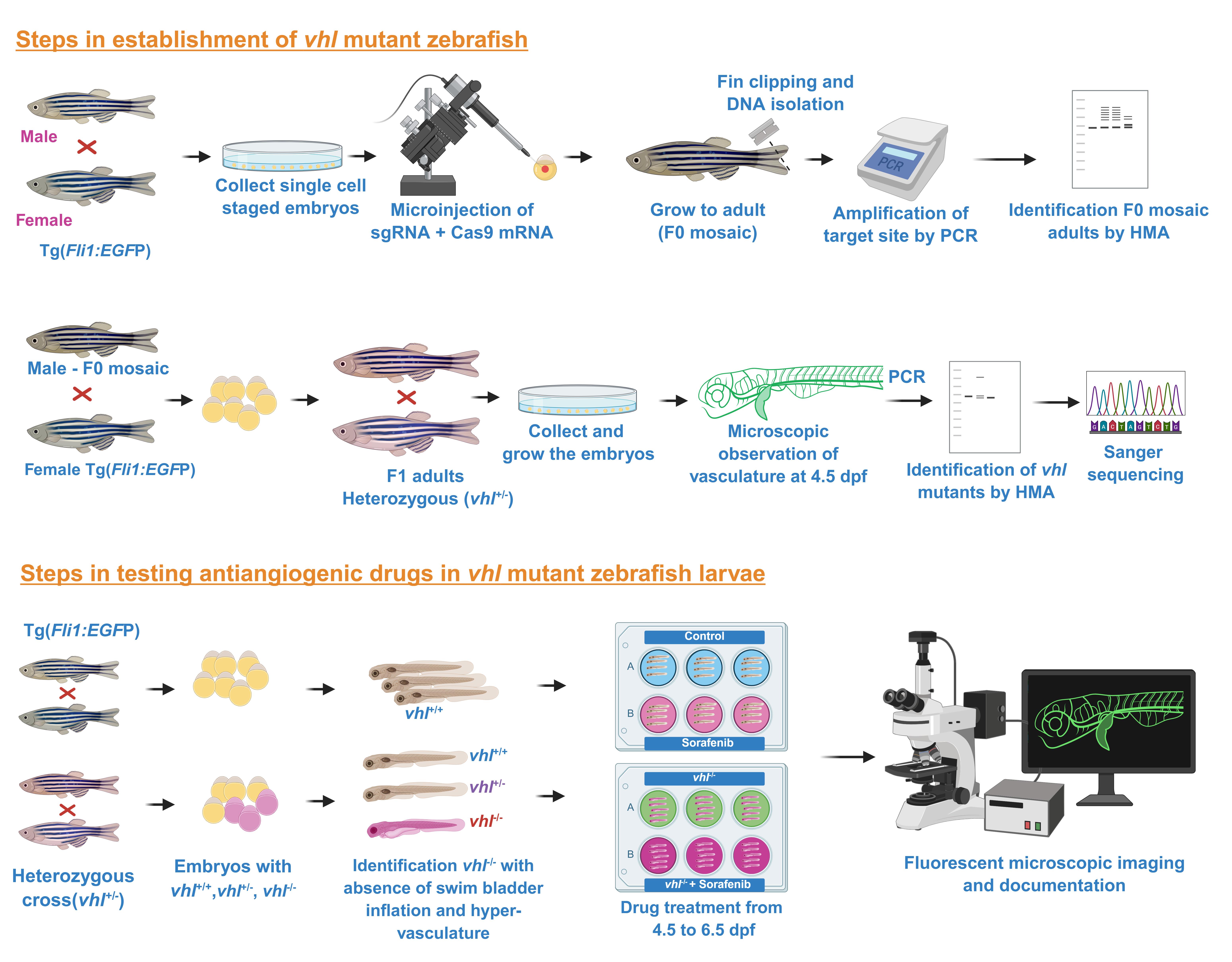
Graphical overview
Background
Cancer angiogenesis is a process of forming abnormal ectopic neovasculatures that are essential for tumor growth and metastasis [1]. Therefore, the identification of novel antiangiogenic compounds has become an important area in cancer therapeutics. Antiangiogenic drugs can be efficiently tested in the high-throughput zebrafish angiogenesis assay [2,3]. The availability of blood vessel–specific transgenic lines such as Tg(fli1:EGFP) and Tg(kdrl:EGFP), which enable real-time visualization of vascular development and angiogenesis in live embryos, and the transparency of early larval stages make zebrafish a suitable model for antiangiogenic drug testing [4]. In zebrafish, the angiogenesis assay has been conducted in wild-type larvae by exposing the embryos to test compounds at 1 dpf and analyzing the vascular changes in the intersegmental vessels (ISVs) and subintestinal vessel (SIV) regions at 2 and 3 dpf, respectively [5,6]. The ISVs are particularly suitable for angiogenesis assessment because they normally form stereotypic secondary vessels without ectopic terminal neovasculatures. Under activated hypoxia signaling, such as in the vhl mutant, abnormal vascular sprouting and ectopic neovascularization can be readily observed in the ISVs [6,7]. Although this conventional model is useful for identifying antiangiogenic compounds that may show efficacy in normal zebrafish larvae, it may not have similar efficacy in patients experiencing hypoxia, a major driver of drug resistance.
To overcome this limitation, we generated a hypoxia zebrafish model by targeting the exon 3 (the alpha domain that helps in the formation of the Vhl complex, which is involved in the negative regulation of Hifα) of the vhl through CRISPR mutagenesis, based on our previous studies in bioinformatics analysis of identifying the most pathological and conserved position of human VHL mutation [6]. The VHL is a tumor-suppressor gene that negatively regulates hypoxia-inducible factor alpha (HIFα) by promoting its proteasomal degradation under normoxic conditions [8]. Loss of Vhl function leads to the stabilization of Hifα and binds with Hifβ, which translocates and activates the transcription of genes involved in angiogenesis, erythropoiesis, and glucose metabolism, which includes genes such as vascular endothelial growth factor (vegfa), erythropoietin (epo), glucose transporter 1 (slc2a1/glut1), and prolyl hydroxylase domain protein 3 (phd3), thereby promoting vascular growth, metabolic adaptation, and cell survival [7]. This constitutive activation of hypoxia signaling induces extensive ectopic vascular branching throughout the larval body, mimicking key features of pathological angiogenesis [6,7].
The generated vhl mutant zebrafish larvae successfully recapitulated the proangiogenic phenotype and showed drug resistance by further elevating hypoxia signaling (more than 200-fold higher expression of phd3) to overcome the antiangiogenic effect of sorafenib. Activation of hypoxia signaling in the vhl mutant can be monitored using levels of phd3 gene expression, a well-established downstream marker of HIF signaling [6,7]. This pseudo-hypoxia zebrafish model clearly showed that the efficacy of antiangiogenic drugs differed under activated hypoxia signaling conditions [6]. Validation of this model using sorafenib, a clinically approved antiangiogenic tyrosine kinase inhibitor, demonstrated significant suppression of ectopic vessel growth, targeting pathological angiogenesis [6].
The vhl mutant larvae survive up to approximately only 12 dpf (due to hyperleaky neovasculatures) [6,9], but this still provides a sufficient time to study angiogenesis development and antiangiogenic drug screening. The transparent larval stages, robust and reproducible proangiogenic phenotype, small size, rapid development, and compatibility with high-throughput screening provide important advantages for drug testing applications. Thus, this model successfully addresses the limitations of previous wild-type zebrafish angiogenesis systems and provides a more physiologically relevant platform for antiangiogenic drug testing in cancer and other angiogenesis-associated diseases.
Materials and reagents
Biological material
1. Zebrafish wild type and vhl mutant in the transgenic line Tg(fli1:EGFP) background
Reagents
1. DMSO (Sigma-Aldrich, catalog number: D2650)
2. Tag DNA polymerase master mix red (Ampliqon, catalog number: A180301)
3. HiScribe T7 High Yield RNA Synthesis kit (NEW ENGLAND Biolabs, catalog number: E2040S) (store at -20 °C)
4. mMESSAGE mMACHINE SP6 kit (Invitrogen, catalog number: AM1344) (store at -20 °C)
5. DNase I (Thermo Fisher Scientific, catalog number: EN0521) (to preserve enzyme activity, it is recommended that DNase I be stored at -20 °C in single-use aliquots to minimize repeated freeze/thaw cycles)
6. Not I (NEW ENGLAND Biolabs, catalog number: R0189S) (store at -20 °C)
7. Monarch NEB RNA Purification kit (NEW ENGLAND Biolabs, catalog number: T2050S) (store at room temperature, 25 °C)
8. Phenol Red solution (Sigma-Aldrich, catalog number: P0290) (store at room temperature)
9. Methyl cellulose (HIMEDIA, catalog number: GRM2257) (store at room temperature)
10. MS222 (Sigma-Aldrich, catalog number: E10521) (store at room temperature)
11. 1 M Tris HCl (Sigma-Aldrich, catalog number: T2819) (store at room temperature)
12. Sorafenib (Sigma-Aldrich, catalog number: SRP0702) (store at -20 °C)
13. Acrylamide/Bis-acrylamide solution 40% (Himedia, catalog number: ML084) (store at 4 °C)
14. Ammonium persulfate (Himedia, catalog number: MB003) (store at 4 °C)
15. TEMED (N,N,N′,N′-Tetramethylethylenediamine) (Himedia, catalog number: MB003) (store at -4 °C)
16. DNA ladder 100 bp (Genei, catalog number: 2653070501730)
17. Tris-base (Himedia, catalog number: MB029)
18. 0.5 M EDTA pH 8.0 (Himedia, catalog number: ML014)
19. Boric acid (Himedia, catalog number: MB007)
20. NaOH (Himedia, catalog number: MB095)
21. NaCl (Himedia, catalog number: MB023)
22. KCl (Himedia, catalog number: MB043)
23. CaCl2·2H2O (Himedia, catalog number: MB034)
24. MgSO4·7H2O (Himedia, catalog number: MB171)
Solutions
1. E3 medium stock solution (see Recipes)
2. Tricaine stock solution (see Recipes)
3. Sorafenib stock (see Recipes)
4. Methyl cellulose (see Recipes)
5. APS stock (see Recipes)
6. TBE stock solution (10×) (see Recipes)
Recipes
1. E3 medium stock solution (60×)
| Components | Volume |
| NaCl | 17.2 g |
| KCl | 0.76 g |
| CaCl2·2H2O | 2.9 g |
| MgSO4·7H2O | 4.9 g |
| Double-distilled water | Up to 1 L |
Store at 4 °C.
2. Tricaine stock solution (40×)
| Components | Volume |
| MS222 | 400 mg |
| 1 M Tris HCl pH 9.0 | 2.1 mL |
| Double-distilled water | Up to 100 mL |
Store at -20 °C in 1 mL aliquots until use.
3. Sorafenib stock (10 mM)
| Components | Volume |
| Sorafenib powder | 4.65 mg |
| DMSO | 1 mL |
Store at -20 °C in 10 μL aliquots until use.
4. Methyl cellulose (3%)
| Components | Volume |
| Methyl cellulose | 3 g |
| 1× E3 medium | 100 mL |
Store at 4 °C.
5. APS stock (10%)
| Components | Volume |
| APS powder | 1 g |
| Distilled water | 10 mL |
Store at 4 °C.
6. TBE stock solution (10×)
| Components | Volume |
| Tris-base | 108 g |
| Boric acid | 55 g |
| 0.5 M EDTA pH 8.0 | 40 mL |
Make up the volume to 1 L with distilled water.
Laboratory supplies
1. Glass slides (Himedia, catalog number: BG004)
2. Pasteur pipettes (Himedia, catalog number: PW1212-1X50NO)
3. Petri dish (Sigma-Aldrich, catalog number: P5481)
4. 6-well plate (NEST, catalog number: 703001)
5. Microinjection needle (Shutter instruments, catalog number: BF100-78-10)
6. Micro loader tips (Eppendorf, catalog number: 5242956003)
Equipment
1. Fluorescent microscope (Olympus, model: BX53 upright fluorescent microscope) (GFP filter excitation ~470 nm, emission ~525 nm)
2. Microinjection unit (World Precision Instruments, catalog number: SYS-PV830)
3. PCR machine (TaKaRa PCR Thermal Cycler, catalog number: TP650)
4. Needle puller (Shutter Instruments, catalog number: P-1000)
5. Nanodrop (Thermo Fisher, catalog number: p2000)
6. BOD incubator (Thermo Scientific, catalog number: 51028132)
7. Mini-PROTEAN® Tetra Vertical Electrophoresis Cell (Bio-Rad, catalog number: 1658001)
8. PowerPacTM Basic Power Supply (Bio-Rad, catalog number: 1645050)
9. Gel DocTM XR+ Imaging System (Bio-Rad, catalog number: 1708195)
Software and datasets
1. ImageJ (https://imagej.net/ij/), CHOPCHOP version 3.0 (https://chopchop.cbu.uib.no)
Procedure
登录/注册后免费查看全文
文章信息
稿件历史记录
提交日期: May 31, 2026
接收日期: Sep 10, 2026
在线发布日期: Sep 23, 2026
出版日期: Oct 20, 2026
版权信息
© 2026 The Author(s); This is an open access article under the CC BY-NC license (https://creativecommons.org/licenses/by-nc/4.0/).
如何引用
Vinoth, S. and Santhakumar, K. (2026). Antiangiogenic Drug Testing Using Proangiogenic and Hypoxia Zebrafish Model. Bio-protocol 16(20): e5846. DOI: 10.21769/BioProtoc.5846.

 Download Citation in RIS Format
分类
癌症生物学 > 血管生成 > 癌症治疗
更新
您对这篇实验方案有问题吗?
在此处发布您的问题,我们将邀请本文作者来回答。同时,我们会将您的问题发布到Bio-protocol Exchange,以便寻求社区成员的帮助。
 发布问题
发布问题


